PET scans guide cancer treatment by showing where the cancer is, how it’s growing, and if the treatment is working. Learn why they matter for cancer care.
PET scans serve as crucial medical tools in cancer detection and monitoring. These advanced imaging systems (using radioactive tracers) create detailed pictures of cells inside the body. The scanner spots areas where cancer might grow faster than normal cells.
During the scan, patients lie still on a table that moves through a large ring-shaped machine. The whole process takes about 30 minutes. The results show doctors exactly where cancer cells are active, making it easier to plan treatments.
Medical teams use these scans to check if cancer treatments work or if the disease has spread to new areas. Early detection through PET scans often leads to better outcomes.
Key Takeaways
- PET scans help doctors learn about cancer and how it spreads.
- They are used to check if treatments are working or if cancer comes back.
- Knowing about PET scans can help patients feel more comfortable during their cancer journey.
What is a PET Scan?
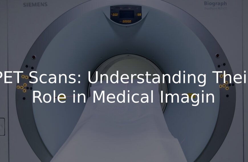
PET scan machines stand out in hospitals, looking like massive white donuts. These medical marvels (Positron Emission Tomography) show doctors how the body works on the inside, not just static images like X-rays do.
The scan uses a special sugar mixture with a tiny radioactive tag that travels through the blood, helping doctors spot potential problems. The process takes about two hours total, but the actual scanning only lasts 30 minutes.
Before a PET scan, patients need to:
- Fast for 4-6 hours (water allowed)
- Get a quick injection
- Rest for an hour
- Lie still during scanning
The scanning room stays quite cool, so warm socks might help. Drinking plenty of water after the scan helps clear the tracer from the body. Most medical centres provide blankets and make the experience as comfortable as possible. The procedure doesn’t hurt – it’s just like getting a very detailed internal photograph while resting.
Why Use PET Scans in Cancer?
PET scans make cancer cells glow like bright spots on a map. The scan uses a special sugar drink mixed with a small amount of radioactive tracer, and cancer cells absorb more of this mixture than normal cells do.
During a PET scan (Positron Emission Tomography), patients lie still on a table that moves through a large ring-shaped scanner. The process takes about 30 minutes, and while it’s painless, the machine makes whirring noises as it works.
These scans help doctors(1):
- Find where cancer is located
- Check if cancer has spread
- Monitor treatment progress
- Look for signs of cancer return
Patients need to fast for 6 hours before the scan, and blood sugar levels must be under 10 mmol/L for accurate results. Most scan rooms stay at 20°C to keep the equipment working properly.
Drinking 2-3 glasses of water after the scan helps clear the tracer from the body faster. Loose, warm clothing makes the experience more comfortable.
Common Cancers That Use PET Scans
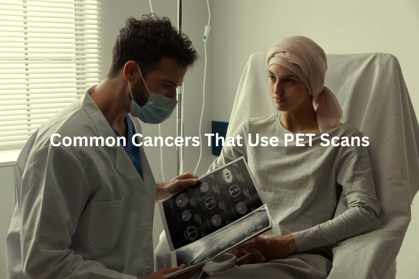
PET scans illuminate the human body like a glowing map, showing details that other imaging tests might miss. These powerful machines detect cancer cells through their unusual appetite for sugar.
During a PET scan, patients receive an injection of fluorodeoxyglucose (a special type of sugar), which cancer cells absorb more quickly than normal cells. After about 60 minutes of waiting, the scanning begins in a large, ring-shaped machine.
Key benefits of PET scans:
- Shows active cancer cells, not just physical structures
- Works for multiple cancer types
- Monitors treatment effectiveness
- Detects cancer spread to other body parts
The entire process takes between 2-3 hours, including preparation and scanning time. The machine captures detailed images as radioactive sugar moves through the body, creating a comprehensive view of cellular activity. Nuclear medicine specialists then analyse these images to identify areas of concern. PET scans serve as essential tools in modern cancer detection and treatment monitoring.
Safety of PET Scans
Sources: ANSTO.
Radiation during PET scans often makes patients nervous in hospitals across Australia. The word itself sounds frightening, but understanding the facts helps put minds at ease.
The human body encounters radiation daily, much like sunshine streaming through windows. A PET scan delivers approximately 7 millisieverts of radiation – equal to about 2 years of natural background radiation from everyday life(2).
Common sources of background radiation include:
• Ground soil
• Air particles
• Certain foods (bananas contain small amounts)
• Cosmic rays from space
Medical centres in Sydney and Melbourne use PET scans as vital diagnostic tools, detecting health issues in their early stages. The benefits typically outweigh the minimal risks for most patients.
Some precautions exist – pregnant women should avoid these scans, and patients need to inform their doctors about any previous reactions to contrast materials. The procedure remains a standard diagnostic tool in Australian healthcare, helping medical teams make informed decisions about treatment paths.
Medicare Rebates and Access in Australia
PET scans in Australia cost patients over $1,000 without proper coverage, making Medicare benefits critical for those who need this cancer-detecting procedure.
Medicare provides benefits for specific PET scan situations:
- Detecting and monitoring lung cancer
- Tracking lymphoma treatment progress
- Selected breast cancer cases
- Pre-surgery bowel cancer assessment
The Medicare system (which updates its coverage rules quarterly) helps reduce these costs at approved facilities(3). Public hospitals often provide more affordable rates compared to private clinics, with some locations offering flexible payment arrangements.
For Medicare-eligible scans, patients might pay nothing out-of-pocket at bulk-billing facilities. The rebate amount varies based on the type of cancer and scanning location (ranging from 400to400 to 400to900 per scan).
Patients should contact Medicare before scheduling a PET scan to check current coverage rules and find nearby bulk-billing providers. Early detection through timely scanning remains crucial for better health outcomes.
Practical Advice for Patients

Patients often show signs of worry in hospital waiting rooms, especially before PET scans. These medical tests don’t need to cause stress with proper preparation.
Key preparation steps:
- No food for 6 hours (water is allowed and encouraged)
- Skip exercise the day before
- Wear loose-fitting clothes without metal zips or buttons
The scan process starts with a small radioactive injection (completely safe at medical doses), followed by about an hour of rest. The actual scanning takes 30-40 minutes, with patients lying still on an open-sided machine.
Unlike MRI machines that look like tubes, PET scanners have open sides that help reduce feelings of being closed in. Many imaging centres offer music during scans, which can help track time and stay relaxed.
Medical staff recommend bringing a calming playlist lasting about 40 minutes – just right for timing the procedure without watching the clock.
FAQ
What is a CT scan and how does it differ from a PET scan?
A CT (Computed Tomography) scan uses X-rays to create detailed images of the body’s internal structures, like organs and bones. In contrast, a PET (Positron Emission Tomography) scan uses a small amount of radioactive tracer to detect changes in the body’s cells and tissues. PET scans can provide information about the function and metabolism of certain areas, which can be helpful in diagnosing and monitoring cancer.
What is FDG PET and how does it work?
FDG PET, or Fluorodeoxyglucose Positron Emission Tomography, is a type of PET scan that uses a radioactive glucose-based tracer to detect areas of high metabolic activity, which can indicate the presence of cancer cells. The tracer is injected into the patient’s vein, and the PET scanner then detects the radioactive emissions from the tracer as it is taken up by the body’s cells.
What is PSMA PET and how is it used for prostate cancer?
PSMA PET, or Prostate-Specific Membrane Antigen Positron Emission Tomography, is a type of PET scan that uses a radioactive tracer that binds to a protein found on the surface of prostate cancer cells. This allows the PET scanner to detect the location and extent of prostate cancer, which can be helpful in staging the disease and guiding treatment decisions.
What is nuclear medicine, and how does it differ from other imaging tests?
Nuclear medicine is a specialised field of medicine that uses small amounts of radioactive materials, called radiopharmaceuticals or radiotracers, to diagnose and treat various medical conditions, including cancer. Unlike other imaging tests like X-rays or CT scans, nuclear medicine imaging techniques like PET and SPECT (Single-Photon Emission Computed Tomography) can provide information about the function and metabolism of the body’s cells and tissues.
What is a C1 trial, and how does it relate to PET scans?
A C1 trial is a type of clinical trial that evaluates the safety and efficacy of a new cancer treatment or diagnostic tool, such as a PET scan. These trials are typically the first step in testing a new treatment or imaging technique in human patients, and they are designed to gather important data on the potential benefits and side effects of the intervention.
How can PET scans be used to assess response to cancer treatment?
PET scans can be a valuable tool for monitoring a patient’s response to cancer treatment. By tracking changes in the uptake of the radioactive tracer, PET scans can provide information about the metabolic activity of cancer cells, which can indicate whether a particular treatment is effectively shrinking or eliminating the tumour. This can help guide treatment decisions and ensure that patients receive the most effective care.
What are the potential side effects of PET scans?
PET scans generally have a low risk of side effects, as they use a small amount of radioactive tracer that is quickly eliminated from the body. However, some patients may experience mild side effects, such as a brief period of nausea or discomfort at the injection site. It’s important for patients to discuss any concerns or potential side effects with their healthcare providers before undergoing a PET scan.
How can PET scans be used to detect cancer in high-risk individuals?
PET scans can be a valuable tool for early detection of cancer in individuals who are considered to be at high risk, such as those with a family history of certain types of cancer or who have been exposed to known carcinogens. By using the radioactive tracer to identify areas of increased metabolic activity, PET scans can sometimes detect the presence of cancer before it becomes visible on other imaging tests or presents with symptoms.
What is the long-tail effect of PET scans, and how does it impact cancer diagnosis and treatment?
The long-tail effect refers to the ability of PET scans to detect small, subtle changes in the body’s cells and tissues that may be indicative of early-stage cancer or other medical conditions. This can lead to earlier diagnosis and intervention, which can ultimately improve patient outcomes and survival rates. By catching cancer in its earliest stages, PET scans can enable more effective and less invasive treatment options.
How can FDG uptake on PET scans be used to guide cancer treatment decisions?
The level of FDG (Fluorodeoxyglucose) uptake on a PET scan can provide important information about the metabolic activity and aggressiveness of a cancer. Higher levels of FDG uptake are often associated with more rapidly growing and aggressive tumours, which can help guide treatment decisions. For example, patients with high FDG uptake may be more likely to benefit from more intensive or targeted cancer therapies.
What is the role of PET scans in the management of lung cancer?
PET scans can play a crucial role in the management of lung cancer, from initial diagnosis to monitoring treatment response. PET scans can help differentiate between benign and malignant lung nodules, stage the cancer, and detect the presence of metastatic disease. Additionally, PET scans can be used to assess a patient’s response to treatment, which can help guide decisions about ongoing care and the need for further interventions.
How do PET scans compare to other imaging tests, such as CT scans, in the evaluation of cancer patients?
PET scans and CT scans are often used together in the evaluation of cancer patients, as they provide complementary information. While CT scans excel at creating detailed anatomical images of the body, PET scans can provide valuable information about the functional and metabolic changes associated with cancer. By combining the strengths of these two imaging techniques, healthcare providers can gain a more comprehensive understanding of a patient’s cancer and make more informed treatment decisions.
What is the role of PET scans in the management of Hodgkin lymphoma (HL) patients?
PET scans play a crucial role in the management of Hodgkin lymphoma (HL) patients. They can be used to stage the disease, assess response to treatment, and detect any residual or recurrent disease. PET scans are particularly valuable in evaluating the efficacy of treatment, as they can identify areas of persistent metabolic activity that may indicate the presence of active cancer cells, even when other imaging tests appear normal.
How can PET scans be used to assess the response to cancer treatment in patients with multiple myeloma?
PET scans can be a useful tool for assessing the response to cancer treatment in patients with multiple myeloma. By tracking the uptake of the radioactive tracer in areas of active disease, PET scans can provide information about the metabolic activity of the myeloma cells, which can indicate whether a particular treatment is effectively reducing the burden of disease. This can help guide treatment decisions and ensure that patients receive the most appropriate and effective care.
What is the Deauville scale, and how is it used to interpret PET scans in cancer patients?
The Deauville scale is a standardised system used to interpret the results of PET scans in cancer patients. It assigns a score from 1 to 5 based on the level of radiotracer uptake in the target lesion, with higher scores indicating a poorer prognosis. Healthcare providers can use the Deauville scale to assess a patient’s response to cancer treatment and make informed decisions about the need for further interventions.
How can PET scans be used to detect and monitor thyroid cancer in adult patients?
PET scans can be a valuable tool for detecting and monitoring thyroid cancer in adult patients. By using a radioactive tracer that is taken up by thyroid cells, PET scans can identify areas of increased metabolic activity that may be indicative of thyroid cancer. PET scans can also be used to monitor the response to treatment and detect any residual or recurrent disease, which can help guide ongoing management decisions.
What is the role of PET scans in the assessment of bone marrow involvement in cancer patients?
PET scans can be useful in assessing the involvement of bone marrow in cancer patients. By detecting areas of increased metabolic activity in the bone marrow, PET scans can help identify the presence of cancer cells that have spread to this location. This information can be critical in staging the disease and determining the appropriate treatment approach, as bone marrow involvement can have significant implications for a patient’s prognosis and management.
How can PET scans be used to detect and monitor lymph node involvement in cancer patients?
PET scans can be a valuable tool for detecting and monitoring lymph node involvement in cancer patients. By using a radioactive tracer that is taken up by metabolically active cells, PET scans can identify areas of increased activity in the lymph nodes, which may indicate the presence of cancer cells. This information can help healthcare providers stage the cancer, assess the extent of disease, and monitor the response to treatment over time.
What is the role of PET scans in the early detection of cancer?
PET scans can play an important role in the early detection of cancer, particularly in high-risk individuals or those with non-specific symptoms. By identifying areas of increased metabolic activity, PET scans can sometimes detect the presence of cancer before it becomes visible on other imaging tests or presents with more obvious symptoms. This can enable earlier diagnosis and intervention, which can ultimately improve patient outcomes and survival rates.
Conclusion
PET scans are really important in cancer care. They help doctors find cancer, see how far it has spread, check if treatments are working, and look for if cancer might come back. Understanding PET scans can make patients feel more ready and less worried about their cancer journey. So, when you hear about a PET scan, remember it’s more than just a test; it’s a helpful tool on the path to better health.
References
- https://gicancer.org.au/news/faq/pet-scan-used/
- https://www.mja.com.au/journal/2005/182/4/clinical-experience-first-combined-positron-emission-tomographycomputed
- https://www.health.gov.au/topics/medicare