Learn about nuclear medicine and how it helps doctors see inside our bodies safely.
Nuclear medicine is a special tool that helps doctors see inside the body. By using small amounts of radioactive materials, it enables them to detect potential health issues early on. For example, a VQ scan can help identify blood clots in the lungs, acting like a treasure map to pinpoint hidden problems.
This method gives doctors a clearer picture of what’s going on, guiding them towards the right treatment. If you’re keen to learn more about how nuclear medicine works and how it can benefit your health, keep reading to discover how it can make a difference in diagnosis and care.
Key Takeaway
- Nuclear medicine uses small amounts of radioactive tracers to see inside the body.
- It helps with scans like thyroid scans and bone scans.
- Safety is important, and doctors know how to keep patients safe during these tests.
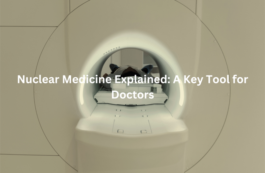
Nuclear Medicine Explained
Nuclear medicine is an exciting way to look inside the body using small amounts of radioactive materials. These materials are tracked by a gamma camera, which shows where they travel within the body.
This helps doctors identify potential health issues, like lung disease or cancer, much earlier than traditional methods might. It’s like a treasure hunt, with the radioactive material acting as a guide to uncover hidden problems.
Key points about nuclear medicine:
- Gamma camera: Tracks radioactive materials inside the body.
- Targeted diagnosis: Helps detect issues like lung disease, cancer, and other conditions.
- Early detection: Enables doctors to find problems before they become serious.
- Safe procedure: The amount of radioactive material used is minimal, making it safe for patients.
This cutting-edge method allows doctors to spot health issues early, giving patients better chances for effective treatment and recovery. Keep reading to explore how it works and the benefits it offers. (1)
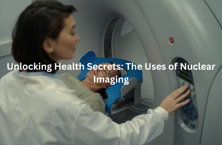
Uses of Nuclear Imaging
Nuclear imaging is a powerful tool that helps doctors get a detailed look at different parts of the body. Using advanced techniques, doctors can track blood flow, spot hot spots, and even see how organs are functioning in real time. This technology is particularly useful for diagnosing serious conditions like lung cancer and blood clots, helping doctors make more informed decisions.
Key benefits of nuclear imaging:
- Tracks blood flow: Helps doctors assess how well blood is circulating throughout the body.
- Detects hot spots: Identifies areas with unusual activity, which could signal issues like cancer.
- Monitors organ function: Provides real-time insights into how organs are performing.
- Lung scans: Shows how air and blood move through the lungs, aiding in the diagnosis of lung conditions.
Nuclear imaging is an invaluable diagnostic tool that gives doctors a clear picture of what’s happening inside the body, ensuring early detection and effective treatment. Keep reading to discover how this technology works and its benefits.
Thyroid Scans
A thyroid scan looks at a small but important gland in the neck that helps control energy in the body. If the thyroid isn’t working properly, the scan can help doctors figure out what’s wrong. This scan is essential because it can detect thyroid problems early, making treatment more effective.
Key points about a thyroid scan:
- Targets the thyroid gland: This small gland plays a key role in regulating energy and metabolism.
- Detects thyroid issues: Helps spot conditions like hyperthyroidism, hypothyroidism, or thyroid cancer.
- Early detection: Identifying thyroid problems early allows for better treatment and management.
- Non-invasive: The scan is quick, simple, and safe, offering a hassle-free diagnostic option.
A thyroid scan gives doctors important information, ensuring thyroid conditions are caught early and managed effectively. Keep reading to find out more about how this scan works and the benefits it offers.
Bone Scans
Bone scans are a useful way for doctors to check for problems with bones. They can detect issues like pain, infections, or even cancer. These scans also show how well blood flows to the bones, which is important for healing and overall bone health. Think of it like checking if a road is clear for cars, ensuring everything is running smoothly.
Key points about bone scans:
- Detects bone problems: Identifies pain, infections, and cancer.
- Checks blood flow: Assesses how well blood circulates to the bones.
- Early diagnosis: Helps catch issues before they become more serious.
Bone scans provide valuable information, helping doctors address bone health concerns quickly.
Cardiac Nuclear Imaging
Cardiac nuclear imaging focuses on the heart, giving doctors a detailed view of how blood flows to the heart muscles. It helps assess the heart’s strength and whether it’s functioning properly. This is particularly important for people experiencing chest pain or those with heart disease, as it provides valuable information for treatment decisions.
Key points about cardiac nuclear imaging:
- Shows blood flow: Allows doctors to see how well blood reaches the heart muscles.
- Assesses heart strength: Indicates if the heart is strong or needs support.
- Crucial for heart disease: Key for diagnosing and managing chest pain or heart conditions.
This imaging technique helps doctors monitor heart health, leading to better treatment and care for those at risk.
Radioactive Tracers
Radioactive tracers are tiny bits of material used by doctors to identify problems inside the body. They’re safe because only very small amounts are used, so there’s no risk to the patient. During a scan, these tracers highlight areas where something might be wrong, giving doctors a clear picture of what’s happening inside.
Key points about radioactive tracers:
- Small amounts: Used in tiny, safe quantities.
- Guides diagnosis: Helps doctors identify areas of concern.
- Non-invasive: Provides valuable insights without the need for surgery or discomfort.
These tracers play a vital role in medical scans, helping doctors gain essential information about the body’s health.
Risks of Nuclear Medicine
Credits: Dr. Paulien Moyaert
Like any medical test, there are some risks, but they are generally minimal. Some people might feel a little off after a scan, though this is usually only temporary. The amount of radiation used in these scans is very low, so it poses very little risk. Doctors carefully consider the potential risks before recommending any test, making sure it’s the safest option for the patient.
Key points to remember:
- Minimal radiation: Only a small, safe amount is used.
- Temporary side effects: Any discomfort usually passes quickly.
- Doctor’s judgement: Doctors assess the risks to ensure safety.
Doctors take all precautions to ensure the test is safe, providing peace of mind for those undergoing the procedure.
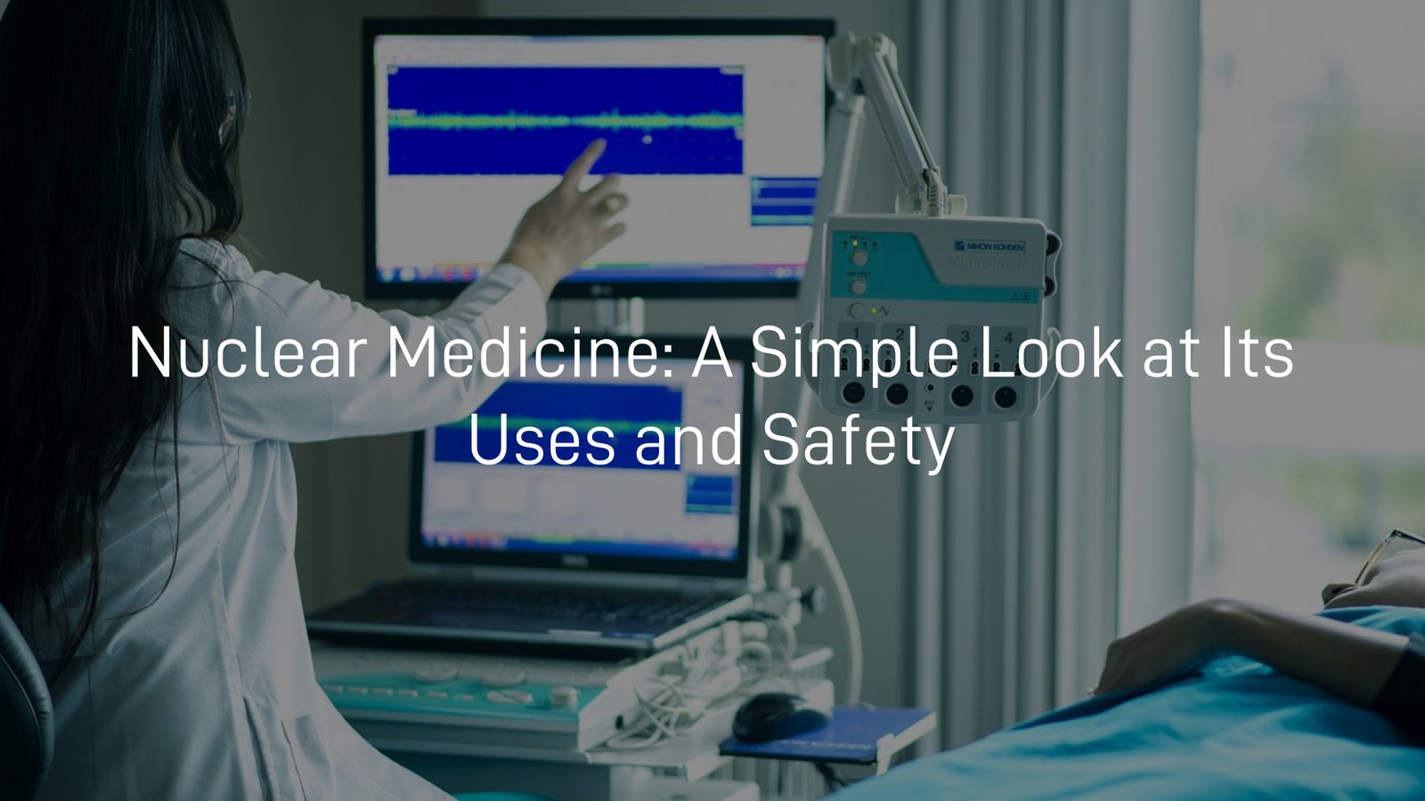
Nuclear Medicine Safety
Safety is a top priority in nuclear medicine, with doctors and nurses trained to ensure patients are well looked after. Before any scan, they carefully check all the details to ensure everything is in order. It’s important to tell the doctor if you’re pregnant or breastfeeding, as this can impact the safety of the test. The healthcare team will make sure the procedure is safe for both the patient and their baby.
Key points to remember:
- Trained professionals: Doctors and nurses are skilled in ensuring safety.
- Check before scanning: All details are reviewed before a test.
- Inform your doctor: Let them know if you’re pregnant or breastfeeding.
By prioritising safety, healthcare teams make sure every procedure is as safe and effective as possible. (2)
SPECT Imaging
SPECT imaging is a specialised scan that shows how blood flows through the body. It’s especially helpful for looking at areas like the heart and brain, allowing doctors to identify problems that might not show up on other types of scans. By highlighting blood flow, SPECT imaging helps uncover underlying issues that could be causing symptoms or health concerns.
Key points about SPECT imaging:
- Shows blood flow: Focuses on how blood circulates through the body.
- Targets heart and brain: Helps doctors examine areas like the heart and brain.
- Detects hidden issues: Identifies problems that may not appear on other scans.
SPECT imaging offers valuable insights, helping doctors diagnose and treat conditions more effectively.
PET and Nuclear Medicine
PET scans are another type of nuclear medicine that assist doctors in detecting diseases like cancer. Unlike other scans, PET uses a unique form of radioactive material to show how the body is functioning in real time. This gives doctors a clearer and more detailed picture of what’s going on inside. (3)
Key points about PET scans:
- Detects diseases: Helps identify conditions such as cancer.
- Real-time function: Displays how the body is working at that moment.
- Unique radioactive material: Uses a different kind of material compared to other scans.
While tests like PET scans might seem a bit daunting, nuclear medicine is a safe way to see what’s happening inside, acting like a flashlight that illuminates the hidden corners of our health.
Conclusion
In conclusion, nuclear medicine plays a key role in helping doctors see inside our bodies, offering valuable insights from thyroid scans to cardiac imaging. These tests are safe, with patient care always the top priority.
By understanding how nuclear medicine works, patients can feel more comfortable and confident when undergoing these procedures, knowing they’re in good hands. It’s an essential tool for diagnosing and managing health conditions effectively.
FAQ
What is a VQ scan, and how is it used?
A VQ scan is a test that helps doctors look at blood flow and air movement in the lungs. It uses a small amount of radioactive material and a gamma camera to take pictures of the lungs. This scan can help diagnose lung conditions such as lung disease or blood clots in the lungs. It’s also useful for detecting hot spots or areas where blood supply is reduced.
What is the difference between a CT scan and a VQ scan?
While both a CT scan and a VQ scan help doctors view the lungs, they do so in different ways. A CT scan uses X-rays and takes detailed CT images of the lungs. A VQ scan, on the other hand, looks at blood flow and lung function using radioactive material. CT scans can help detect conditions like lung cancer, while a VQ scan is typically used to find blood clots or lung disease.
How does a CT scan help with lung imaging?
A CT scan is often used to create CT images of the lungs, allowing doctors to see the detailed structure of the lung tissue. It can be used to check for conditions such as lung cancer, chest pain, or lung disease. The scan takes multiple images and puts them together to give a clearer picture. CT scans are often combined with other tests, like a VQ scan, to provide more information about blood flow and lung function.
References
- https://www.prpimaging.com.au/nuclear-medicine-uses-benefits-applications/
- https://pch.health.wa.gov.au/Our-services/Medical-Imaging/Nuclear-Medicine
- https://aanms.org.au/what-is-nuclear-medicine/