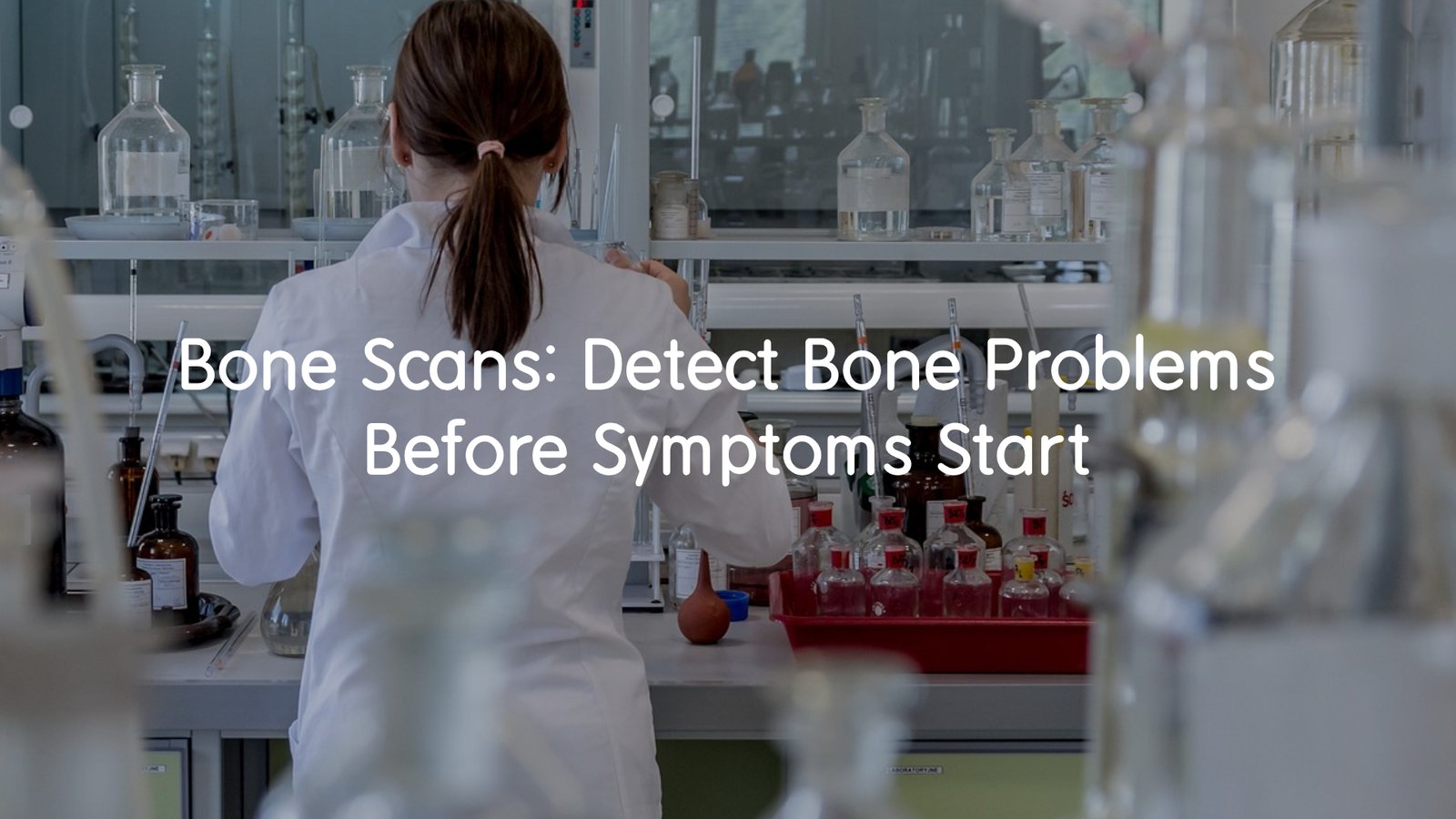
A bone scan can detect fractures, infections, and cancer early—before symptoms appear. Learn how it works and what to expect.
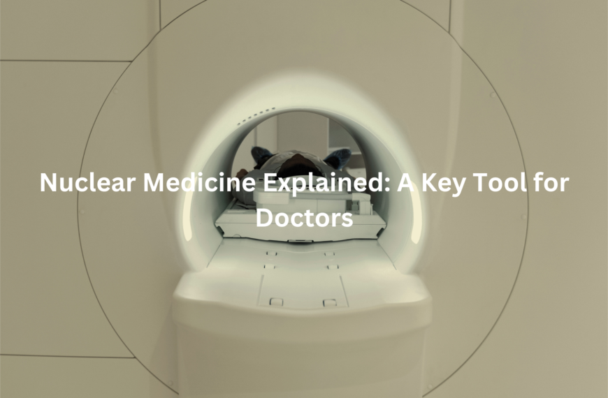
A bone scan is a nuclear medicine imaging test used to detect bone abnormalities, often before symptoms develop. It helps diagnose fractures, infections, cancer, and other conditions by tracking how a small amount of radioactive material moves through the bones. This scan is a valuable tool for early diagnosis and treatment planning, ensuring better health outcomes.
Key Takeaways
- Early detection of fractures, cancer, and bone infections before symptoms arise.
- Safe and effective with minimal radiation exposure.
- Guides treatment by identifying changes in bone activity and metabolism.
Purpose of a Bone Scan

Someone who had been dealing with nagging hip pain for weeks. After trying rest and painkillers with no success, the doctor recommended a bone scan to investigate further. It turns out that bone scans are powerful tools that help uncover hidden issues in bones.
A bone scan works like a deep dive into your skeletal health, revealing problems that might not show up on regular X-rays. This includes:
- Detecting bone cancer and its spread (metastases).
- Spotting fractures and injuries that aren’t visible on standard imaging.
- Diagnosing infections like osteomyelitis.
- Monitoring conditions like osteoporosis and metabolic bone disorders.
- Assessing blood flow and bone metabolism for a clearer diagnosis.
It’s like giving your bones a thorough check-up, helping doctors catch issues early and start treatment before things get worse. If persistent pain doesn’t ease up, a bone scan might be worth considering!
How Bone Scan Works
Here’s how it works: first, you get a small injection of a radioactive tracer into a vein, usually in your arm. This tracer travels through your bloodstream and eventually settles in your bones, where it’ll give doctors the information they need.
After the injection, they might take a few early images to assess blood flow to your bones. These initial images help check how well your blood is circulating through your bones, which can offer valuable clues about your bone health. Then, after about 2 to 4 hours, you’ll return for a second round of imaging.
- Early imaging: Helps assess blood flow to your bones.
- Delayed imaging (2-4 hours later): Shows how the tracer is absorbed by your bones.
- Gamma camera: A special camera that captures detailed images of your bones, helping doctors get a clear view of any potential issues.
This method gives a thorough picture of your bone health, revealing problems that may not be visible on regular X-rays. (1)
Understanding Scan Results
Once the results are in, the images are carefully examined. If certain areas show increased tracer uptake, it could point to issues like cancer, fractures, or infections. The more active these areas are, the more likely there’s something happening.
On the flip side, decreased tracer uptake might suggest conditions like avascular necrosis (when bone tissue dies due to lack of blood flow) or bone loss, often associated with diseases like osteoporosis.
- Increased uptake: Could indicate cancer, fractures, or infections.
- Decreased uptake: May point to avascular necrosis or bone loss.
- Radiologist review: A trained radiologist will review the images to make the final diagnosis.
It’s a detailed process, but one that offers valuable insights into your bone health. By catching problems early, doctors can recommend the right treatment, helping to prevent further damage.
Safety and Risks
I get it, the thought of radiation can be a bit unsettling, but here’s the good news: bone scans use minimal radiation and are considered safe for most people. The amount is quite small, and the benefits usually outweigh the risks. Side effects are rare, but it’s a good idea to drink plenty of water afterward to help flush the radioactive tracer out of your system.
- Minimal radiation: The exposure is low, making it generally safe for most individuals.
- Stay hydrated: Drinking plenty of water will help eliminate the tracer from your body.
- Consult your doctor: If you’re pregnant or breastfeeding, make sure to speak with your healthcare provider about any potential radiation risks.
So, while radiation may sound concerning, the amount used in bone scans is very small, and it’s all about keeping you safe. If you’re worried, don’t hesitate to chat with your doctor for peace of mind.
Preparing for the Scan & After
Credits: Mayfair Diagnostics
Before your bone scan, there are a few things to keep in mind. Wear comfortable clothing, as you’ll be lying down during the procedure. You’ll also be asked to sign a consent form to acknowledge that you understand the process. If you’re on any medications or have any medical conditions, make sure to tell your doctor so they can provide the right advice for your situation.
- Wear comfy clothes: Comfort is key, as you’ll be lying down for a while.
- Sign the consent form: This lets you confirm that you’re aware of the process.
- Inform your doctor: Let them know if you’re taking any medications or have any medical conditions.
Once the scan is over, it’s easy. Drink plenty of water to help your body clear out the radioactive tracer. You should be able to go back to your usual routine, unless your doctor advises otherwise. Simple steps, but they help make sure everything goes smoothly.
Alternative Imaging Tests
Depending on your situation, your doctor may recommend other imaging tests to get a clearer picture of your bone health. These tests are used for specific conditions, and your doctor will choose the most appropriate one based on your needs.
- Bone density scan (DXA scan): This test measures bone mineral density and is typically used to assess conditions like osteoporosis or other bone thinning issues.
- CT scan / PET scan: These provide detailed images and are particularly helpful for detecting tumours or infections.
- MRI scan: MRIs use magnetic fields rather than radiation and are ideal for looking at soft tissue around the bones.
Each test has its advantages, and your doctor will determine which one is best suited for you. Whether it’s a bone scan or another type of imaging, the aim is always to provide the best insights into your bone health. (2)
Expert Tips & Advice
If you’re over 50 or have risk factors for osteoporosis, a bone density test could be worth considering. It’s a straightforward way to check if your bones are thinning and assess your risk of fractures. Having a chat with your doctor about it can help you stay on top of your bone health.
When it comes to minimising radiation exposure, bone scans are generally safe, but it’s always a good idea to let your healthcare provider know about any recent imaging tests you’ve had. This helps avoid unnecessary duplication and ensures you’re only exposed to what’s needed.
Before you go in for a bone scan, here are a few questions you might want to ask your healthcare provider:
- Why is this test necessary for me?
- Are there alternative tests I could consider?
- What should I expect before, during, and after the procedure?
Being proactive and informed can help you feel more at ease and ensure you get the best care possible.
Conclusion
In conclusion, bone scans are essential for detecting and monitoring a range of bone conditions. By understanding how the procedure works and what to expect, you can approach it with confidence.
This knowledge allows you to take an active role in your healthcare, ensuring that any issues are detected early and treated appropriately. Being informed helps you make the best decisions for your bone health and overall well-being.
FAQ
What is a bone scan and how does it work?
A bone scan is a type of medical imaging that uses a radioactive tracer to help detect problems in the bones. The tracer is injected into your vein, travels through your bloodstream, and settles in the bones. A gamma camera takes pictures of the bone health to identify any issues like fractures, infections, or diseases. The scan helps check your bone condition before symptoms start to appear.
How is a bone density scan different from a bone scan?
A bone density scan or bone density test measures the density of your bones to detect conditions like bone loss or osteoporosis. It uses a technique like dual energy X-ray absorptiometry (DXA scan) to provide detailed images. A bone scan, on the other hand, looks for infections, fractures, or other bone problems using a radioactive tracer.
Are there any side effects from a bone scan?
Bone scans involve a small amount of radiation exposure, but the levels are very low, making the procedure safe for most people. Side effects are rare. It’s always a good idea to drink plenty of water after the scan to help flush out the radioactive material from your body. If you’re pregnant or breastfeeding, consult with your healthcare provider before the scan.
Who might need a bone density scan?
If you’re over 50 or have risk factors for conditions like osteoporosis or bone loss, you may want to talk to your doctor about having a bone density scan to check for any issues. Health conditions like a history of fractures or certain medications can increase your risk factors for low bone density.
How long does a bone scan take?
A bone scan typically takes a short time, around 2-4 hours. After the injection of the radioactive tracer, you may need to wait for a few hours before the gamma camera can take the images of your bones. The process is non-invasive, and you’ll usually be able to resume normal activities afterward unless advised otherwise.
References
- https://www.healthdirect.gov.au/bone-density-scan
- https://www.insideradiology.com.au/nuclear-medicine-bone-scan/