Understand how PET and nuclear medicine scans work, their benefits, and why they’re essential for early and accurate diagnosis.
PET (Positron Emission Tomography) and nuclear medicine scans are advanced imaging techniques that help detect diseases like cancer, heart conditions, and brain disorders. Unlike standard scans, they show how organs function in real-time, allowing for earlier detection and more precise treatment planning.
Key Takeaways
- PET and nuclear medicine scans provide detailed insights into organ function, improving early and accurate diagnosis.
- Commonly used for cancer staging, heart disease assessment, and neurological conditions like Alzheimer’s.
- Safe, widely available across Australia, and essential in modern medical imaging.
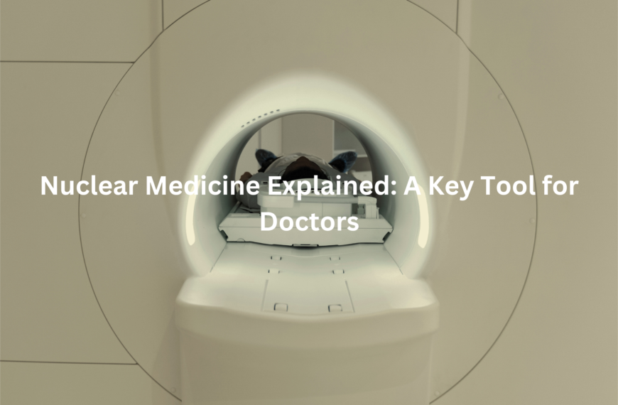
PET Scans and Nuclear Medicine: What You Need to Know

PET (Positron Emission Tomography) scans and nuclear medicine are advanced imaging techniques that use small amounts of radioactive substances to diagnose and treat health conditions.
Both methods involve the use of radioactive material, known as a “tracer,” to observe how well organs, tissues, or bones are functioning. This helps doctors detect problems early, often before symptoms appear.
PET scans, a specific type of nuclear medicine, focus on revealing activity within cells, providing insight into how active different tissues are. (1)
Key points include:
- Radioactive tracers: These substances help track the function of organs and tissues by emitting small amounts of radiation.
- PET scan function: PET scans highlight cell activity, making them particularly useful for detecting conditions like cancer, heart disease, and brain disorders.
- Early detection: By visualising cellular activity, these techniques enable earlier diagnosis and more effective treatment planning.
How Do They Work
In nuclear medicine, a radiologist (a specialist in medical imaging) injects a small amount of radioactive material into the body to track how it moves and behaves. Unlike X-rays or CT scans, which send radiation through the body, nuclear medicine uses this material to create digital images observed by special cameras. The radiologist typically analyses these images within hours after the scan.
PET scans enhance this process by detecting tiny particles (photons) released from the radioactive substance in the organ or tissue. This substance, such as fluorodeoxyglucose (FDG), mimics substances the body naturally uses. The more of this substance that accumulates, the brighter the scan appears, indicating higher activity in that area. (2)
Key points include:
- PET-CT fusion: Most PET scanners are combined with CT scanners, merging structural images with functional information for more accurate diagnoses.
- Fluorodeoxyglucose (FDG): Commonly used for brain scans to track activity levels.
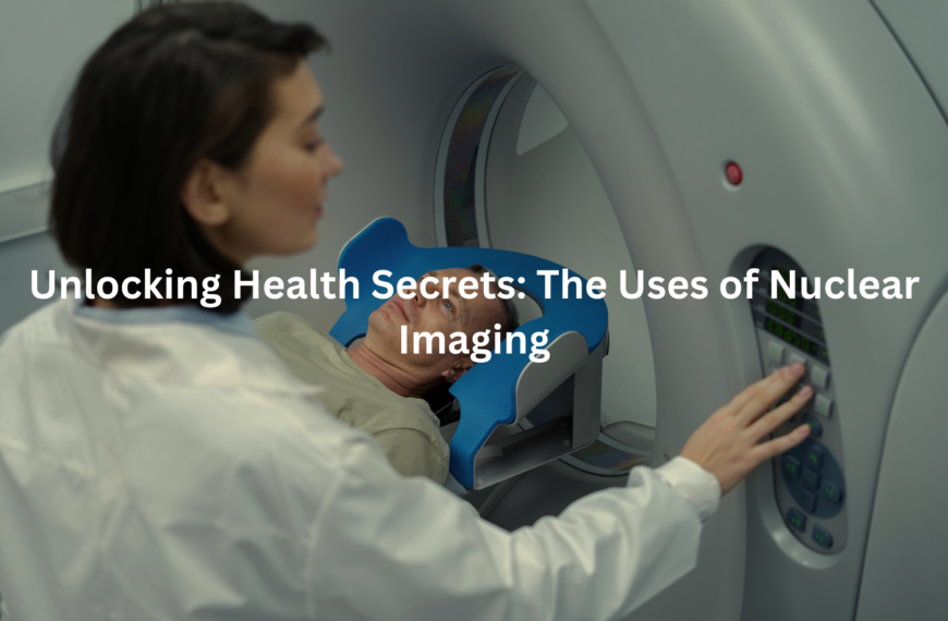
What Problems Can They Find
PET scans and nuclear medicine are essential for diagnosing and managing a broad range of health conditions, including cancers, heart issues, and brain disorders. These advanced imaging techniques enable early detection, allowing for more effective treatment options.
PET scans and nuclear medicine are particularly useful for detecting:
- Brain blood flow issues: Identifying irregularities in brain blood flow, which could point to strokes or other neurological concerns.
- Heart disease: Detecting heart conditions in the early stages before they become more serious.
- Epilepsy: Helping pinpoint the source of seizures in the brain.
- Dementia: Including conditions like Alzheimer’s disease.
- Cancers and organ health: Offering valuable insights into cancer progression and the overall health of organs.
Beyond diagnosis, nuclear medicine is also used in treating conditions such as:
- Cancer spread (e.g., prostate cancer spreading to bones)
- Liver and thyroid cancers
- Neuroendocrine tumours
- Overactive thyroid
Why Are PET Scans Useful
Credits: HealthTree University for Multiple Myeloma
PET scans are especially useful because they can often pick up problems earlier than other types of imaging. This is due to their ability to detect the chemical activity happening in tissues, offering a clearer picture of the body’s function. As a result, doctors can spot potential issues before they’re visible on other scans.
The key benefits of PET scans include:
- Early cancer detection: PET scans can identify and stage many cancers, sometimes even before they’re visible with other imaging methods.
- Heart disease insights: They can highlight issues with blood flow, offering early warning signs of heart disease.
- Neurological disorder diagnosis: PET scans are essential for diagnosing conditions affecting the brain and nerves, such as Alzheimer’s, epilepsy, and other neurological disorders.
By detecting problems early, PET scans allow for more accurate diagnosis and treatment, helping doctors create more effective plans to improve patient outcomes.
Who Oversees Nuclear Medicine Services
In Australia, PET and nuclear medicine scans must be supervised by a qualified specialist to ensure both patient safety and the accuracy of the results. These procedures are governed by strict rules and guidelines, which are designed to maintain the highest standards of care and ensure that scans are performed correctly.
Key guidelines for PET and nuclear medicine scans:
- Scans must be managed by a qualified specialist to guarantee safety and accuracy.
- Stringent regulations are in place to protect patients and uphold the integrity of scan results.
- Specialists are responsible for interpreting the images and guiding treatment decisions.
These regulations help ensure that patients receive high-quality care while assisting doctors in making informed decisions. With expert supervision, these advanced imaging techniques continue to be essential for diagnosing and managing various health conditions.
Training in Nuclear Medicine (RANZCR)
In Australia and New Zealand, the Royal Australian and New Zealand College of Radiologists (RANZCR) plays a central role in training radiologists, ensuring they meet the high standards required in their field.
Aspiring nuclear medicine specialists, including those focusing on PET scanning, must undergo rigorous training to master the skills necessary for accurate diagnoses and patient care. This training is no small feat, requiring a minimum of 24 months of hands-on experience to build expertise.
Key points about RANZCR’s training for nuclear medicine specialists:
- A minimum of 24 months of training is required for nuclear medicine specialisation.
- PET scanning specialists must first be fully qualified nuclear medicine specialists.
- The training ensures radiologists gain deep knowledge of both the technical and clinical aspects of nuclear medicine.
This extensive training helps radiologists develop a well-rounded skill set, empowering them to deliver high-quality imaging that aids in precise diagnosis and treatment planning for various medical conditions.
Conclusion
PET scans and nuclear medicine are vital in healthcare, providing insights into organ and tissue function that other imaging can’t. Using radioactive tracers, they help detect issues early, making treatments more effective.
Whether diagnosing cancer, heart disease, or neurological disorders, these techniques offer a crucial tool for informed decisions. With skilled radiologists and continuous advancements in imaging, the future of nuclear medicine is promising, delivering more accurate and precise diagnoses for various health conditions.
FAQ
What is the difference between a PET scan and a CT scan?
A CT scan uses X-rays to take detailed pictures of the body’s organs and structures, while a PET scan involves using a small amount of radioactive material, known as a radioactive tracer, to track how organs and tissues are functioning. PET imaging is useful for detecting diseases like lung cancer, heart disease, and Alzheimer’s disease by showing the activity of cells in real-time.
How is a PET scan used in diagnosing lung cancer?
A lung scan with fdg pet involves injecting a small amount of a substance called 18f fdg pet, which is a type of radioactive glucose. The fdg pet scans help doctors see which parts of the lungs are more active, indicating potential cancer growth or other issues. Nuclear medicine plays a key role in identifying abnormalities early, which is critical for effective treatment planning.
Are there any side effects from PET scans?
PET scans generally have very few side effects because they use small amounts of radioactive material. Most people tolerate the procedure well, but some may feel slight discomfort from the injection of the radioactive tracer. However, the radiation dose used is carefully controlled to minimise risks.
What is a nuclear medicine scan used for?
Nuclear medicine scans, such as mol imaging or psma pet, help diagnose a wide range of conditions, including heart disease, Hodgkin lymphoma, and pulmonary embolism. They work by injecting a radioactive tracer into the body, which is tracked by a special camera to show how organs and tissues are functioning.
Do I need a referral letter for PET services?
Yes, to receive PET services like nuclear medicine imaging, you typically need a referral letter from your doctor. This letter should outline why the imaging tests are necessary for your diagnosis or treatment.
References
- https://www.healthdirect.gov.au/pet-scan
- https://www.prpimaging.com.au/what-is-nuclear-medicine-used-for/