Safe imaging matters—paediatric radiology uses specialised techniques to minimise risks and maximise accuracy for growing bodies.
Paediatric radiology is a key part of healthcare that focuses on imaging techniques to check children’s health. Doctors use scans like X-rays, MRIs, and CT scans to get important details about a child’s body. These images help find problems like broken bones or conditions that affect development.
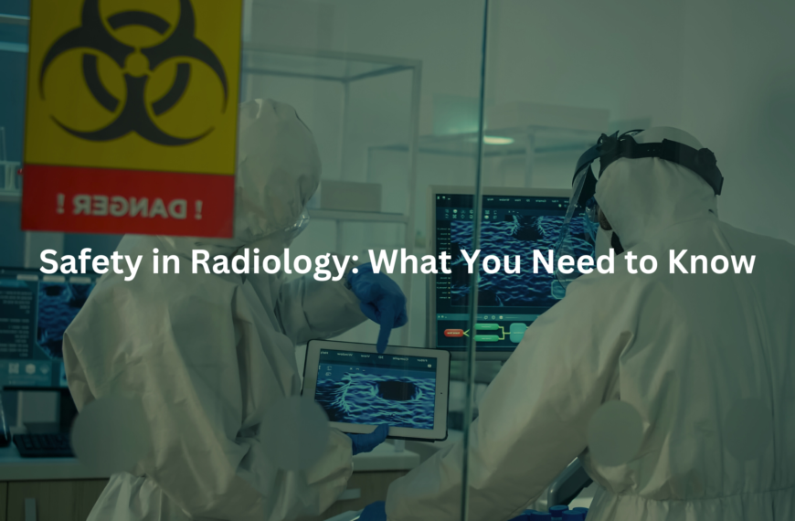
Kids’ bodies differ from adults, which is why their medical needs are also unique. Their growing bones, organs, and tissues require special attention when it comes to imaging. Also, young children are more sensitive to radiation, making safety protocols crucial.
Key Takeaway
- Paediatric radiology uses special methods to keep children safe during imaging.
- There are many types of imaging techniques that work well for kids.
- Understanding how imaging works can help reduce fear and anxiety in children.
Pediatric Radiology
Paediatric radiology is all about imaging designed just for children. Since kids have smaller bodies, the techniques need to be adjusted to get the right images while keeping them safe. Radiologists don’t just change their equipment settings—they also change how they work depending on the child’s age and size.
The biggest difference in paediatric radiology is the focus on radiation safety. Some imaging techniques don’t use radiation at all, but X-rays and CT scans, do. These scans help diagnose fractures, internal bleeding, and more serious conditions like tumours. Because children are more sensitive to radiation, strict safety rules are in place. Australian hospitals follow guidelines to make sure the radiation dose is kept as low as possible while still getting clear, useful images.
Pediatric Imaging Techniques
Paediatric radiologists use different imaging methods to check a child’s health. These include:
- X-rays – Fast and effective, X-rays help find fractures and bone injuries. They are often used in emergency cases.
- MRI (Magnetic Resonance Imaging) – Uses magnetic fields and radio waves to create clear images of soft tissues like the brain, spine, and muscles.
- CT Scans – Combines multiple X-ray images to create a detailed view inside the body. This helps doctors diagnose internal injuries or complex medical conditions.
- Ultrasound – A safe test that doesn’t use radiation. Instead, it uses sound waves to capture images of organs like the heart and liver.
Each of these techniques follows strict safety rules to protect children. X-rays and CT scans use the lowest possible radiation dose, while MRIs and ultrasounds rely on child-friendly methods to keep young patients comfortable.
Ionizing Radiation in Children
Ionising radiation is useful in medical imaging, but it must be used carefully—especially for children. Since their bodies are still growing, they are more sensitive to radiation’s effects. To keep them safe, doctors follow the ALARA principle—which means using the lowest radiation dose possible while still getting clear images.
In Australia, hospitals follow strict rules to limit radiation exposure. For example, paediatric imaging doses can be up to 68% lower than those used for adults. This reduction is especially important for children who need multiple scans over time. Keeping radiation levels low helps protect their health and supports proper growth. (1)
Magnetic Resonance Imaging (MRI) in Pediatrics
MRI scans are an important tool in paediatric radiology. They create detailed images of soft tissues and are often used to check the brain, spinal cord, and joints. But there’s a challenge—MRI machines are loud, and the small space inside can feel scary for young children. Staying still for a long time can also be hard, making the experience stressful.
To help, many hospitals have found ways to make MRIs less intimidating. Some use virtual reality (VR) glasses to keep kids distracted in a fun environment. Others play music or offer child-friendly activities to ease anxiety. The goal is simple—keeping kids as calm and comfortable as possible during the scan.
Computed Tomography (CT) for Children
CT scans are a fast and detailed way to check for problems, especially in emergencies like head injuries or severe stomach pain. They provide clear images, but they also use more radiation than X-rays. Because children are more sensitive to radiation, doctors are extra careful about when and how CT scans are used.
In Australia, radiologists adjust CT settings based on a child’s size and weight. Smaller children need lower doses than older kids or adults. By fine-tuning the settings, doctors make sure they use just enough radiation to get the right images while keeping exposure as low as possible.
Ultrasound in Pediatric Diagnostics
Ultrasound is a great imaging tool for children because it doesn’t use radiation. Instead, it relies on sound waves to create pictures of organs and tissues inside the body. This makes it a safe choice for babies, young kids, and even pregnant women.
Doctors use ultrasound to check organs like the liver, kidneys, and heart. It’s painless, non-invasive, and usually doesn’t require sedation. For newborns, an anterior fontanelle ultrasound can examine the brain through the soft spot on their skull. Since there’s no radiation, it’s a preferred method for diagnosing conditions like appendicitis and monitoring how organs develop over time.
Fluoroscopy in Pediatric Care
Fluoroscopy is an imaging technique that captures real-time, moving images of the body, much like a video. It’s used to study things like the digestive system, where doctors can observe how food moves through the body or monitor the function of organs like the heart or lungs.
In paediatrics, fluoroscopy can help diagnose issues like swallowing problems or even aid in placing a catheter for medical treatment. Yet, fluoroscopy does involve radiation, so it’s used with caution. Hospitals follow strict guidelines, adjusting the radiation dose to suit the size and age of the child. They also make sure to keep the exposure time as short as possible to further minimise any risks. (2)
Pediatric Neuroradiology
Paediatric neuroradiology is all about imaging the brain and spinal cord. It’s an important part of paediatric radiology because a child’s brain is still developing, and finding issues early can help with treatment and care.
Doctors often use MRI and CT scans for brain and spine imaging. MRIs are especially helpful for looking at soft tissues, giving a clear view of the brain and spinal cord. They can help diagnose conditions like brain tumours, developmental disorders, or injuries. In Australia, radiologists follow strict safety rules, using special MRI techniques to get detailed images while keeping radiation exposure as low as possible.
Fetal Radiology
Fetal radiology is a specialised type of imaging that looks at a baby before birth. Using ultrasound, doctors can track the baby’s growth and check for any health concerns that might need attention after birth. Catching these issues early helps doctors and families prepare for any challenges.
Ultrasound is the main tool used in fetal radiology because it’s completely safe for both the mother and baby. It creates clear images of the baby’s organs, like the heart, kidneys, and brain, helping doctors spot any problems early. Since it’s non-invasive, it’s a simple and effective way to monitor a baby’s health.
Pediatric Musculoskeletal Imaging
Credit: RadiologyACR
Pediatric musculoskeletal imaging is vital for detecting bone and joint problems in children. Doctors use it to diagnose injuries, growth issues, and developmental abnormalities that affect a child’s mobility. Take, for instance, the case of a young child who falls off a swing and complains of leg pain. X-rays or an MRI might be needed to confirm if there’s a fracture or a problem with growth plates.
Growth plates are soft areas of cartilage found near the ends of long bones. They play a big role in how bones grow. If a growth plate gets injured, it can affect how the bone develops.
When taking images of children’s bones, radiologists must be extra careful because kids are still growing. They also try to limit radiation exposure. MRI is a great option because it doesn’t use radiation and gives clear images of soft tissues. This makes it useful for spotting issues like joint inflammation or injuries that could affect a child’s bone growth.
Here are the imaging methods commonly used in pediatric musculoskeletal diagnostics:
- X-rays: Common for diagnosing fractures and structural problems.
- MRI: Effective for imaging soft tissues and assessing joint health.
- Ultrasound: Sometimes used for more superficial injuries.
When imaging children, radiologists always aim to limit exposure while ensuring they get the necessary information to treat the child properly.
Managing Growth Plate Imaging
Growth plates are areas of cartilage at the ends of long bones, crucial for bone growth in children. These plates are where new bone cells form, allowing bones to lengthen as the child grows. Doctors use imaging to check growth plates, especially if they think there’s an injury. A fracture near a growth plate can make the bone grow unevenly. This might cause long-term problems with bone alignment or how a joint works.
Radiologists usually recommend X-rays or MRIs to monitor these areas. The key is to get accurate images without exposing children to unnecessary radiation. MRI is often preferred for deeper or more complex injuries. With an X-ray, the radiologist can quickly check if the bone is healing correctly, and they can track how the growth plate is developing.
Here’s how doctors use imaging to manage growth plate health:
- X-rays: To check for fractures and alignment near growth plates.
- MRI: Used when soft tissue around the growth plate needs further investigation.
- Follow-up Imaging: Often needed to track growth and healing progress.
Proper growth plate management helps ensure that children grow up healthy, and any injuries or abnormalities are addressed early on.
Pediatric Chest Radiography
Paediatric chest X-rays help doctors see a child’s lungs and heart. They’re useful for spotting problems like pneumonia or heart defects. To get clear images with as little radiation as possible, radiologists adjust how the child is positioned.
Sometimes, kids need to stand or sit upright to check for fluid or infection in the lungs. For heart problems, doctors may take images from different angles to see the heart’s size and shape more clearly. These careful adjustments help make sure the images provide the right information for diagnosis while keeping the child safe.
Here’s how pediatric chest radiography typically works:
- Chest X-ray: To look for pneumonia, lung infections, and heart abnormalities.
- Special positioning: Used to achieve the best imaging with low radiation.
- Fluoroscopy: Occasionally used to observe moving organs like the heart or lungs.
Minimising radiation exposure in these situations is essential for ensuring that children’s developing bodies are not harmed during necessary imaging.
Abdominal Imaging in Children
Abdominal imaging helps doctors find problems in a child’s stomach, intestines, or other organs. Since kids can’t always explain their symptoms clearly, imaging gives a better look at what’s happening inside.
Ultrasound and CT scans are common tools. Ultrasound is a favourite because it’s fast, painless, and doesn’t use radiation. One of its main uses is diagnosing appendicitis. CT scans give a more detailed view and are used for complex cases when doctors need a clearer picture to make a diagnosis. (3)
Here are the common methods used for abdominal imaging:
- Ultrasound: Great for assessing appendicitis or checking for abdominal masses.
- CT Scan: Used when more detailed images of the organs are needed.
- X-rays: Sometimes used to check for obstructions or constipation.
Using these methods safely ensures that any abdominal issues are detected early, leading to faster and more effective treatment.
Pediatric Tumor Imaging
When doctors think a child might have a tumour, imaging is key to diagnosing and planning treatment. MRI and CT scans are the most common ways to see tumours inside the body. Tumours can grow in different areas, so imaging helps doctors understand their size, location, and whether they might spread.
Radiologists use images to plan surgeries or other treatments. Since children are more sensitive to radiation, reducing exposure is a top priority. MRI is often the best option because it doesn’t use radiation and provides very detailed images of soft tissues.
Here’s how pediatric tumor imaging works:
- MRI: Ideal for imaging soft tissue tumours, providing high-resolution images.
- CT scans: Sometimes used for clearer pictures of tumours in bones or lungs.
- Follow-up imaging: Necessary for tracking tumour growth or treatment effectiveness.
Imaging is essential for making sure the right treatment plan is in place while protecting the child from unnecessary harm.
Neonatal Brain Imaging
Neonatal brain imaging helps doctors check a newborn’s brain health, especially if there are worries about development. MRI and ultrasound give clear images that can show problems or complications early. Finding issues early can make a big difference in how babies are cared for. If a baby is at risk of hydrocephalus (fluid buildup in the brain), an MRI can spot it quickly, helping doctors start treatment sooner.
Premature babies often have brain scans to check for bleeding or other concerns. Ultrasound is commonly used in the first few days because it’s gentle and works well with babies’ soft skulls.
4o
Here’s what neonatal brain imaging often involves:
- Ultrasound: Often the first imaging tool for premature infants, helping to detect conditions like bleeding in the brain.
- MRI: Offers more detailed images to monitor brain development or detect abnormalities.
- Routine follow-ups: To ensure proper brain growth and catch issues early.
By using safe, effective imaging methods, doctors can ensure that babies receive the best care possible, even if they need special monitoring after birth.
FAQ
What is pediatric radiology and how is it different from adult radiology?
Paediatric radiology is all about diagnosing and treating health conditions in children using medical imaging. Unlike adult radiology, it considers key differences in a child’s body, such as growing bones and smaller size. Different imaging techniques like X-rays, MRI, and CT scans are used, but since children are more sensitive to radiation, doctors take extra care to keep exposure as low as possible. This means radiologists work to optimise doses while ensuring diagnostic accuracy.
What are common pediatric imaging techniques used in diagnostics?
Common paediatric imaging techniques include ultrasound, MRI, and CT scans. These tests help doctors diagnose conditions like birth defects and bone injuries. MRI is especially useful for getting clear images of soft tissues, while ultrasound is a safe, non-invasive way to check for infections or examine organs in the abdomen. Each technique plays a key role in helping doctors understand what’s happening inside a child’s body.
How is ionizing radiation managed in pediatric radiology?
Ionizing radiation in children needs careful management due to their radiosensitivity. To protect young patients, pediatric radiology uses techniques like dose optimization in pediatric radiology, the ALARA principle (As Low As Reasonably Achievable), and advanced imaging systems like photon-counting detector CT for pediatrics. Radiologists also carefully evaluate each case to ensure that the benefits of imaging outweigh any potential risks.
Why is growth plate imaging important in pediatric radiology?
Growth plate imaging is crucial for assessing bone growth and development in children. These plates, located at the ends of long bones, help doctors evaluate bone injuries or conditions that may affect growth. Pediatric musculoskeletal imaging, including growth plate imaging, helps identify problems like fractures or delayed bone development. Early detection can help guide treatment to ensure healthy development.
What is neonatal brain imaging used for in pediatric radiology?
Neonatal brain imaging is used to check the brain development of newborns. Doctors use MRI and ultrasound for this purpose, as they provide detailed images of the brain. Neonatal brain imaging can help detect conditions like hydrocephalus, brain bleeds, or congenital abnormalities. Early diagnosis allows for timely intervention, improving outcomes for newborns with neurological concerns.
How does pediatric tumor imaging work?
Pediatric tumor imaging is essential for diagnosing and monitoring cancers in children. Techniques like MRI and CT scans help identify the location, size, and type of tumor. For example, neuroblastoma diagnosis with MRI can be used to evaluate suspected tumours in the adrenal glands or abdomen. Pediatric tumor imaging ensures that doctors can create the best treatment plan for young patients with cancer.
How are congenital abnormalities diagnosed using imaging in children?
Congenital abnormalities diagnosis often involves imaging techniques like X-rays, ultrasounds, and MRI scans. For example, fetal radiology helps identify structural abnormalities before birth. In children, imaging can help diagnose conditions such as heart defects, skeletal dysplasia, or brain malformations. Early imaging plays a crucial role in treatment planning and early intervention, especially in conditions like vascular pathologies in children.
What role does sedation play in pediatric imaging procedures?
Sedation may be used in pediatric imaging procedures to help young patients stay still during scans like MRIs or CTs. This is especially helpful in preventing movement during scans, which can distort images. For example, sedation for pediatric imaging procedures can be necessary for younger children who can’t stay still during longer scans. It’s a careful decision made by doctors and radiologists to ensure both comfort and safety during imaging.
What are some ethical considerations in pediatric radiology?
Ethical considerations in pediatric radiology include ensuring minimal exposure to ionizing radiation and obtaining parental consent before imaging. Radiologists must always consider the long-term effects of radiation on children’s health. Ethical practices also include balancing diagnostic accuracy with the potential risks, especially in cases like emergency CT scans for traumatic brain injuries in children. Decisions should always be made with the child’s best interests in mind.
How does artificial intelligence (AI) help in pediatric radiology?
Artificial intelligence (AI) is making significant advancements in pediatric radiology, particularly in the areas of machine learning applications and deep learning image reconstruction for children. AI helps radiologists improve diagnostic accuracy by analysing large amounts of imaging data. For example, AI can be used for soft tissue differentiation with MRI in pediatrics or even helping with teleradiology for children’s diagnostics, making remote evaluations more efficient.
Conclusion
To wrap up, paediatric radiology plays a vital role in keeping children safe and healthy. With special techniques and safety protocols, doctors can diagnose problems while minimizing risks. Parents can help by understanding these processes and asking questions. Remember, the goal is always to provide the best care, and paediatric radiology is a key part of that journey.
References
- https://www.ohsu.edu/school-of-medicine/diagnostic-radiology/pediatric-radiology-normal-measurements
- https://www.iaea.org/publications/8727/radiation-protection-in-paediatric-radiology
- https://pmc.ncbi.nlm.nih.gov/articles/PMC7980103/