
Safe imaging for infants matters. Learn how to protect your little one while ensuring they get the medical help they need—without unnecessary risks.
Medical imaging for infants needs careful thought, since their small bodies grow fast and remain extra sensitive to radiation. X-rays and CT scans (computed tomography) help doctors spot health issues, but these tools must be used with caution. The radiation from medical scans might affect growing cells in babies more than in adults.
Smart choices in infant imaging include using ultrasound or MRI when possible, as these don’t use ionising radiation. Doctors should only order scans when the benefits outweigh any risks, and parents can ask about alternative imaging methods that might work just as well for their baby’s case. It’s all about balancing safety with health – keep reading!
Key Takeaways
- MRI is safe because it doesn’t use harmful radiation.
- CT scans can help but come with some risks.
- Parents play an important role in keeping their children calm during imaging.
MRI: A Safe Choice for Young Kids
MRI machines create detailed images of the body using strong magnets and radio waves. The large, tunnel-shaped device measures about 2 metres long and 1.5 metres high, making it look big to young patients.
Children need special preparation for an MRI scan(1). The process requires them to:
- Remove all metal items (including some clothes with metal threads)
- Stay completely still during the scan
- Sometimes receive contrast dye through an IV line
The scan typically takes 20-30 minutes, though some might be shorter. Unlike CT scans that use radiation, MRIs offer a safer option for young patients. The machine makes loud humming noises, but hospitals provide headphones for comfort.
Some medical centres now use child-friendly MRI machines with wider openings and shorter tunnels. For children who struggle to stay still, doctors might suggest sedation options – from light relaxation to full sleep. Practice sessions at home, like playing “statue” games, can help children prepare for the actual scan.
CT Scans: Weighing the Risks
CT scanners create detailed 3D pictures of bones, organs, and blood vessels by taking multiple X-ray images from different angles. While these machines don’t feel as tight as MRIs, they do use ionising radiation during the scanning process.
In Australia, medical statistics show that more than 80,000 children undergo CT scans yearly. The radiation exposure, though minimal, requires careful consideration, particularly in young patients where the risk might be slightly higher.
Australian medical guidelines for CT scans in children include:
- Medical necessity must be proven
- Lowest possible radiation dose
- Alternative imaging (MRI, ultrasound) considered first
Parents should discuss these points with doctors:
- Reason for choosing CT over other options
- Expected radiation exposure
- Potential alternatives
Medical professionals can often find different ways to diagnose problems without using radiation. Sometimes, watchful waiting or simpler tests might give the needed answers.
Keeping Radiation Exposure Low
Sources: DocsTalk.
X-ray machines are vital tools in medical clinics across Australia. These machines use radiation to capture images of bones and organs inside the body, working a bit like a camera that sees through skin.
Medical centres follow the ALARA rule (As Low As Reasonably Achievable) to keep patients safe. Modern X-ray equipment uses 50% less radiation than older machines, and technicians use special shields to protect sensitive body parts.
Safety measures in Australian clinics include:
- Lead aprons to shield organs
- Thyroid covers for neck protection
- Precise positioning to limit retakes
- Quick exposure times (under 10 seconds)
Sometimes, doctors might suggest different options like ultrasounds or MRIs that don’t use radiation at all. Each scan type serves different purposes, and healthcare providers choose the most suitable one.
Parents can discuss with radiographers about radiation doses and alternative imaging options. The goal is getting clear images while keeping radiation exposure low.
Ultrasound: A Gentle Approach
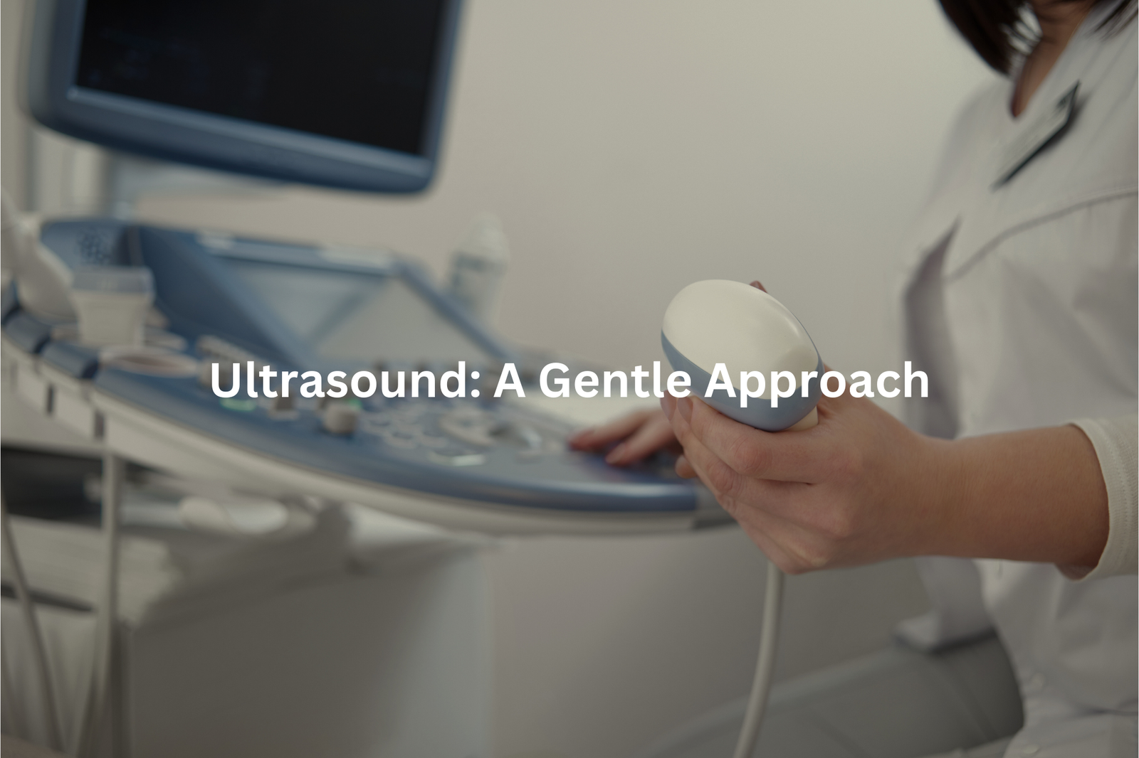
Ultrasound machines create detailed pictures of the body’s inside using sound waves (frequencies above 20,000 Hz). Unlike X-rays, these waves don’t use any radiation, making them safe for checking babies and sensitive body parts.
The process starts when a doctor puts some cold gel on the skin and moves a small device called a transducer across it. This device sends sound waves that bounce back from organs and tissues, creating black-and-white images on a screen.
Different types of ultrasound scans serve different purposes:
- Standard scans check pregnancy, muscles, and organs
- Doppler scans look at blood flow
- Transvaginal scans examine female reproductive organs
A typical ultrasound takes 15 to 45 minutes, and patients don’t feel any pain during the scan. The gel might feel a bit cold and sticky, but it wipes off easily. For many medical checks, ultrasound works better than X-rays, especially when doctors need to see organs moving in real-time.
The Role of Technology: AI in Pediatric Imaging
Artificial intelligence transforms pediatric imaging in Australian hospitals, creating a shift in medical practices. The technology works alongside medical professionals, not replacing them. AI assists radiologists in three main areas:
Image Enhancement
- Sharpens unclear scans
- Makes details more visible
- Reduces noise in images
Diagnostic Support
- Analyses patterns in scans
- Flags potential concerns
- Compares with existing cases
Safety Improvements
- Enables lower radiation doses
- Maintains image quality
- Protects young patients
Machine learning systems (a branch of AI) process thousands of medical images, helping spot signs of infection or fluid build-up in chest scans(2). The software highlights areas needing review, though radiologists always make final decisions.
Like any medical tool, AI has limits. Data quality affects its performance, making specialist oversight essential. Current AI systems support healthcare teams rather than work alone, adding an extra layer of accuracy to paediatric imaging.
Being Prepared: What Parents Can Do

Medical imaging can feel scary for kids in hospitals. Parents need a few simple steps to make the process smoother.
Questions about the procedure help both parents and children know what’s coming. Most imaging centers in Australia let parents stay close during the scan (except for X-rays, which require distance for safety). A parent’s presence often keeps children relaxed through the 15-30 minute procedures.
The Royal Australian College of Radiologists suggests these steps(3):
- Talk to kids about the scan the day before
- Bring a comfort item like a stuffed toy
- Ask about available distraction tools
- Learn about radiation safety measures
Different scans need different prep work. Ultrasounds use cold gel and take about 20 minutes. CT scans need kids to lie still for 5-10 minutes. MRIs make loud noises but don’t use radiation. Parents who understand the process can better support their children through medical imaging tests.
FAQ
What is a CT scan and how does it work for infants?
A CT (computed tomography) scan is a type of imaging test that uses X-rays to create detailed images of the body. For infants, a CT scan can help doctors see issues with the brain, organs, or bones. The scan takes multiple X-ray images from different angles, and a computer puts them together to create a 3D image. CT scans are quick and provide high-quality images, but they do use a small amount of ionising radiation.
How do VQ scans monitor blood flow in infants?
A VQ (ventilation-perfusion) scan is a type of nuclear medicine imaging test that looks at how air and blood are flowing through the lungs. For infants, a VQ scan can help doctors see if there are any problems with blood flow or lung function. The test involves injecting a small amount of radioactive material into the bloodstream, which then shows up on the scan. VQ scans are useful but do involve a small radiation dose.
What is the role of ultrasound in paediatric imaging?
Ultrasound uses high-frequency sound waves to create images of the body’s internal structures. For infants and children, ultrasound is a great choice because it doesn’t use any ionising radiation. Ultrasound can be used to check the heart, abdomen, hips, and other areas. It’s often used to guide procedures like liver biopsies. Ultrasound is a safe, real-time imaging method that provides high-quality images of a child’s developing body.
How do MRI scans work for brain imaging in young children?
Magnetic resonance imaging (MRI) uses strong magnetic fields and radio waves to generate detailed images of the body’s internal structures, including the brain. For infants and young children, MRI can provide very high-quality images of the developing brain without using any ionising radiation. MRI scans take longer than other imaging tests and may require sedation for young patients, but they offer unparalleled views of the brain and nervous system.
What are the benefits of using artificial intelligence in paediatric imaging?
Artificial intelligence (AI) is increasingly being applied to medical imaging to help analyse scans and detect potential issues more efficiently. In paediatric imaging, AI can help identify abnormalities, reduce motion artefacts, and optimise image quality – all while minimising radiation exposure for young patients. AI-powered tools can assist radiologists in making faster, more accurate diagnoses, leading to improved healthcare outcomes for infants and children.
Conclusion
Safe medical imaging stands as a critical part of infant care. MRI and ultrasound scans (both using non-ionising radiation) let doctors check babies without added risks. Parents should stay close during these 15–30 minute procedures, asking questions about the process.
Medical staff often explain each step, which helps create a secure setting for the tiny patients. Active involvement from parents, plus clear communication with doctors, supports better health outcomes for infants needing diagnostic scans.
References
- https://www.childrens.health.qld.gov.au/health-a-to-z/mri
- https://espace.curtin.edu.au/handle/20.500.11937/90826
- https://www.arpansa.gov.au/understanding-radiation/radiation-sources/more-radiation-sources/ct-imaging-and-children-parents