Pregnancy and Imaging: What Scans Are Safe for You and Baby? Learn Which Tests Doctors Recommend, What to Avoid, and How to Keep Both Mum and Bub Healthy.
Pregnancy and imaging go hand in hand for many expectant mums. When pregnant, women often need imaging tests like ultrasounds to check their health and the baby’s health. Generally, ultrasounds are safe; they use sound waves, not radiation.
Doctors recommend avoiding X-rays and CT scans if possible, since they use radiation, which can be a concern for an unborn baby. Always talk to your doctor if you have questions about imaging during pregnancy. They’ll guide you on what’s best for you and your baby. Keep reading to learn more about safe imaging options during pregnancy!
Key Takeaway
- Imaging tests like ultrasounds are safe during pregnancy.
- X-rays and CT scans should be used carefully due to radiation risks.
- Always talk to your doctor about the best imaging options during pregnancy.
Radiology During Pregnancy
Medical imaging helps doctors check inside a pregnant woman’s body. Different types of scans serve different purposes, and each comes with its own set of considerations.
Ultrasound stands out as the safest choice during pregnancy, using harmless sound waves to create pictures. The procedure takes about 30 minutes (depending on what needs checking) and shows the baby’s development clearly.
MRI scans, which use magnetic fields instead of radiation, provide detailed views of soft tissues. These scans run longer, sometimes 45-60 minutes, and require the patient to lie still.
X-rays and CT scans use radiation but remain essential tools when doctors need to spot serious problems like broken bones or dangerous infections. Medical staff use protective lead shields and careful positioning to keep radiation exposure as low as possible.
For most pregnancy-related concerns, doctors start with ultrasound. They choose other imaging methods only when the benefits outweigh any potential risks to mother and baby.
Safety of X-rays During Pregnancy
X-rays deliver a quick burst of radiation to capture internal images, raising concerns for pregnant women about safety. The medical community measures radiation exposure in millisieverts (mSv), with different scans producing varying levels of exposure.
Common X-ray exposure levels:
• Dental X-rays: 0.005 mSv
• Chest X-rays: 0.1 mSv
• Abdominal X-rays: 1.0 mSv
Medical professionals assess the necessity of X-rays during pregnancy by weighing potential risks against diagnostic benefits(1). While a developing foetus shows sensitivity to radiation, the exposure from a single X-ray typically falls below harmful levels.
Healthcare providers often implement protective measures:
• Lead aprons to shield the abdomen
• Alternative imaging like ultrasound or MRI when possible
• Postponing non-urgent scans until after pregnancy
Pregnant women should inform their healthcare providers about their pregnancy before any radiological procedures. This allows the medical team to consider safer alternatives or implement additional protective measures when X-rays prove medically necessary.
Imaging Alternatives for Pregnancy
Sources: Queensland X-Ray.
Medical imaging goes beyond standard X-rays. The ultrasound machine, a handheld device that glides across skin, creates pictures from sound waves without any radiation exposure.
Different scans serve different needs:
Ultrasound
- Uses harmless sound waves
- Best for soft tissue and pregnancy
- Shows real-time movement
- Takes about 15-20 minutes
MRI (Magnetic Resonance Imaging)
- Uses magnetic fields, not radiation
- Shows detailed organ images
- Requires 45-60 minutes
- Costs more than ultrasound
Low-dose CT
- Minimal radiation exposure
- Reserved for urgent cases
- Takes 5-10 minutes
- Shows bones and organs
Pregnant patients should discuss alternatives with their doctor. While X-rays might be needed for broken bones or chest problems, ultrasound often provides the necessary information. The choice depends on what needs examining – gallstones show up well on ultrasound, while joint issues might need an MRI. Each scan type has its place in modern medicine, giving doctors clear views without unnecessary radiation.
MRI During Pregnancy

MRI machines create detailed body images through magnetic fields and radio waves (1.5 to 3 Tesla strength). These scans help doctors see inside the body without using radiation, making them safer than X-rays or CT scans.
During pregnancy, MRI scans serve several purposes:
- Finding issues with the placenta
- Checking the baby’s growth
- Looking at the mother’s organs
- Spotting possible complications
Most doctors suggest MRI scans after the first trimester, around week 13 and beyond. The second and third trimesters are considered more suitable for these tests. Research shows no harmful effects on the growing baby, but medical staff still take extra care with pregnant patients.
The scan takes about 30 to 60 minutes. Patients lie still in a tunnel-shaped machine while it captures images. The machine makes loud tapping noises, so patients wear earplugs or headphones for comfort.
Medical teams only order MRIs when the benefits outweigh any concerns, always putting both mother and baby’s safety first.
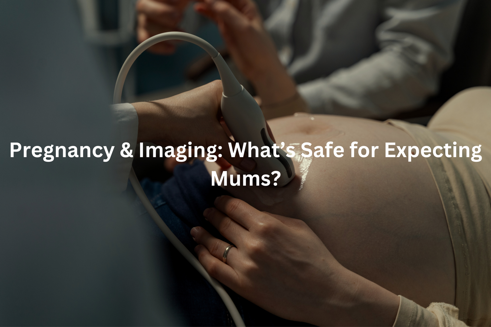
Ultrasounds in Pregnancy
Ultrasound scans create pictures of unborn babies using sound waves that bounce off body tissues. These waves, moving at speeds higher than human hearing can detect (about 20,000 cycles per second), help doctors check how babies grow inside the womb.
Most pregnant women get two main scans:
- Dating scan (6-9 weeks): Shows when the baby might arrive
- Morphology scan (18-22 weeks): Checks baby’s growth and organs
Some might need extra scans if doctors want to watch:
- The amount of fluid around the baby
- Blood flow through the umbilical cord
- Baby’s size and position
The sonographer puts gel on the belly and moves a small probe across it. The probe sends sound waves that create black-and-white images on a screen. Unlike X-rays, ultrasounds don’t use radiation, making them safe for both mum and baby(2).
Each scan takes about 20-30 minutes, giving doctors key information about the baby’s health and growth patterns.
Risks of Radiation to Fetus
Medical imaging during pregnancy raises questions about radiation safety. The amount of radiation exposure varies with different procedures, measured in millisieverts (mSv).
Common medical scans produce different radiation levels:
- Dental X-rays: 0.01 mSv
- Chest X-rays: 0.1 mSv
- CT scan of the abdomen: 8 mSv
The first trimester (12 weeks) needs extra caution since the baby’s organs are developing. Most diagnostic X-rays don’t reach harmful levels, which start above 100 mSv. Still, medical staff take precautions by using lead shields and limiting exposure.
Non-radiation options like ultrasounds and MRIs offer safer alternatives for pregnant patients. These scanning methods provide detailed images without any radiation risks to the developing baby.
Medical professionals carefully balance the need for diagnostic imaging against potential risks(3). When X-rays become necessary during pregnancy, the benefits typically exceed any small risks involved. Pregnant patients should discuss their concerns with healthcare providers who can explain specific safety measures.
Imaging Precautions for Pregnant Women
Medical imaging during pregnancy requires careful thought. The right scan at the right time makes all the difference (though doctors must weigh risks against benefits).
Ultrasound scans stand out as the safest choice, using harmless sound waves to create pictures. MRI scans come second – they’re safe but doctors skip the contrast dye in early pregnancy. X-rays work fine for teeth or broken bones, with proper lead shielding over the belly.
CT scans pack more radiation punch, so doctors use them sparingly. During the first 12 weeks, when tiny organs form, protection matters most. The medical team can:
- Lower radiation doses
- Use protective lead covers
- Put off non-urgent scans
Most single X-rays (even tummy ones) don’t reach harmful levels. The safe limit sits at 50 mSv – anything higher needs expert review. Pregnant patients should ask their doctor about scan options. There’s always a medical reason behind each recommendation.
Trimester-Specific Imaging Guidelines
Medical imaging follows specific rules for pregnant women, with different guidelines for each stage of pregnancy(4). The main concern? Radiation exposure to the developing baby.
During the first trimester (weeks 1-12), doctors avoid X-rays when possible. This period needs extra care since the baby’s organs are developing. The second trimester (weeks 13-26) allows more flexibility with proper shielding, while the third trimester (weeks 27-40) sees regular ultrasound scans to monitor growth.
Ultrasound scans use sound waves (not radiation) and remain safe throughout pregnancy. Most standard X-rays deliver less than 5 mSv of radiation – well below the 50 mSv safety threshold. MRI scans, though safe, are only used when needed.
For any imaging test, doctors consider these factors:
- Type of scan needed
- Stage of pregnancy
- Medical necessity
- Alternative options
Pregnant women should discuss concerns with their healthcare provider and ask about radiation levels for specific procedures.
Doctor’s Recommendations for Pregnancy Imaging

Medical imaging during pregnancy helps doctors check both mum and bub’s health. These scans serve different purposes throughout the nine months.
Common reasons for pregnancy scans:
- Regular growth checks (measuring bub’s size and organs)
- Monitoring health concerns (like high blood pressure or diabetes)
- Emergency situations (unexplained pain or bleeding)
Ultrasound scans, which use sound waves, are the most frequent type. They don’t use radiation and create clear pictures of the growing baby. MRI scans might be needed for more detailed views of certain areas.
X-rays aren’t typical during pregnancy, but doctors can use them safely when needed. The radiation dose gets adjusted to protect mum and bub.
Pregnant women should discuss scan options with their healthcare provider. Questions about timing, necessity, and safety help create better understanding. Most imaging centres in Australia follow strict guidelines to ensure safe, effective scans for expectant mums.
Pregnancy-Safe Imaging
Medical imaging during pregnancy requires careful planning. Different scans help doctors check both mum and baby’s health throughout the nine months.
Ultrasound stands as the primary choice for pregnancy scans. It uses sound waves instead of radiation, making it safe for regular use. Doctors rely on ultrasounds to:
- Check the baby’s growth
- Confirm due dates
- Look at the baby’s anatomy
- Monitor the placenta
MRI scans come next in line, offering detailed views when needed. They don’t use radiation but rather magnetic fields and radio waves.
X-rays and CT scans sit at the bottom of the list. While these tests give off small amounts of radiation, doctors use lead shields and limit exposure. The radiation from a single chest X-ray measures less than what someone gets from natural background radiation in 10 days.
Pregnant women should discuss any concerns about imaging with their healthcare team. Different options might work better at different stages of pregnancy.
FAQ
What is a CT scan during pregnancy?
A CT (computed tomography) scan uses X-rays to create detailed images of the body. During pregnancy, a CT scan may be used to check for conditions like pulmonary embolism (blood clot in the lungs) or other emergencies. The radiation dose is carefully monitored to minimise risks to the foetus.
What is a low-dose CT scan?
A low-dose CT scan uses a smaller amount of radiation compared to a standard CT scan. This can help reduce radiation exposure for pregnant patients and their developing baby. Low-dose CT scans may be used when the benefits outweigh the small risks.
What is a perfusion scan during pregnancy?
A perfusion scan looks at blood flow in the lungs. It may be used to check for pulmonary embolism in pregnant patients who have shortness of breath or other symptoms. The procedure involves injecting a small amount of radioactive tracer to assess lung function.
How can I reduce radiation risks during pregnancy imaging?
To reduce radiation risks, healthcare providers often recommend using the lowest possible radiation dose and limiting the number of imaging tests. Alternatives like ultrasound or MRI may be preferred when appropriate. Discussing concerns with your doctor can help ensure safe and informed decisions about pregnancy imaging.
What are the risks of foetal radiation exposure?
Foetal radiation exposure, even at low levels, may potentially increase the risk of childhood cancer or other health issues. The specific risks depend on factors like the type of imaging, radiation dose, and gestational age. Your healthcare team will carefully weigh the benefits and risks for your situation.
When should I have a dating scan or morphology scan?
Dating scans are typically done in early pregnancy to confirm the gestational age. Morphology scans, usually around 18–22 weeks, assess the baby’s development and screen for certain conditions. These routine ultrasound scans provide important information without radiation exposure.
What blood tests may be done during pregnancy?
Routine blood tests, such as checking for anaemia, infections, or genetic conditions, are a normal part of prenatal care. These simple blood draws help monitor your health and the baby’s growth throughout the pregnancy.
Conclusion
Imaging during pregnancy needs careful thought. Expecting mothers have several safe options to check their baby’s growth. Ultrasound scans (using sound waves instead of radiation) stand out as the most common choice, while MRI offers detailed views without any radiation risks.
X-rays and CT scans use radiation, so doctors limit these unless absolutely needed. The right choice depends on what needs checking – some scans work better for bones, others for soft tissue. Doctors match each test to the mother’s specific needs.
References
- https://ris.com.au/how-safe-are-x-rays-during-pregnancy/
- https://www.qld.gov.au/health/children/pregnancy/antenatal-information/your-care-during-pregnancy/ultrasounds-during-pregnancy
- https://www.pregnancybirthbaby.org.au/radiation-exposure-during-pregnancy
- https://aci.health.nsw.gov.au/networks/eci/clinical/ed-factsheets/medical-imaging-in-pregnancy