Discover how radiologists use advanced tools to capture images of bones, organs, and tissues, helping patients feel better with accurate diagnoses.
Radiologist tools are super important for doctors to look inside our bodies. They help to diagnose problems when we’re feeling unwell. For instance, X-rays can show our bones, while MRIs and CT scans help see soft tissues like organs. These machines use special technology to create detailed images that guide doctors in their decisions.
Each tool has a unique way of capturing images, making them essential for patient care. Understanding these tools can help you appreciate how doctors find answers. So keep reading to learn more about how radiologist tools really work and why they matter!
Key Takeaway
- Radiologists use machines like X-rays and MRIs to see inside the body.
- These tools help doctors find problems like tumors and fractures.
- New technology makes it easier for doctors to share and understand medical images.
X-ray Machines: Quick Snapshots of Our Bones
X-ray machines are a bit like magic cameras, aren’t they? They let doctors see inside our bodies without needing to make a single cut(1). They work by using special rays (called X-rays, of course) that can pass through soft tissues like skin and muscles but get blocked by denser stuff like bones. That’s why bones show up so clearly on the X-ray images, making it easier for doctors to spot things like fractures or infections.
Just last week, my mate Liam broke his wrist falling off his bike. At the hospital, they took him straight to the X-ray room. The radiologist (that’s the person trained to take these images) had the whole thing done in under a minute. Liam said he barely had time to blink before they had the pictures ready. Turns out, he had a clean break, and they knew exactly how to treat it because of the X-ray.
But X-rays aren’t just for broken bones. They’re also used to check for tumours, lung infections, joint issues, and even dental problems like cavities. It’s pretty incredible how much information doctors can get from just one image. If you ever need one, don’t worry—it’s quick, painless, and super helpful.
MRI Machines: Seeing Soft Tissues Clearly
MRI stands for Magnetic Resonance Imaging. It’s a machine that uses strong magnets and radio waves to take detailed pictures of the soft tissues in our body. Soft tissues are things like muscles, organs, and the brain—basically, anything that isn’t bone.
My cousin once had an MRI when she injured her knee playing soccer. She told me it was loud, with all these banging noises, but it didn’t hurt at all. The doctors used the pictures to figure out what was wrong with her knee, and she didn’t even need an X-ray. That’s pretty impressive, right?
Doctors use MRIs for all sorts of things. They can check for brain injuries, heart problems, or even find tumours. Sometimes, they even use it to guide treatments like tumour ablation (that’s when they destroy tumours without surgery). It’s like having X-ray vision but without any harmful radiation.
Here’s what MRIs are great for:
- Brain scans
- Torn muscles
- Tumours
- Heart conditions
- Organ problems
It’s wild to think a machine can do all that while keeping patients safe and comfortable.
CT Scanners: A 3D View of Our Bodies
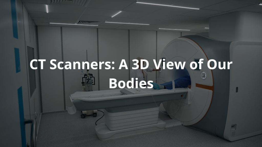
CT scanners, or Computed Tomography scanners, are pretty amazing machines. They take a bunch of X-ray pictures from all sorts of angles and then combine them to make a 3D image of what’s inside our bodies. This makes it easier for doctors to spot things like tumours or bleeding. It’s like having a super-detailed map of your insides.
Just the other week, my neighbour had to get a CT scan because he wasn’t feeling too great. The doctors needed answers quickly, and CT scans are perfect for that. They’re fast and often used in emergencies when every second counts. It’s kind of fascinating how they can show soft tissues, organs, and even bones all in one go.
But there’s a catch—CT scans use ionising radiation. That means they expose you to a small amount of radiation, so doctors try not to use them too often unless it’s really necessary.
Here’s what CT scans can help with:
- Tumours
- Bleeding
- Organ damage
- Bone fractures
- Heart problems
So, while they’re not something you’d want every day, CT scanners are like lifesaving cameras that help doctors figure out what’s wrong and how to fix it. If you ever need one, just remember—it’s all about getting the right answers to keep you healthy.
Ultrasound Machines: Using Sound Waves
Ultrasound machines are pretty unique compared to other medical tools(2). Instead of using X-rays or radiation, they rely on sound waves to create pictures of the inside of our bodies. These sound waves are completely safe, which is why ultrasounds are used so often, especially during pregnancy.
I remember when my mate’s mum had an ultrasound to check on her baby. She showed us the screen, and there was this tiny little person moving around—it was amazing! But ultrasounds aren’t just for babies. They can also check on organs like the liver, kidneys, and heart. They’re even used to see how blood flows through veins and arteries. It’s all in real-time too, so doctors can watch things as they happen.
Ultrasounds are also handy for guiding procedures. For example, they can help with needle biopsies or treatments like artery embolization (that’s a way to stop bleeding).
Here’s what ultrasounds can do:
- Monitor pregnancy
- Check organs for problems
- Find cysts or tumours
- Guide medical procedures
- Watch blood flow
It’s like having a live video of what’s going on inside us. Pretty cool, right? If you ever need one, don’t worry—they’re quick, painless, and super helpful!
Digital Imaging Systems: Sharing Made Easy
Source: AtlanticMedical.
In Australia, radiology is going digital, and it’s changing how doctors work together. Digital imaging systems let radiologists share scans quickly with other doctors. If a GP or specialist needs a second opinion, they can send the images in seconds. No waiting for films to be mailed or carried around. Just click, send, and done.
These systems also store images safely, so doctors can pull up old scans whenever they need. It’s like having a health timeline saved on a computer. Plus, the images can be adjusted—zoomed in, brightened, or sharpened—making it easier to spot something small or tricky.
Radiologists now use advanced software to help analyze scans. Some programs even use artificial intelligence (AI) to find patterns or highlight areas that might need attention. It’s like having an extra set of super-sharp eyes.
Why does this matter?
- Doctors can share and discuss scans faster.
- Images are stored securely for future use.
- Technology improves accuracy.
- It helps teams work together better.
This all leads to better care for patients. If you ask me, it’s a big step forward for healthcare in Australia.
Radiation Safety Equipment: Keeping Everyone Safe
Radiologists use machines that work with radiation, like X-rays or CT scanners, to help see inside the body. But radiation can be risky if there’s too much exposure. That’s why radiologists and other medical staff wear special protective gear, such as lead aprons, gloves, and even lead glasses. These items are designed to block harmful rays and keep everyone safe.
Safety is a big deal in radiology. Doctors always calculate the smallest amount of radiation needed to get clear images. They carefully balance the benefits of the test (like finding out what’s wrong) with any potential risks. It’s all about protecting both the patient and themselves.
Here’s what the safety gear does:
- Shields patients from unnecessary radiation.
- Protects doctors and staff during procedures.
- Helps lower long-term risks of exposure.
- Ensures imaging is done safely and responsibly.
So, if you see your doctor wearing a heavy apron or standing behind a glass screen, it’s not just for show. It’s part of keeping everyone safe while getting the best results.
Continuous Education Tools: Always Learning
Radiology isn’t just about using machines or taking pictures of bones—it’s about learning all the time. Radiologists (the doctors who read and understand medical images) spend years in medical school, but even after that, their education doesn’t stop. They go to workshops, attend conferences, and use online courses to stay sharp and up to date with the latest technology and methods. It’s like they’re students forever, but in a good way.
Why? Because medical tools and imaging techniques are always changing. New machines like MRI or CT scanners get better every year, and radiologists need to know how to use them properly. They also learn new ways to spot diseases earlier or more clearly, which helps patients get the right treatment faster(3).
Here’s what all this learning does for them:
- Keeps them updated on new technology.
- Makes their skills sharper.
- Helps them work better with other doctors.
- Improves care for patients.
I reckon it’s pretty amazing how much effort they put into learning just to help others. If you’re thinking about radiology, know that it’s a career for curious minds who never stop asking questions.ven more!
FAQ
What is a ray tube and how is it used in radiological imaging?
A ray tube is a key component in many radiological imaging machines. It generates the X-rays or other types of radiation that are used to capture detailed images of the body’s internal structures, including bones, organs, and soft tissues. The ray tube produces a focused beam of radiation that passes through the patient and is detected by the imaging equipment, allowing doctors to see what’s happening inside.
What is a ray beam and how does it work in medical imaging?
A ray beam refers to the concentrated stream of radiation that is emitted from the ray tube and directed at the patient during an imaging scan. This ray beam allows the imaging equipment to capture high-quality, detailed images of the body’s internal structures, from bones to organs to soft tissues. The way the ray beam interacts with the body’s tissues produces the contrast needed for the diagnostic images.
What is real-time imaging and how is it used in radiology?
Real-time imaging, also known as fluoroscopy, allows radiologists to view continuous, live images of the body’s internal structures as they move. This is particularly useful for guiding minimally invasive procedures, such as inserting catheters or performing biopsies, by allowing the doctor to see exactly where the medical instruments are located within the body in real-time.
What is a ray source and how does it differ from a ray tube?
The ray source is the part of the imaging equipment that generates the radiation beam used for medical imaging. While the ray tube is a specific component that produces the X-rays or other types of radiation, the ray source refers more broadly to the entire system that creates and controls the radiation beam directed at the patient. Understanding the differences between the ray tube and the overall ray source is important for comprehending how imaging machinery works.
What is a ray machine and what are its key features?
A ray machine, such as an X-ray or CT scanner, is the primary piece of equipment used to capture detailed diagnostic images of the body. These machines contain components like the ray tube, ray source, and imaging detectors that work together to generate high-quality images. Key features of ray machines often include the ability to adjust radiation doses, produce real-time images, and integrate with other medical systems for comprehensive patient care.
What is ray imaging and how does it differ from other medical imaging modalities?
Ray imaging, such as X-rays and CT scans, uses ionizing radiation to capture detailed images of the body’s internal structures. This differs from other modalities like MRI, which uses magnetic fields and radio waves, or ultrasound, which uses sound waves. Each imaging technique has its own strengths and is suited for visualizing different types of tissues and conditions, so radiologists often employ a variety of imaging modalities to get a complete picture of a patient’s health.
How do soft tissues show up in ray imaging compared to other imaging tests?
Soft tissues, like muscles, organs, and blood vessels, can be more challenging to visualize using ray imaging techniques like X-rays compared to other modalities. While ray imaging excels at capturing bone and dense structures, the contrast between soft tissues is often lower. Techniques like CT scans and MRI can provide much clearer and more detailed images of soft tissues, making them valuable complements to traditional ray imaging for comprehensive diagnostic evaluations.
What is an MRI machine and how does it work differently than a ray machine?
An MRI (magnetic resonance imaging) machine uses strong magnetic fields and radio waves, rather than ionizing radiation, to produce detailed images of the body’s internal structures. This non-invasive technique allows for excellent visualization of soft tissues like the brain, muscles, and organs, which may not show up as clearly on X-ray or CT scans. MRI machines are an important tool in the radiologist’s toolkit, providing a different imaging perspective compared to ray-based modalities.
How can high-quality imaging improve patient outcomes?
High-quality, detailed medical imaging plays a crucial role in helping radiologists and other healthcare providers make accurate diagnoses and develop effective treatment plans. Advances in imaging technology, from higher resolution scanners to more sophisticated computer-assisted analysis, can provide doctors with a clearer, more comprehensive view of a patient’s condition. This, in turn, supports better-informed clinical decisions that can lead to improved patient outcomes, such as earlier detection of disease, more targeted therapies, and enhanced monitoring of treatment progress.
What role does ray imaging play in the diagnosis and treatment of conditions like heart disease and breast cancer?
Ray imaging techniques like CT scans and mammograms are invaluable tools for the early detection and management of serious conditions like heart disease and breast cancer. CT scans can provide detailed images of the heart and blood vessels, helping identify blockages or other abnormalities, while mammograms use low-dose X-rays to screen for breast tumors. These imaging tests play a critical role in allowing radiologists and other medical professionals to quickly and accurately diagnose these diseases, enabling prompt, appropriate treatment that can significantly improve patient outcomes.
How do radiologists use imaging equipment and techniques like CT fluoroscopy for guided medical procedures?
Radiologists often use advanced imaging technologies like CT fluoroscopy to guide minimally invasive medical procedures. CT fluoroscopy provides real-time, continuous X-ray imaging that allows the radiologist to precisely visualize the location of surgical instruments or catheters as they are being inserted into the body. This “live” imagery helps the radiologist navigate to the correct target area and monitor the procedure, leading to more accurate and successful interventions with less risk to the patient.
How do radiologic technologists play a crucial role in medical imaging and patient care?
Radiologic technologists are highly trained medical professionals who operate the complex imaging equipment used in hospitals, clinics, and other healthcare settings. They are responsible for properly positioning patients, adjusting imaging parameters, and ensuring the safe, effective use of radiation during procedures. Radiologic technologists also play a vital role in patient communication and education, helping to ease any concerns and ensure patients understand the imaging process. Their expertise and dedicated patient care are essential for producing high-quality diagnostic images that support accurate diagnoses and effective treatments.
Conclusion
When doctors need to look inside our bodies, they use special machines called imaging technologies. These include X-ray machines, MRIs, CT scanners, and ultrasounds. Each one is different and helps doctors see things clearly and safely. Radiologists, the experts who use these machines, keep learning about new tools and methods to improve care. Next time you hear about these machines, remember they help keep us healthy in a big way.
References
- https://medicine.umich.edu/dept/radiology/research/basic-radiological-sciences-brs/medical-physics-quality-control-services/equipment
- https://www.siemens-healthineers.com/en-id/ultrasound
- https://www.carestream.com/blog/2018/12/11/continuing-education-in-radiology/