Learn how medical imaging for treatment enhances diagnosis and care, transforming healthcare into a precision science.
Seeing inside the human body without making a single cut—now that’s something straight out of a storybook, isn’t it? But it’s real. Medical imaging, like X-rays, CT scans, and MRIs, gives doctors a way to look deep inside, spotting problems and planning treatments. It’s not just about fixing broken bones or finding illnesses; it’s about understanding how the body works and staying ahead of health issues.
These tools make healthcare feel a little less scary and a lot more precise. Curious about how these machines work and what they reveal? Keep reading to uncover the details behind the images!
Key Takeaway
- Medical imaging helps doctors see inside the human body.
- There are many types of imaging tests like CT scans and MRIs.
- These images help diagnose problems and plan treatments.
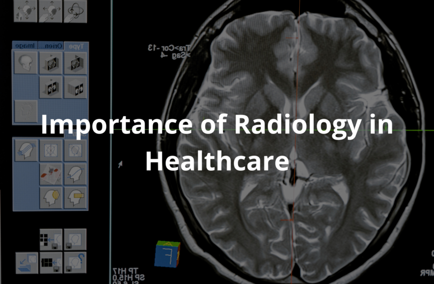
What is Medical Imaging?
Medical imaging is how doctors take pictures of the inside of the body using special technology. These pictures help them figure out what’s wrong or how to treat someone. There are a few main types of medical imaging, each with its own job:
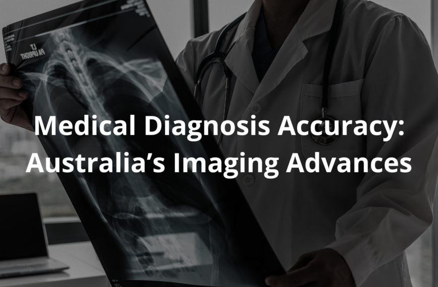
- X-rays: These are used to look at bones. If someone breaks their arm falling off a bike, an X-ray can show exactly where the break is. It’s like peeking inside without opening anything up.
- CT Scans (Computed Tomography): A CT scan takes lots of X-rays from different angles and puts them together to make a detailed picture. Doctors use these to look at organs or find things like tumours. In hospitals across Australia, CT scans are used every day to check for serious conditions. They’re fast and give a lot of information.
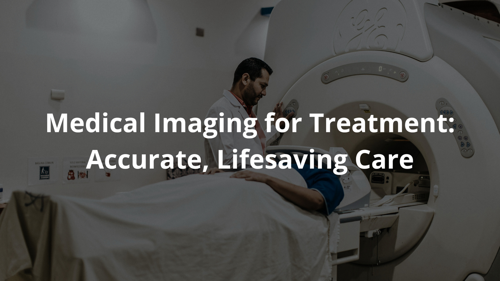
- MRI Scans (Magnetic Resonance Imaging): MRIs use magnets and radio waves to make clear pictures of soft tissues, like the brain or muscles. If someone has a head injury, an MRI can show what’s happening inside.
- PET Scans (Positron Emission Tomography): PET scans are different because they show how organs are working, not just what they look like. They’re often used to check for cancer or see how well a treatment is working.
- Ultrasound: This uses sound waves to create images. It’s great for looking at babies in the womb or checking organs like the liver or kidneys.
Each of these imaging tools helps doctors understand what’s happening in the body. They’re like detectives, using these pictures to solve medical mysteries.
Why Do We Need Medical Imaging?
Imagine feeling sick and not knowing why. Medical imaging helps doctors figure it out. For example, if someone has a bad stomach ache, a doctor might use an ultrasound to see what’s going on. It could show if the appendix is inflamed or if it’s just a simple stomach bug.
Imaging is also used to check if treatments are working. If a patient is being treated for a tumour, scans can show if the tumour is shrinking. It’s like checking the scoreboard during a match—are we winning?
Doctors also use imaging to plan surgeries. Knowing exactly where a problem is means they can be precise, which often makes surgeries quicker and safer. Patients feel more confident when they know their doctor has a clear plan.
Safety in Medical Imaging
Some people worry about the safety of imaging, especially with tests that use radiation, like X-rays or CT scans. But the risks are very small. Doctors always use the lowest amount of radiation needed to get a clear picture, especially for children or pregnant women.
In Australia, strict rules make sure imaging is safe. The Australian Radiation Protection and Nuclear Safety Agency (ARPANSA) checks that radiation levels stay low and safe. Imaging centres must follow these rules to protect patients. [1]
Doctors also explain the risks and benefits before ordering a scan. For example, if a CT scan is needed, they’ll explain that while it uses some radiation, the information it provides is worth it. This helps patients feel more comfortable and informed.
The Role of Technology in Imaging
Medical imaging keeps improving thanks to technology. For example, artificial intelligence (AI) is now being used to help doctors read scans. AI can spot problems quickly and sometimes even catch things a human might miss. It’s like having an extra set of very sharp eyes.
Some machines can also create 3D images, which are incredibly helpful for planning surgeries. Imagine a surgeon preparing to operate on a brain tumour. A 3D image can show exactly where the tumour is, helping the surgeon be more accurate.
Portable imaging machines, like small ultrasound devices, are also making a big difference. These can be used in remote areas or during emergencies, bringing life-saving care to people who might not otherwise get it.
The Impact of Medical Imaging on Health Care

Medical imaging has changed health care in Australia and around the world. It helps doctors make better decisions and provide better treatments. With clear images, they can see details like blood vessels or tumours, which is crucial for diagnosing conditions like heart disease or cancer.
In Australia, organisations like the Royal Australian and New Zealand College of Radiologists (RANZCR) set high standards for imaging practices. This ensures patients get safe and reliable care.
The numbers show how important imaging is. Between 2000 and 2021, over 436 million imaging tests were done in Australia. That’s millions of times doctors used these tools to help patients. [2]
FAQ
How do magnetic fields and radio waves work together in an mri machine to create images?
When you get an mri scan, the mri machine uses powerful magnetic fields and radio waves to take pictures of your body parts. The machine’s magnetic field lines up water molecules in your body, and radio waves make these molecules release energy that creates detailed images. This lets doctors see soft tissues like muscles and organs really clearly.
What’s the difference between common imaging types like ray ct, pet scans, and mri scans?
Each type of medical image helps doctors in different ways. Ray ct uses x-rays to see bones and dense tissues, mri scans show soft tissues using magnetic fields, and pet scans track blood flow and cell activity. These imaging types give doctors a wide range of tools to help diagnose different conditions.
How does functional mri help us understand the human brain?
Functional mri is a special kind of brain imaging that shows which parts of your brain are active when you do different tasks. It measures blood flow changes in your brain, creating high quality images that help doctors and researchers understand how your brain works. This is super helpful for studying things like memory and learning.
What are contrast agents and why are they sometimes used in medical images?
Contrast agents, like iron oxide or other special materials, help make certain body parts show up more clearly in medical images. When doctors need to see specific areas better, they might use these agents to create high resolution pictures. Different contrast agents work best for different types of scans.
What should I know about radiation dose and small risk in medical imaging?
While some imaging types like ray imaging use radiation, doctors always aim for a low dose that balances good quality images with safety. There’s a small risk from radiation, but medical centers carefully control the radiation dose. The benefits of detecting health issues usually far outweigh these risks.
How is deep learning changing medical image analysis?
Deep learning is making it easier and faster to analyze imaging data. This technology helps doctors spot things like bone fractures, breast cancer, or bone tumor more accurately. It’s improving early detection of health issues by finding patterns in medical images that might be hard for human eyes to see.
What role do sound waves play in certain medical imaging techniques?
Sound waves are used to create images of body including your spinal cord and other deep structures. Unlike gamma rays or magnetic fields, sound waves don’t use radiation, making them especially safe. They bounce off different tissues to create high frequency signals that turn into detailed pictures of what’s inside your body.
How is bone density measured and what can it tell us?
Specialized medical imaging helps measure bone density, which shows how strong your bones are. This helps diagnose conditions like osteoporosis and tracks bone health over time. The imaging creates clear pictures of your bones using small amounts of radiation to check their strength and structure.
What should I bring to my medical imaging appointment?
Bring your medical history and any previous imaging records if you have them. Wearing comfortable clothes helps, and you might need to remove metal items for certain scans. Your doctor might also ask you not to eat before some types of scans, especially if you’ll need contrast agents.
How long do different types of medical imaging take?
Timing varies based on what part of your patient’s body needs scanning. A quick ray ct might take just a few minutes, while some specialized imaging types like mri scans can take 30-60 minutes to get high quality images. Your doctor will let you know how to prepare and what to expect.
How do doctors choose between imaging types to help diagnose conditions?
Doctors pick the best type of scan based on what they need to see – like using mri scanner for soft tissues or ray imaging for bones. They consider things like image quality, side effects, and what they’re looking for. Sometimes they might use multiple types of scans to get the most complete picture.
Conclusion
Medical imaging is a brilliant tool in healthcare, letting doctors see inside the body safely. With X-rays, CT scans, and MRIs, they can find and treat all sorts of problems. It’s pretty amazing how these machines work, and they’re always getting better. Patient safety is key, so the tech is designed to minimise risks. Next time someone mentions medical imaging, think about how it’s helping doctors and keeping people well. It’s a lifesaver, really.
References
- https://www.health.gov.au/topics/diagnostic-imaging/about
- https://pubmed.ncbi.nlm.nih.gov/37797195/