Unlock the power of radiology diagnostic techniques to understand the unseen and support better healthcare outcomes.
Radiology diagnostic techniques play a crucial role for doctors in Australia. They help uncover the mysteries of our bodies, especially when someone feels unwell. The machines and tools used create images that show bones, organs, and soft tissues, almost like a superpower.
From X-rays to MRIs, every imaging technique has a unique purpose. It’s fascinating how these technologies work together to help doctors understand what’s going on inside us. If you’re keen to discover more about these amazing techniques and their impact on health, keep reading. There’s much more to uncover about how they help save lives every day!
Key Takeaway
- Radiology techniques help doctors see inside the body.
- Different imaging techniques are best for different problems.
- Safety and quality are very important in medical imaging.
X-rays: Quick and Easy
X-rays are a common way to peek inside the body. When someone gets an X-ray, a special machine sends out a beam of rays. This beam goes through the body and creates images on film or on a computer screen.
- What Can X-rays See? They work wonders for checking broken bones, spotting lung issues, and helping dentists look at teeth. X-rays can show exactly how bones look, and if they’re in the right place. For example, if someone has a persistent cough, an X-ray can help figure out if there’s a lung problem.
- How Fast Are They? They’re really quick! A complete X-ray usually takes just a few minutes. The whole process goes faster than waiting for a delicious snack at the school canteen.
- Are There Risks? Yes, X-rays involve a small amount of ionizing radiation. It’s safe when used wisely, but doctors always try to keep them to a minimum. This means they won’t keep taking X-rays if it’s not necessary.
One time, a kid named Sam had a rough fall while playing at school. His arm hurt so much that it was hard to move. The doctor advised an X-ray. It was finished super fast, and thankfully, the X-ray showed his arm was only bruised—not broken! That definitely lifted his spirits.
X-rays are like magic windows into our bodies. They reveal what’s hidden, which helps doctors find out what’s wrong.
CT Scans: Detailed Images
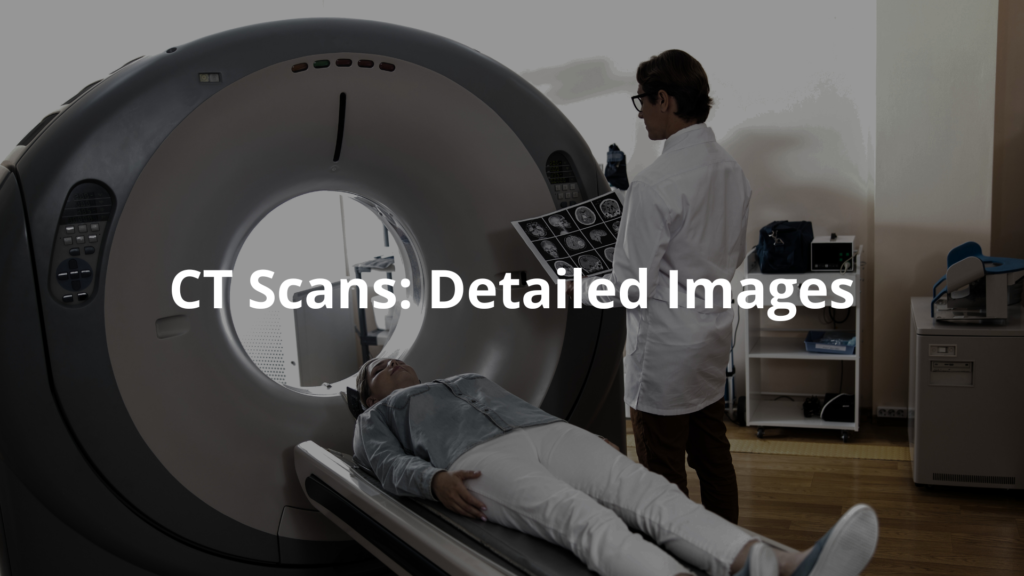
CT scans, or computed tomography scans, are like super X-rays. They create detailed pictures of what’s inside the body. The CT scanner takes multiple X-ray images from different angles and puts them together to create cross-sectional images.
- When Are They Used? CT scans are helpful for doctors to see problems with internal organs, like the lungs or liver. They’re also used for spotting serious problems, such as cancers. If someone has a severe injury, a CT scan can let doctors gauge how serious it is.
- What’s the Good Part? They provide a very detailed look at both soft tissues and bones. This helps doctors see things like blood clots or tumors that might be hidden away.
- What About Safety? CT scans involve more radiation than regular X-rays, so they’re not used lightly. Doctors carefully consider the risks and benefits. If a CT scan is needed, it’s usually because the doctor believes it’s very important.
Once, a man named Tom had to get a CT scan when he wasn’t feeling well. Though it took a bit longer, it helped the doctors understand his condition. They found a small issue with his liver, and thanks to that CT scan, the doctors could help him feel better.
CT scans are like taking a slice of bread and peering inside. They let doctors see all the layers, making it easier to understand what’s happening.
MRI Scans: No Radiation
MRI, or magnetic resonance imaging, is a special method for taking pictures of soft tissues inside our bodies. Rather than using X-rays, it uses powerful magnets and radio waves. [1]
- What Can MRI See? It’s excellent for checking the brain, spinal cord, and muscles. It can reveal details that other scans might miss. For instance, if someone has back pain, an MRI can show if there’s a problem with the muscles or discs in the spine.
- Why is it Safe? MRIs don’t use any radiation, making them a safer option for many patients. This is especially important for pregnant women and children.
- How Long Does It Take? MRIs usually take longer than CT scans—sometimes up to an hour. Patients have to lie still while the machine works, which can feel a bit odd.
A woman named Lisa once shared how she had an MRI to look at her back. She found it a bit noisy, but felt safe knowing there was no radiation involved. The images turned out so clear that the doctors could see exactly what was wrong.
MRI scans are like taking a super clear photograph of the inside of the body. They help doctors spot things that are hard to see with other imaging tests.
Ultrasound: Using Sound Waves
Ultrasound is a pretty neat technique. It uses sound waves to create images of what’s happening inside the body. The doctor puts a special jelly on your skin and then moves a small device called a transducer over it.
- When is Ultrasound Used? It’s often used during pregnancy to see the baby. It’s also great for looking at organs like the heart and abdomen. Doctors can check the shape and size of these organs using ultrasound.
- What’s Cool About It? Ultrasound is very safe and doesn’t involve any radiation. You can even see real-time images, which is quite amazing! For instance, if a woman’s having a baby, she can see her little one moving around in her tummy!
- What Can’t It Do? Ultrasound can’t go very deep, so it might miss some things. If a person has a puzzle inside their bones, other imaging tests might be needed.
A girl named Mia remembers seeing a video of an ultrasound when her cousin was about to have her baby. It was truly exciting to see the baby moving inside her tummy! Everyone was keen to find out if it was a boy or a girl.
Ultrasound acts like a live movie of the inside of the body. It shows doctors how things are working in real-time, which is very helpful!
Nuclear Medicine: Seeing Inside with Tracers
Nuclear medicine is slightly different. It uses small amounts of radioactive materials, called tracers, to see how organs are working. Techniques like PET scans and SPECT scans fall under this category.
- What Do They Show? These scans can reveal how well organs like the heart or brain are functioning. They can also help find cancers. If someone’s heart isn’t working right, a nuclear scan can show how well it’s performing.
- What’s the Benefit? They provide doctors with important functional information. It helps clinics see not just how things look, but how they work too.
- Is It Safe? There’s some radiation exposure, but the amount is very small and is deemed safe for most people. Doctors are careful to use just the right amount.
One time, a kid named Jake learned how doctors can see blood flow using nuclear medicine. It’s truly fascinating how these tiny tracers help resolve big health mysteries!
Nuclear medicine is like shining a light inside the body. It helps doctors see how everything functions in real-time.
The Importance of Safety and Quality
In Australia, there are strict rules to keep patients safe during imaging tests. The Australian Radiation Protection and Nuclear Safety Agency (ARPANSA) ensures all facilities stick to safety guidelines. [2]
- Why is This Important? It helps ensure patients don’t get too much radiation and that they receive quality images. This is important for keeping everyone safe during medical tests.
- Who Makes the Rules? The Royal Australian and New Zealand College of Radiologists (RANZCR) sets standards for imaging practices to keep everyone safe. They help ensure the images are clear and useful for doctors.
It’s comforting to know there are guidelines protecting patients. Knowing that doctors follow these rules helps everyone feel better about getting imaging tests they might need.
These safety standards ensure patients receive the best care possible. It’s nice to know there are people dedicated to looking after everyone’s health!
FAQ
What are MRI and CT scans, and how do they differ in medical image creation?
MRI scan and CT scanning work differently to produce images of your body. MRI uses magnetic field and radio waves, while CT uses ray beams to create detailed pictures. MRI is great for looking at soft tissue and the spinal cord, while CT provides good quality views of bones.
How do doctors use 3d imaging and image guided techniques in modern health care?
Medical teams now use amazing 3d images to see inside your body in real time. This helps them spot problems more accurately. Image guided procedures let doctors see exactly where they’re working, making treatments safer and more precise for patient care.
What role do PET scan and cardiac PET play in diagnosing heart disease?
Pet scan and cardiac pet help doctors check how well your heart is working. These scans can spot heart disease early by showing blood vessels and how your heart uses energy. They often use a small amount of contrast agent to get clearer pictures.
How do radiation dose and side effects impact patient safety during ray imaging?
While ray imaging uses radiation dose to create pictures, doctors keep it as low dose as possible. Side effects are usually mild, but patient safety is always the top priority. That’s why pregnant women and others with risk factors get special attention.
What makes breast MRI different from other imaging tests for breast cancer?
Breast MRI uses magnetic waves to look at breast tissue in detail. It helps find breast cancer that might be hard to spot with other tests. The image quality is very high, letting doctors see even tiny changes in the tissue.
How do gamma rays and gamma camera work in bone scan procedures?
A bone scan uses a gamma camera to spot problems like bone tumor or bone fractures. It involves using gamma rays and a tiny bit of safe tracer. The scanner tracks how your bones take up this tracer, showing areas that might need attention.
What advances in image analysis help doctors interpret medical images of the human brain?
New ways to study medical images let doctors better understand the human brain. Brain imaging technology keeps getting better at showing both big and small problems. High frequency scanning gives sharp, detailed views doctors can trust.
What makes dual energy imaging special for looking at the human body?
Dual energy scans are clever – they use two different ray strengths to see both bones and soft bits at once. This helps doctors spot problems they might miss with regular scans. It’s especially good at showing how different parts of your body work together.
How do doctors check bone density and what can it tell us?
Bone density scans use special ray equipment to check how strong your bones are. These simple tests use iron oxide or similar materials to create detailed pictures. They’re great for spotting early signs of bone weakness, which plays a vital role in preventing future problems.
What should patients know about the patient’s body position during scans?
Getting comfy is key for good scans. Whether you’re in an mri machine or mri scanner, staying still helps create sharp medical images. Most scans are quick, and the united states has strict rules about making them as comfortable as possible.
Conclusion
Radiology diagnostic techniques are super important for doctors to see what’s going on inside us. With tools like X-rays, CT scans, MRIs, ultrasounds, and nuclear medicine, each one has its own special purpose. Safety and quality really matter, and strict rules make sure patients get top-notch care. So, if you ever need an imaging test, just remember, these clever technologies work hard to help you heal. It’s like a cool superhero power for keeping us healthy!
References
- https://www.prpimaging.com.au/medical-imaging-techniques/
- https://www.health.gov.au/topics/diagnostic-imaging/about
Related Articles
- https://radiologyscan.com.au/diagnostic-radiology/
- https://radiologyscan.com.au/diagnostic-radiology-meaning/
- https://radiologyscan.com.au/medical-imaging-for-treatment/