Imaging modalities help doctors see inside your body without surgery. Learn how X-rays, MRIs, and more keep us healthy and safe with non-invasive technology.
Imaging modalities are really cool tools used in healthcare. They let doctors look inside our bodies without any cuts or pain! It’s like having superpowers, just with science. There are different types of imaging, like X-rays, CT scans, MRIs, and ultrasounds, each helping in its own way.
For example, X-rays show bones clearly, while MRIs help doctors see soft tissues like muscles and brains. Knowing about these techniques can make you understand how doctors keep us healthy. So, if you wanna learn more about how these amazing imaging tools work, keep reading!
Key Takeaway
- Imaging modalities are used to create pictures of our insides.
- Each type of imaging has unique benefits and uses.
- Understanding these can help us make better health choices.
Imaging Modalities Explained
Doctors use imaging tools like X-rays, CT scans, and MRIs to see inside our bodies without surgery(1). Each method has its strengths. X-rays are best for bones, while MRIs are better for soft tissues like the brain or muscles.
In Australia, these tests are very common. Between 2000 and 2021, over 436 million imaging studies were done. In just 2022-2023, Medicare funded more than 27 million of them. That’s a lot! CT scans are popular because they’re quick, taking 10–30 minutes, and give detailed results. But they use radiation—about as much as 200 chest X-rays per scan. Hospitals follow strict safety rules to keep it safe.
MRIs, though, don’t use radiation. They rely on magnets and radio waves instead. They take longer, 15–60 minutes, but are great for soft tissues like the spine or knees. Each test has a job. Doctors choose the one that fits best. Simple as that.
X-Ray Overview
X-rays are one of the oldest and most common ways to look inside the body. They’ve been used for over 100 years, which is pretty amazing when you think about it. When someone gets an X-ray, they usually stand or lie still while a big machine (kind of like a camera) takes a picture of their bones or organs. It uses a small amount of radiation—basically, energy that can pass through the body—to create the image.
Doctors often use X-rays to check for broken bones (called fractures) or problems in the chest, like lung infections. The process is super quick, and most people don’t feel a thing. But here’s the thing: too many X-rays can be harmful because of the radiation. That’s why doctors only order them when necessary.
It’s pretty wild to think we can see our skeletons without surgery. Science, huh? Always feels a bit like magic.
MRI Guide
An MRI, or Magnetic Resonance Imaging, is a machine shaped like a big donut that you lie inside. It might look a bit odd, but there’s nothing to be scared of. It uses radio waves and a strong magnetic field to take detailed pictures of the soft tissues in your body (like your brain, spinal cord, or muscles).
The process can take a while—sometimes 30 minutes or even longer—because the machine is working hard to get really clear images. You’ll hear some loud clicking and whirring noises, but that’s just the machine doing its job.
What’s amazing is that MRIs don’t use radiation like X-rays or CT scans(2). This makes them safer for looking at things like breast cancer, heart problems, or injuries. Some people say it feels like being in a spaceship while it scans you. It’s pretty cool how this technology helps doctors figure out what’s going on inside!
CT Scan Guide
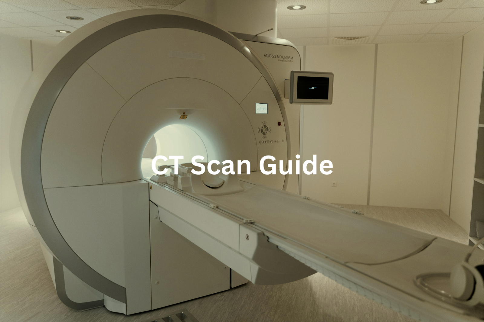
CT scans, or Computed Tomography scans, are like super-smart X-rays. They take lots of pictures from different angles and then piece them together to make a 3D image of the inside of your body. It’s a bit like building a model, but instead of a castle or a plane, it’s your organs.
Doctors often use CT scans to look at areas like the lungs, liver, or even to check for cancer. The images are much clearer than regular X-rays, which makes them great for spotting problems that might be hard to see otherwise.
But here’s the thing—CT scans use more radiation than normal X-rays. So, they’re only done when it’s really needed. Kind of like saving your best tools for the toughest jobs. I always think of CT scans as a kind of high-tech magic, letting doctors see what’s hidden inside. Pretty cool, but also something to use wisely.
PET Scan Benefits

A PET scan, which stands for Positron Emission Tomography, is a special medical test that helps doctors see how your organs are working inside your body. It uses a small amount of radioactive material mixed with a special dye (don’t worry, it’s safe). This mixture is injected into your body, and then a machine takes pictures of how your organs are doing their jobs.
PET scans are especially useful for spotting cancer. Cancer cells often use more energy than normal cells, so they light up on the scan. They’re also great for checking blood flow in the heart, which is really important for keeping your heart healthy.
What’s amazing is that PET scans can catch problems early—like finding a crack in a dam before it bursts. It’s like giving doctors a superpower to see things before they get worse. Pretty cool, right? Always follow your doctor’s advice if they suggest one.
Nuclear Medicine Explained
Nuclear medicine’s a bit different from other imaging techniques(3). It uses tiny amounts of radioactive materials (called radiotracers) to see how organs and tissues are working. This isn’t like X-rays that just show pictures of bones or structures—it’s more about function. For example, doctors can check how well your heart pumps blood or if your kidneys are filtering properly.
What’s really interesting is how nuclear medicine can help find cancer. Those radiotracers sort of “light up” areas where cells are growing too fast, which might mean cancer. It’s amazing how such a small amount of radiation can give doctors so much information. Like, a few milligrams—less than a pinch of salt—can make a big difference.
I think of it like a tiny investigator inside your body, looking for clues to keep you healthy. If you ever need a scan, don’t stress—it’s safe and helps doctors figure out what’s going on.
Ultrasound Imaging
Sources: Deakin University.
Ultrasound imaging is a fascinating way to look inside the body using sound waves. It works like this: a machine sends out high-frequency sound waves (too high for our ears to hear), and those waves bounce off organs and tissues. The echoes are turned into pictures by the machine. Pretty clever, right?
One of the most common uses is during pregnancy. It lets doctors see the baby growing inside the womb, and it’s completely safe and painless. But ultrasounds aren’t just for pregnancies. They’re also used to check organs like the liver, kidneys, or heart. If something’s not working quite right, an ultrasound can help doctors figure it out.
I like to think of it as a kind of invisible flashlight, shining sound waves into the body to see what’s happening. It’s amazing how something as simple as sound can give us such detailed images.
Fluoroscopy Applications
Fluoroscopy is a type of X-ray that works in real-time, almost like a live movie of the inside of your body. It’s used by doctors to watch how things move or function, like swallowing or blood flow. Pretty fascinating, right? Instead of just a still picture, fluoroscopy shows the action as it happens.
During the test, you might be asked to drink a special liquid (called a contrast agent) that makes certain areas of your body easier to see on the screen. This is especially helpful for looking at the digestive system—like spotting issues with swallowing or digestion.
I reckon it’s amazing how this technology lets doctors see what’s happening inside without needing surgery. It’s like having a peek through a window into the human body. If you ever need one, just remember it’s painless and helps doctors figure out what’s going on. Pretty handy tool, I’d say.
Doppler Ultrasound Uses
Blood flow is a bit like traffic on a busy road. It needs to move smoothly to keep everything working properly. That’s where Doppler ultrasound comes in. It’s a special type of ultrasound that checks how fast blood moves through your veins and arteries (those are the blood vessels in your body). Doctors use it to spot problems like blood clots or heart issues.
The best part? It’s non-invasive, which means no needles or cutting, and it doesn’t hurt a bit. It’s just a simple scan that uses sound waves to create images and measure blood flow. Kind of like a radar gun for your bloodstream.
This tool helps doctors see if there’s a blockage or if blood isn’t flowing the way it should. It’s quick, safe, and super helpful. If you ever need one, don’t worry—it’s just another way to keep your body’s “traffic” running smoothly.
Imaging Comparison
Each type of medical imaging has its own job. X-rays are quick and work well for looking at bones (like spotting fractures). MRIs, on the other hand, are better for soft tissues, such as muscles or the brain, because they show more detail. CT scans are great for creating 3D pictures of organs, which helps doctors see things like tumours or injuries. PET scans are different—they show how organs are working, not just what they look like.
Ultrasounds are super handy too. They’re safe and let doctors see real-time images, which is why they’re often used during pregnancy. Nuclear medicine is another option, helping find diseases by looking at how organs function. It’s like a toolbox. Each tool has its purpose. Together, they give doctors a full picture of what’s happening inside the body. That teamwork is what makes patient care so effective.
FAQ
What is CT (Computed Tomography) scanning and how does it work?
CT scanning, also known as a “CAT scan,” is a type of medical imaging that uses X-rays and computer technology to produce detailed images of the body’s internal structures. It allows doctors to see inside the body without making any incisions. CT scans can be used to diagnose a wide range of conditions, from cancer to lung disease, and are often used to guide medical procedures.
How is PET (Positron Emission Tomography) imaging used in healthcare?
PET imaging is a type of nuclear imaging that uses a small amount of radioactive material, called a tracer, to help detect and monitor diseases. The tracer is injected into the patient’s body and travels to the area of interest, where it emits positrons that are detected by the PET scanner. PET scans are often used to detect and monitor cancer, heart disease, and brain disorders.
What are the benefits of low-dose CT scans?
Low-dose CT scans use a lower amount of radiation than traditional CT scans, making them a safer option for patients who need multiple scans or for those who are particularly sensitive to radiation, such as children. Low-dose CT scans can be used to screen for lung cancer, detect heart disease, and diagnose other conditions with a reduced risk of radiation exposure.
How do MRI (Magnetic Resonance Imaging) scans work and what are they used for?
MRI scans use strong magnetic fields and radio waves to create detailed images of the body’s internal structures. MRI is particularly useful for imaging soft tissues, such as the brain, muscles, and organs, and is often used to diagnose conditions like tumours, joint injuries, and neurological disorders. MRI scans are safe and do not use ionising radiation like X-rays or CT scans.
What is the difference between CT and MRI scans?
CT and MRI scans are both types of medical imaging, but they use different technologies to create images of the body. CT scans use X-rays to produce detailed images of the body’s internal structures, while MRI scans use strong magnetic fields and radio waves. CT scans are often better for imaging bones and dense tissues, while MRI scans are better for imaging soft tissues like the brain, muscles, and organs.
How do 3D imaging and 3D printing technologies enhance medical care?
3D imaging technologies, such as CT and MRI scans, can create detailed, three-dimensional models of the body’s internal structures. These 3D images can be used to plan complex medical procedures, create custom medical devices, and even 3D print models of a patient’s anatomy for educational or surgical planning purposes. 3D imaging and printing can improve patient outcomes and reduce the time and cost of medical procedures.
What are the potential side effects of contrast media used in medical imaging?
Contrast media, also known as contrast agents, are substances that are injected into the patient’s body to enhance the visibility of certain tissues or organs during medical imaging. While generally safe, contrast media can sometimes cause side effects, such as nausea, headaches, or allergic reactions. Patients with kidney problems or other medical conditions may be at a higher risk of experiencing side effects from contrast media.
How does SPECT (Single-Photon Emission Computed Tomography) imaging work and what is it used for?
SPECT is a type of nuclear imaging that uses gamma rays to create three-dimensional images of the body’s internal structures. SPECT scans are often used to diagnose and monitor a variety of conditions, including heart disease, brain disorders, and certain types of cancer. SPECT imaging works by injecting a small amount of radioactive tracer into the patient’s body, which is then detected by the SPECT scanner.
How are artificial intelligence and deep learning technologies being used in medical imaging?
Advancements in artificial intelligence (AI) and deep learning algorithms are transforming the field of medical imaging. These technologies can be used to analyse medical images more quickly and accurately than human experts, helping to detect and diagnose a wide range of conditions. AI-powered image analysis can also be used to guide medical procedures, monitor patient progress, and personalise treatment plans based on individual needs.
Conclusion
Imaging tools are important in healthcare, helping doctors see inside our bodies. There are many types, like X-rays, which are quick and useful for bones, and MRIs, which show soft tissues in great detail. Each type has its own benefits. Learning about these tools can help us understand our health better. So, if you hear about an imaging test, you’ll know how it works and why it matters for your health decisions.
References
- https://www.nps.org.au/consumers/imaging-explained
- https://www.ranzcr.com/college/document-library/mri-safety-guidelines
- https://www.ansto.gov.au/education/nuclear-facts/nuclear-medicine-and-health#:~:text=Nuclear%20medicine%20uses%20small%20amounts,show%20how%20your%20organs%20work.