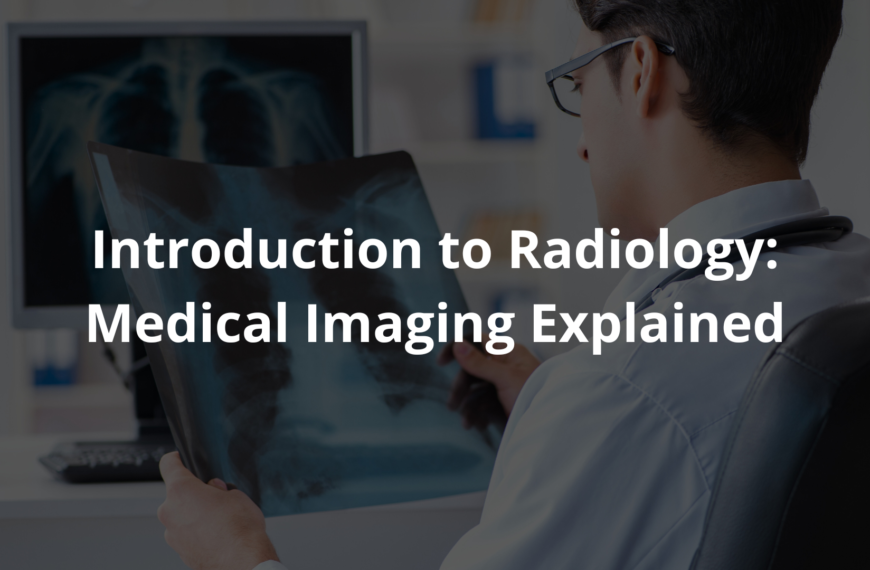
Imaging modalities explained: Discover how doctors see inside your body without surgery to find problems early and improve your health.
Imaging modalities are like special cameras that let doctors see inside our bodies without surgery. They use different techniques to create detailed pictures of organs and tissues, which helps find problems early. For example, X-rays show bones, while MRIs and CT scans provide clearer images of soft tissues.
This early detection is very important for treatment and recovery. When I first learned about these amazing technologies, I thought it was incredible how they help keep us healthy. If you wanna know more about how these machines work and their different types, keep reading!
Key Takeaway
- Imaging modalities help doctors see inside our bodies without surgery.
- There are different types like X-rays, CT scans, MRIs, and PET scans.
- Safety is important, and doctors use low doses of radiation to keep patients safe.
Understanding Imaging Modalities
Doctors have some pretty amazing tools to look inside our bodies without needing to cut us open. In Australia, there are over 8,000 imaging facilities (that’s a lot!) where people can get tests like X-rays, ultrasounds, and MRIs. In the past 20 years, Australians have had about 436 million imaging studies done. That’s more than 20 million every year. Imagine that!
Ultrasound is the most popular type of scan, making up 43% of all imaging tests. For every 100 Australians, about 106 imaging services are performed. That’s more than one scan per person! But it’s not always the same everywhere. People in cities usually have better access than those in the country. Still, Medicare helps a lot, with nearly 39% of Australians using these services in 2022-23.
Safety is a big deal too. The Australian Radiation Protection and Nuclear Safety Agency makes sure everything is done carefully(1). With new tech like artificial intelligence, these scans are getting even better. They help doctors find problems early and treat them faster. It’s pretty cool how much this has changed healthcare in Australia. If you ever need a scan, know it’s all about keeping you healthy.
X-rays: The Quick Snapshots
X-rays are probably the first thing people think of when it comes to medical imaging. They’re like taking a photo, but instead of capturing your smile, they show what’s inside—mainly your bones. Doctors use X-rays to check for fractures, infections, or even things like arthritis. The process is quick, usually just a few minutes, and it’s painless.
The machine sends out a small amount of electromagnetic radiation, which passes through your body to create an image. It might sound a bit worrying, but the radiation dose is very low, so it’s safe. I remember breaking my arm when I was nine.I had to sit still while the doctor positioned the X-ray machine.
The hardest part was not moving! After a short wait, the doctor showed me the image. “There’s the fracture,” she said, pointing to a thin line on the screen. It was fascinating to see my own bones. X-rays are a key tool in healthcare. They’re often the first step when diagnosing injuries or illnesses. If you ever need one, just stay still, and it’ll be over in no time.
CT Scans: The 3D View
The CT scan, or computed tomography scan, is like an upgraded version of an X-ray. It’s a bit more advanced because instead of just taking one image, it takes many X-ray pictures from different angles. Then, a computer puts all those images together to make a detailed 3D picture of what’s inside your body. It’s clever, really. This kind of scan helps doctors see things like organs, bones, and tissues much more clearly.
Picture this: if someone has a bad fall or is in a car accident, a CT scan can show if there’s internal bleeding or broken bones. The machine itself looks like a giant donut (a very serious donut). You lie on a table that moves through the middle while it takes the images. It’s quick and not scary, though it can be a bit noisy.
CT scans do use more radiation than regular X-rays, but doctors are careful about how much they use. They only do it when it’s really needed and keep the dose as low as possible. It’s pretty amazing how this technology works to help people heal and stay healthy. If you ever need one, just think of it as a high-tech way to get a peek inside without any cutting!
MRI: The Soft Tissue Expert

Magnetic Resonance Imaging, or MRI, is a pretty amazing tool. It’s like a camera for your insides, especially good at showing soft tissues like your brain, muscles, and even organs. What’s cool is that it doesn’t use radiation like X-rays do. Instead, it relies on strong magnets and radio waves (sounds fancy, right?).
I remember my first MRI. I was a bit nervous, to be honest. The machine is this big, tube-shaped thing, and it makes these loud, clunky noises. But the technicians were really kind. They told me exactly what to expect. “Just lie still,” they said, “it’ll take about 30 minutes.” They even talked to me through a little speaker, which made me feel less alone in there.
Some people might feel a bit squished or claustrophobic because the space is tight. But they’ve got ways to help, like giving you headphones or even a mirror to see out. MRIs are super useful for spotting injuries or checking how your organs are doing. They help doctors figure out the best way to treat you. If you ever need one, just remember to breathe and stay still—it’s worth it.
PET Scans: The Metabolism Detective
PET scans, or Positron Emission Tomography, are a type of medical imaging that helps doctors see how your body is working on the inside. They’re not just about looking at structures like bones or organs; they actually show activity, like how cells are using energy. This makes them super useful for spotting things like cancer, where cells might be growing or working faster than they should.
Before a PET scan, you’ll get a small amount of radioactive material (called a tracer). It’s injected into your body, and it travels to areas that are more active. Don’t worry—it’s a tiny amount, and it’s very safe. The tracer helps the scanner pick up on those active spots. Sometimes, PET scans are combined with CT scans (that’s Computed Tomography) to give a clearer, more detailed picture. Kind of like layering two maps to see all the details.
Doctors use PET scans to find problems early, which can make treatments work better. The radiation dose is small, and they always take care to keep it as low as possible. If you ever need one, just remember—it’s a tool to help you get the right care.
Why Are Imaging Modalities Important?
Medical imaging is like a window into the body, helping doctors see what’s going on inside without needing to cut anything open. It’s pretty amazing if you think about it. For example, mammograms (a special kind of X-ray) are used to spot breast cancer early, sometimes even before a lump can be felt. Early detection like this gives patients a much better chance at successful treatment.
There are heaps of imaging tools out there. X-rays, ultrasounds, MRIs, and CT scans are just a few. Each one works differently, but they all help doctors figure out what’s wrong. An ultrasound, for instance, uses sound waves to create pictures, which is super handy for checking on babies during pregnancy. MRIs, on the other hand, use magnets and radio waves to get detailed images of soft tissues, like the brain or muscles.
These tools don’t just help find problems—they also reduce the need for surgeries or other invasive procedures(2). Less poking around means less risk for patients. So, if you ever need an imaging test, it’s probably because your doctor wants the clearest picture to help you get better. Always ask questions if you’re curious; understanding what’s happening can make things less scary.
Safety Comes First
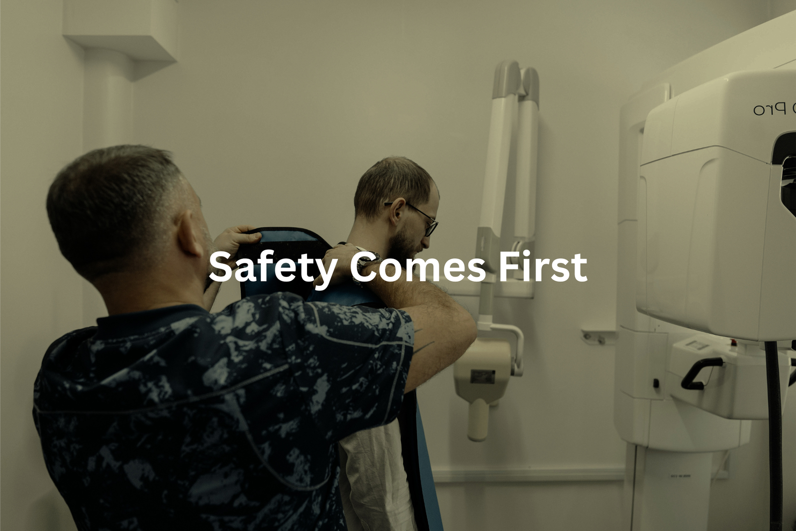
Imaging technologies, like X-rays and CT scans, are really helpful for doctors to see inside your body and figure out what’s going on. But safety is always the most important thing. In Australia, there are strict rules made by ARPANSA (that’s the Australian Radiation Protection and Nuclear Safety Agency) to protect patients during these procedures.
Doctors and radiologists are trained to use the smallest amount of radiation possible while still getting clear images. This is called the “ALARA principle,” which stands for “As Low As Reasonably Achievable.” It’s a fancy way of saying they’ll only use what’s absolutely needed. For example, a chest X-ray might use about 0.1 millisieverts of radiation, which is less than what you’d get from natural background radiation in a month.
If you ever feel unsure or nervous about having an imaging test, it’s okay to ask questions. Your doctor can explain why the test is needed and how they’ll keep you safe. Always speak up if you’re curious—it’s your health, after all.
Keeping Quality High
In Australia, there’s something called the Diagnostic Imaging Accreditation Scheme, or DIAS for short. It’s a program that makes sure imaging facilities, like those that do X-rays, MRIs, or CT scans, meet strict safety and quality rules. This is really important because it helps people feel confident that the care they’re getting is safe and reliable.
When a facility is accredited under DIAS, it means they’ve passed a bunch of checks and tests. These checks make sure the equipment is working properly, the staff are trained well, and the procedures follow national standards. It’s not just about the machines either—it’s about patient care too. For example, making sure images are clear and accurate so doctors can make the right decisions.
Facilities don’t just get accredited once and forget about it. They have to keep everything up to date, like renewing their accreditation every four years. It’s a way to keep improving and staying safe. If you’re ever getting an X-ray or scan, you might notice a DIAS certificate on the wall—that’s a good sign.
Trends in Imaging Modalities
Source: Sydney Health.
The way we use imaging in medicine is growing heaps. From 2000 to 2021, more than 436 million imaging studies were done. That’s a staggering number of scans! These include things like X-rays, MRIs, CT scans, and ultrasounds. Each one helps doctors see inside the body without needing surgery (which is pretty amazing if you think about it).
Now, with new technologies like artificial intelligence (AI), imaging is getting even better. AI can help spot tiny details in scans that might be easy to miss. For example, it can help detect early signs of diseases like cancer or heart problems. This means doctors can make faster and more accurate diagnoses, which could save lives.
The future of imaging looks bright. We might soon have tools that give us clearer, sharper pictures than ever before. And that means better care for patients. If you’re curious about this field, keep an eye on how AI keeps changing the game in medical imaging!
FAQ
What is an X-ray and how does it work?
X-rays are a type of high-energy radiation that can pass through the human body and create images of internal structures such as bones, organs, and blood vessels. An X-ray machine generates these X-rays using a specialised X-ray tube, which produces a beam of X-rays that are directed at the patient. The X-rays pass through the body, and the amount that is absorbed or scattered is detected and used to create a two-dimensional image on film or a digital screen.
How do CT scans differ from regular X-rays?
CT (computed tomography) scans use X-rays to create detailed, three-dimensional images of the human body. Unlike a regular X-ray that captures a single image, a CT scanner rotates around the patient, taking multiple X-ray images from different angles. These images are then assembled by a computer to generate a 3D representation of the internal structures, providing much more detailed information than a standard X-ray.
What is PET imaging and how is it used?
PET (positron emission tomography) is an imaging technique that uses a radioactive tracer, usually FDG (a glucose-based compound), to detect changes in the body’s metabolism and blood flow. PET scans are particularly useful for diagnosing and monitoring certain conditions, such as cancer, heart disease, and neurological disorders, by identifying areas of the body with abnormal activity or functioning.
How do MRI scans work and what do they show?
MRI (magnetic resonance imaging) uses powerful magnetic fields and radio waves to create detailed, three-dimensional images of the human body’s internal structures, including soft tissues such as the brain, organs, and muscles. MRI scans can provide high-quality, clear images without using ionising radiation, making them a valuable tool for diagnosing a wide range of medical conditions.
What are the different types of medical imaging techniques?
Medical imaging encompasses a wide range of technologies, each with its own unique capabilities and applications. In addition to X-rays, CT scans, PET, and MRI, other common imaging techniques include ultrasound, which uses sound waves to create images, and SPECT (single-photon emission computed tomography), which captures gamma rays emitted by radioactive tracers.
Each imaging type provides different information about the body’s structure and function, allowing healthcare professionals to make accurate diagnoses and develop effective treatment plans.
How can I minimise the risks of medical imaging?
While medical imaging is generally safe, there are some potential risks, such as exposure to radiation or side effects from contrast dyes. To reduce these risks, healthcare providers may recommend lower-dose imaging techniques, such as low-dose CT scans, or use alternative imaging methods when appropriate.
Patients can also play a role by asking questions about the imaging process, any potential risks, and ways to minimise exposure, especially for repeat or long-term imaging needs.
Conclusion
Imaging tools like X-rays, CT scans, MRIs, and PET scans are amazing parts of modern healthcare. They let doctors see inside our bodies without needing surgery. This means they can find problems early and keep us safe. Every time you hear about these tests, remember they play a big role in looking after our health. Understanding how they work shows us how important they are for helping doctors care for patients effectively. It’s all about keeping us well!
References
- https://ccdcare.com/resource-center/radiology-modalities/
- https://www.sciencedirect.com/topics/computer-science/imaging-modality