Explore how radiomics applications are changing cancer treatment in Australia, offering insights into benefits, challenges, and future directions.
Sometimes, medical breakthroughs seem to hide in plain sight, waiting to be uncovered. Radiomics is one of those quiet revolutions, giving doctors in Australia a deeper look into the human body. It’s not just about scans anymore—CTs and MRIs are now tools for extracting layers of data, turning images into valuable insights.
This technology is especially powerful in cancer care, helping doctors personalise treatments and predict outcomes. It’s like giving them a clearer map to navigate complex diseases. Curious about how this works and what it means for patients? Keep reading to explore the impact of radiomics on cancer care.
Key Takeaway
- Radiomics helps doctors understand cancer through detailed image data.
- It can improve how quickly and accurately doctors treat patients.
- There are challenges, but the future looks bright for radiomics in medicine.
Understanding Radiomics
Radiomics is a clever way for doctors to get more out of medical images, like CT or MRI scans (the ones that show detailed pictures of the inside of the body). It’s not just about spotting a tumour or a problem—it’s about digging deeper. Radiomics pulls out loads of details, like the texture, shape, and size of a tumour. These details can tell doctors a lot about what’s happening inside a patient’s body, especially with diseases like cancer.
Take lung cancer, for example. Radiomics can help doctors figure out whether a tumour is growing quickly or slowly. That information can guide them to choose the best treatment. In Australia, where cancer treatment is a big focus, this kind of technology is making a real difference.
There’s a story about a doctor who used radiomics to treat a patient with a brain tumour. The scans didn’t just show the tumour itself—they also revealed how it was affecting the tissue around it. With that extra information, the doctor could plan a treatment that was more precise and effective. Radiomics is like having a detailed map that helps doctors find the best path forward.
The Role of RANZCR
The Royal Australian and New Zealand College of Radiologists (RANZCR) plays a key role in how radiomics is used in hospitals and clinics. They’re the ones making sure this technology is safe and works well for patients.
RANZCR’s guidelines focus on two main things:
- Safety First: They set rules to make sure patients are protected when new radiomics tools are used.
- Quality Care: They help doctors learn how to use radiomics properly, so patients get the best possible treatment.
These guidelines are followed by many hospitals in Australia, and they give patients confidence that their care is in good hands. RANZCR also runs training sessions for doctors and technicians, teaching them how to read and use radiomic data correctly. It’s a team effort, with everyone working together to improve patient care. [1]
Challenges in Using Radiomics
Radiomics is exciting, but it’s not perfect. There are some challenges that doctors and researchers are still working through:
- Reproducibility: The results from radiomics can vary depending on the machine used to take the images. It’s a bit like how photos look different depending on the camera. This can make it tricky for doctors to trust the data completely. [2]
- Training Needed: Not all doctors and technicians are experts in radiomics yet. If they don’t follow the same steps, the results can be inconsistent.
- Ongoing Research: Scientists are still testing and improving radiomics. There’s more to learn before it can be used for every patient.
One doctor shared how their hospital struggled with reproducibility. They found that scans from different machines gave slightly different results. To fix this, they created a standard process for taking and analysing images. It took time, but now their results are much more reliable. It shows that while there are hurdles, they’re not impossible to overcome.
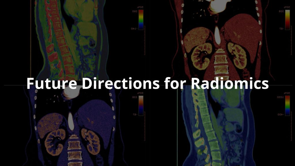
Future Directions for Radiomics

The future of radiomics looks promising, with plenty of room to grow. Here’s what’s on the horizon:
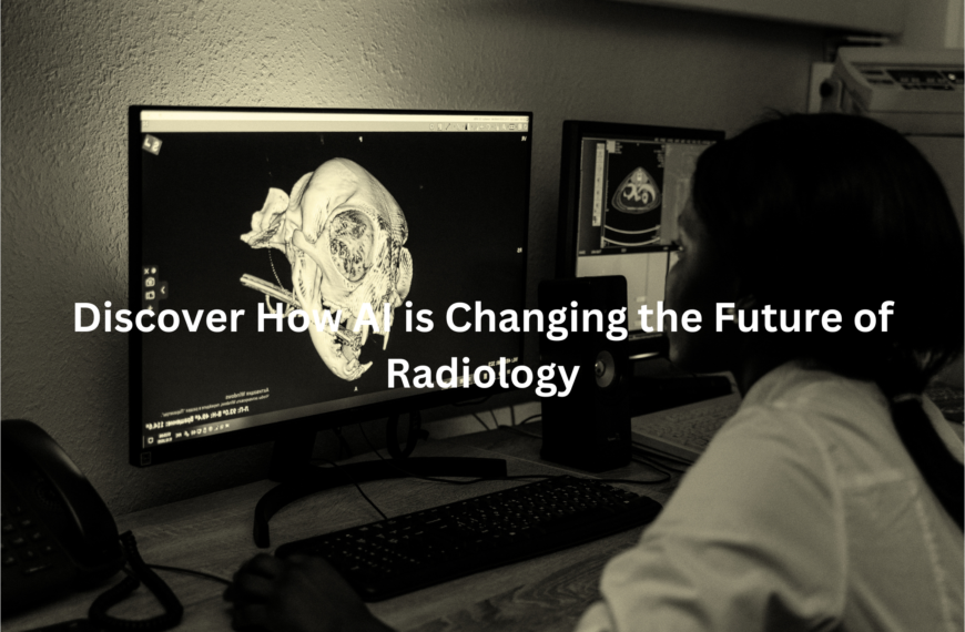
- Better AI Integration: Computers that can learn (artificial intelligence or AI) are already helping doctors analyse radiomics data. AI can sort through huge amounts of information quickly, spotting patterns that might be missed otherwise.
- More Applications: Right now, radiomics is mostly used for cancer, but researchers are exploring how it could help with other conditions, like heart disease or brain disorders.
- Collaboration: Hospitals and researchers are sharing data to build better radiomic models. By pooling their information, they can make the technology more accurate and useful for patients.
Collaboration is especially exciting because it’s like putting together a giant puzzle. Each hospital or researcher adds a piece, and together they create a clearer picture. This teamwork could make radiomics even more effective in the years to come.
For now, radiomics is still growing, but it’s already changing the way doctors look at medical images. If you’re curious about it, keep an eye on how hospitals and researchers are working to make it even better. It’s a field that’s definitely worth watching.
FAQ
What role does deep learning play in analysing MRI features and CT images for cancer diagnosis?
Deep learning helps doctors find patterns in medical images that humans might miss. The technology can look at both MRI and CT scans at once, making it easier to spot cancer early. It’s especially helpful when looking at complex cases where traditional methods might not be enough.
How do imaging data and tumour volume measurements help determine risk factors?
Doctors use detailed imaging measurements to figure out how risky a tumour might be. By looking at the size and shape of tumours in scans, they can better predict if a patient has low risk or high risk cancer. This helps them choose the right treatment plan.
What makes second order texture analysis useful in early stage cancer detection?
This special way of looking at medical images helps find subtle patterns in CT texture that regular analysis might miss. It’s particularly good at finding early stage cancers, which can lead to better free survival rates for patients.
How do radiomics applications benefit head and neck and breast cancer diagnosis?
Radiomics helps doctors study both head and neck and breast cancer by turning medical images into useful data. It can spot small changes in lymph nodes and tumour tissue, which helps create better treatment plans and improve patient outcomes.
What are the key quality score factors in model based radiomics research?
A good study design and proper sample size are crucial for reliable radiomics research. Random forest methods often help validate findings. Open access to imaging data lets other researchers check and improve on the work.
What’s the connection between tumour imaging and squamous cell cancer detection?
Advanced tumour imaging techniques help doctors spot and track squamous cell cancers. These methods look at gray level patterns in scans, which helps tell the difference between cancer types and normal tissue.
How are rectal cancer and lower grade tumours analysed using radiomics?
Radiomics helps doctors study both rectal cancer and lower grade tumours by looking at detailed patterns in medical scans. This technology can spot tiny differences that help predict how well treatment might work.
What makes pilot study results important in mri radiomics research?
Pilot studies help test if new MRI radiomics methods actually work in real life. They’re like trial runs that show if bigger studies are worth doing, especially for medical imaging based research.
How do doctors use PET scans and special medical images to find cancer?
When doctors use FDG PET and other PET images, they can see where cancer is active in your body, just like using a special flashlight to find things in the dark. These tools help spot cancer early and show if medicines are working right.
Why do doctors look at both MRI and CT scans together?
DCE MRI and CT based scans work like a team – one shows how blood moves through your body, while the other takes detailed pictures of your insides. It’s like having both a video camera and a regular camera to get the whole story.
How does molecular imaging enhance cancer-based diagnosis?
Molecular imaging helps doctors see how cancer behaves at a tiny level. This helps them understand why some treatments work better than others and lets them spot problems early, which can lead to better patient outcomes.
What makes radiomics valuable for brain tumour analysis?
Radiomics helps doctors study brain tumours by turning detailed scan images into useful information. This helps them tell different types of brain tumours apart and choose the best treatment path for each patient.
How do physicians use imaging-based biomarkers to track treatment progress?
Doctors use special markers they can see in medical images to check if treatment is working. These imaging-based signs help them know early on if they need to change their approach, rather than waiting for other symptoms to show up.
What role do nuclear medicine techniques play in modern cancer diagnosis?
Nuclear medicine techniques give doctors a unique way to see how cancer cells are behaving in the body. These methods help spot cancer early and show whether treatment is working, especially when combined with other types of scans.
How do radiomics applications help with determining sample size for clinical trials?
Radiomics helps researchers figure out how many patients they need for their studies to be reliable. This is crucial for making sure their findings about cancer treatments are trustworthy and can help other patients.
What makes this field important for understanding tumour heterogeneity?
By looking at detailed patterns in medical images, doctors can better understand how tumours vary within themselves. This helps them spot which parts of a tumour might be more dangerous and need different treatments.
Conclusion
Radiomics is changing how cancer is treated in Australia. By converting medical images into detailed data, it helps doctors create personalised treatment plans. Groups like RANZCR ensure it’s used safely and properly.
While challenges remain, radiomics shows promise for better patient care. It’s not just a buzzword—it’s a tool with real potential. With ongoing research and teamwork, radiomics could transform cancer care, making it more accurate and helpful for patients across the country.
References
- https://www.tga.gov.au/sites/default/files/submissions-received-draft-clinical-evidence-guidelines-medical-devices-ranzcr.pdf
- https://pmc.ncbi.nlm.nih.gov/articles/PMC8472571/


