Learn about biopsies guided by radiology, what they involve, and how they help doctors diagnose health issues.
Walking into a hospital can feel a bit scary for anyone. There are bright lights, nurses rushing, and that distinct smell of antiseptic in the air. But there’s something comforting about how doctors and machines work hand-in-hand to care for patients. A crucial part of this teamwork is a procedure known as a biopsy.
Biopsies with radiology are unique because they allow doctors to take a small piece of tissue from the body, helping to check for diseases. If you’re curious about how these imaging techniques aid in patient care and the steps involved in a biopsy, keep reading.
Key Takeaway
- Biopsies guided by radiology use imaging techniques like CT scans or ultrasounds.
- These procedures help doctors get tissue samples for testing.
- It’s important to follow the guidelines for safety during these procedures.
What is a Biopsy with Radiology?
A biopsy with radiology is a way doctors take a little piece of tissue from the body for testing. They use special imaging tools, like CT scans or ultrasounds, to see exactly where to collect the sample. This helps make the whole process safer and more accurate.
For instance, if a doctor sees a lump in a person’s breast or a strange area in the liver, they might suggest doing a biopsy to find out what’s going on. With the imaging, the doctor can guide a needle right to the trouble spot. This way, they get a good sample for testing.
Biopsies are often done when doctors see something concerning. They want to find out if a lump is something serious, or if it’s nothing to worry about. They might say, “We need to check this further.” Radiology is like having a treasure map in this search. Instead of groping around in the dark, doctors can see exactly where to go.
The needle is carefully inserted to collect the tissue, and the doctors need to be sure they get enough. Sometimes they might even take several samples, depending on what they find. This careful approach helps doctors make the best decisions about what to do next.
Types of Biopsies
There are different types of biopsies doctors might do with the help of radiology. Let’s look at a few:
Core Biopsy
A core biopsy uses a thicker needle to extract a small piece of tissue, usually about 1–3 mm in diameter. It’s often used for checking trouble in the liver or breast. The needle is guided in by imaging, which helps the doctor make sure it’s in the right spot.
Core biopsies are popular because they provide a bigger tissue sample. This allows the doctors to see more details when they look closely under a microscope. The radiologist will study the images carefully before taking the sample to be sure they’re on target. It’s crucial because if the wrong tissue is taken, it could lead to wrong diagnoses.
Fine Needle Aspiration (FNA)
FNA uses a very thin needle, kind of like a tiny straw, to take out smaller samples of tissue. This is really good for things like swollen lymph nodes. It’s also guided by imaging to make sure it goes smoothly.
When a doctor thinks there might be an issue in the lymph nodes or thyroid, they might suggest FNA. It’s quick, often done in a clinic, and gives doctors quick answers without needing a big surgery. [1]
Surgical Biopsy
If the other types of biopsies can’t provide enough information, a surgical biopsy may be necessary. This is when doctors make a larger cut to get the sample. This type usually takes longer to heal.
Surgical biopsies are needed when doctors are really unsure about what’s wrong. They might say, “We need to take a closer look inside.” This type is more invasive, but it can give a lot of detailed information about what’s happening.
Procedure Details
Before getting a biopsy, patients usually need to prepare a little bit. Here’s a general step-by-step view:
Preparation
First, the doctor does an imaging test to pinpoint where the sample will be taken from. They explain everything that will happen and make sure the patient agrees to have the biopsy done. The doctor might also ask about medications, especially blood thinners, which could affect the procedure.
Anesthesia and Sample Collection
Doctors usually use a local anaesthetic, which numbs the skin, so the patient doesn’t feel pain during the procedure. This step helps patients feel more relaxed. After that, the doctor makes a small cut and inserts the biopsy needle while watching the images on a screen to make sure they’re getting it right.
Post-Procedure Care
After the biopsy, the doctor presses on the area to stop any bleeding and keeps an eye on the patient for a short while to check that everything is okay. The biopsy results will be sent to the doctor who ordered it.
Patients might feel a little sore afterward, but that’s normal. They may be advised to take it easy for a few days as they heal properly.
Role of Radiologists
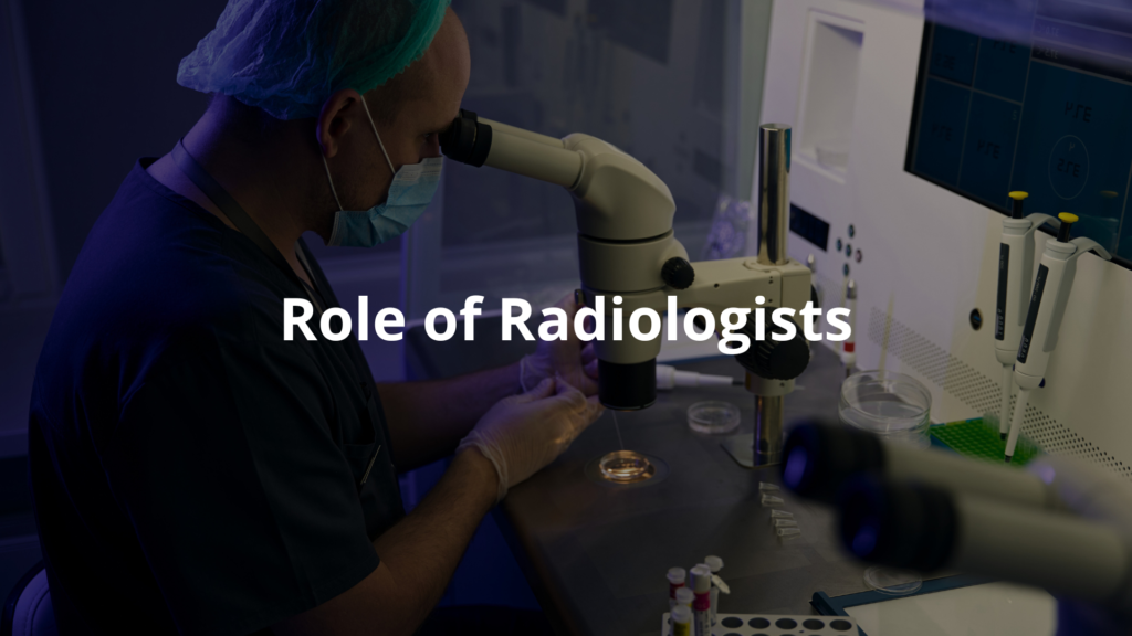
Radiologists are doctors who specialize in using imaging to find out what’s wrong. They play a key role in the biopsy process. Their focus is on ensuring that everything is done safely and correctly.
These specialists often have a special set of skills. They operate imaging machines, like CT or ultrasound machines, which help them see inside the body. Sometimes, they work with pathologists who examine the tissue samples under a microscope looking for diseases.
The teamwork between radiologists and pathologists is really important. One takes the sample, while the other interprets it, forming a clear understanding of what’s happening in a patient’s body.
Guidelines and Standards
In Australia, the Royal Australian and New Zealand College of Radiologists (RANZCR) offers guidelines to help doctors carry out biopsies safely. [2] They focus on:
- Keeping patients safe during the procedure.
- Using the right imaging tools.
- Having clear ways to report the findings so everyone understands the results.
These guidelines help standardize the process everywhere, ensuring that patients receive high-quality care. When patients go in for a biopsy, they can rest easy knowing there are strict protocols in place to keep things running smoothly.
Why are Biopsies Important?
Biopsies are a really important tool for diagnosing diseases. They help find out if a lump is cancerous or something else. By using imaging techniques, doctors make sure they get the right tissue sample, leading to better diagnoses and treatments for patients.
Without biopsies, doctors would have to guess, which isn’t safe or effective. By getting a tissue sample, they can make informed decisions that might mean starting treatment sooner or avoiding unnecessary surgery. It’s like having a map guiding them to the right conclusions.
FAQ
What happens during a breast core biopsy and how is imaging guidance used?
During this minimally invasive procedure, your specialist doctor uses imaging guidance (like ultrasound guidance) to locate the area of concern in your breast. They’ll numb the skin and use a special needle to take small samples of tissue. The whole process typically takes about 30 minutes.
How do doctors perform image guided liver biopsy procedures?
Your doctor uses ultrasound or CT guidance to safely access your liver tissue through your abdominal wall. They’ll numb the skin before the needle is inserted. This type of procedure is more precise than open surgical methods and helps your doctor get the exact sample of tissue needed.
What should I know about different types of needle biopsies?
There are several kinds, including fine needle and core needle biopsies. The choice depends on the area being tested. Guided biopsies use medical imaging like computed tomography to help doctors perform the biopsy accurately and safely.
What preparation is needed before going to the radiology clinic for a biopsy?
You’ll need to sign a consent form and talk with your referring doctor about the test or procedure. Tell them about any medications you take. Med radiology staff will inform you about specific preparations needed, including whether you should avoid strenuous activity beforehand.
How do doctors determine if deep tissue or lymph node biopsies are needed?
Your specialist uses imaging guided procedures to examine soft tissues and determine whether tissue samples look cancerous or benign. The biopsy site is chosen carefully using imaging guidance to ensure accurate sampling.
What happens during ultrasound guided breast biopsies?
The biopsy is performed using real-time ultrasound to guide the needle precisely to the area of concern. Nuclear medicine or other imaging techniques might be used depending on the area being examined. This helps ensure accurate sampling while keeping you comfortable.
Conclusion
Biopsies with radiology are really important for getting tissue samples that help doctors figure out what’s wrong with patients. Using imaging techniques, these procedures are safe and effective. Following guidelines from groups like RANZCR makes sure people get the best care during their biopsy.
If you ever need one, remember it’s a common procedure and don’t be shy to ask your doctor questions. Knowing what’s going on is important. It helps you feel more in control of your health.
References
- https://www.melbourneradiology.com.au/interventional-radiology/core-biopsy/
- https://www.ranzcr.com/images/Pathology_A_Guide_to_Candidates.pdf