Learn the essentials of diagnostic X-rays and their importance in medical diagnosis.
Diagnostic X-rays are fascinating, truly. They allow doctors to look inside our bodies without a single cut. Imagine a child with a broken arm; the worry in their eyes is replaced by wonder when an X-ray is taken. The images reveal the bones in a way that has the power to calm fears.
Doctors use these X-rays for all sorts of reasons, like spotting broken bones or checking for illnesses. So, why are these pictures so important for our health? You’re about to find out. Keep reading to uncover the magic and significance of diagnostic X-rays in everyday life!
Key Takeaway
- Diagnostic X-rays help doctors see inside our bodies to find problems.
- They use a small amount of radiation, which is usually safe.
- There are different types of X-rays, like CT scans and mammograms.
What Are Diagnostic X-Rays?
Diagnostic X-rays are sort of like magic cameras that take pictures inside our bodies. They help doctors peek at our bones, organs, and soft tissues. For example, if someone hurts their leg, a doctor might use an X-ray to check for a broken bone. It’s like having a superpower that shows hidden things. With these images, doctors can spot infections or even something serious like cancer.
There are different types of X-rays:
- Plain X-rays: The most common type. They help find broken bones and check if something’s wrong with the chest.
- CT Scans: These use many X-ray beams and snap lots of pictures to create a detailed image of the inside of your body. They can show your brain, organs, and blood vessels!
- Mammograms: These special X-rays check for breast health. They help catch breast cancer early when treatment can be better.
X-rays do a lot more than just show bones. It’s pretty cool how a simple machine can help with so much!
How Do X-Rays Work?
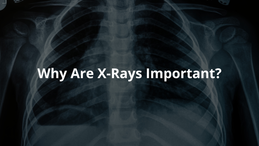
X-rays work by sending invisible rays through the body. Some parts, like bones, take in more of these rays, which makes them show up white on the X-ray image. Softer tissues, like muscles and organs, let more rays pass through, making them look darker.
Think of shining a flashlight through your fingers onto a wall – the light shows where your fingers are. Just like that, X-rays create pictures showing bones and organs hiding inside the body.
Key Points:
- X-rays pass through the body, capturing images on special film or a digital plate.
- How body parts absorb the rays makes clear images.
- These images help doctors find issues like fractures, infections, or other problems.
- X-rays give a ‘map’ of what’s inside the body, helping doctors figure things out accurately.
This technology plays a big part in healthcare, showing clear pictures of internal structures to lead to better medical choices.
What Happens During an X-Ray?
When someone gets an X-ray, they usually wear a special gown to keep the picture clear. Patients have to take off their jewelry and metal objects like belts because those can block the rays. The actual imaging takes just a few minutes, but it feels like nothing at all!
The technician, who knows exactly how to take X-ray images, will ask the patient to stand or lie down in a certain way. They may need to hold their breath for a tiny bit, but it’s super quick! The machine makes a buzzing sound, and then it’s all done. If patients feel a little nervous, that’s okay—there’s usually nothing to worry about.
Safety and Risks
You might be curious about whether X-rays are safe. They do use a small amount of radiation, but doctors are very careful. An average person in Australia gets about 1.7 mSv (milliSieverts) of radiation each year from medical procedures, so it’s pretty low. [1] Most of the time, the benefits of finding out what’s wrong are way more important than the tiny risk from the rays.
If someone’s pregnant or thinks they might be, it’s super important to tell the doctor. They have special ways to keep an unborn child safe.
What to Expect After an X-Ray
After the X-ray, a special doctor called a radiologist looks at the pictures. They check for anything unusual. Then, they write a report. Patients usually get their results from their doctor, and it’s often within a day or so unless it’s really urgent.
Sometimes, the doctor might want to do more tests based on what they see. This can be normal, so there’s no need to feel scared. It’s all part of making sure everything is okay.
Why Are X-Rays Important?

X-rays are super important in healthcare. They help doctors find problems like broken bones, infections, and tumors. By giving clear images of what’s inside the body, X-rays help doctors diagnose things faster and more accurately, which means patients can get the right treatment sooner.
Key Points:
- X-rays help identify fractures, infections, and tumors.
- In Australia, millions of X-rays are done each year, showing how essential they are in medical care.
- For example, a chest X-ray can quickly diagnose issues like chest pain.
- Proper imaging often leads to life-saving treatments.
X-rays are a crucial tool, helping improve healthcare for everyone.
Advancements in X-Ray Technology
X-ray technology keeps improving. Now, there are digital X-rays that offer clearer images with less radiation. [2] This makes it easier for doctors to see what’s going on inside a person’s body without putting them at risk.
New machines can create 3D images, which helps doctors plan treatments better. It’s exciting to think about how technology can help us stay healthy! This means more accurate diagnoses and safer exams for everyone.
If you or someone you know is going to have an X-ray, just remember it’s a simple and important part of staying healthy!
FAQ
What are diagnostic x-rays and how do they create images of inside your body?
X-ray imaging uses ray beams to generate images of what’s happening inside your body. Unlike visible light, these ray beams pass through your body and are picked up by a ray detector. The machine can then create an image showing your bones and other structures clearly.
How do different type of imaging tests like CT scans and plain radiograph work?
While regular x-ray procedures take single pictures, CT scans take many pictures from different angles. Both use ray beams, but CT scans create more detailed images. A plain radiograph is what most people think of as a regular x-ray – it’s great for looking at bones and some soft tissues.
What should I know about radiation safety and radiation doses during medical exposure?
Healthcare providers follow strict radiation safety guidelines and reference levels to keep radiation doses as low as possible while still getting good image quality. The form of radiation used is carefully controlled, and ray equipment is regularly checked to ensure patient safety.
When might I need contrast media or contrast dye during ray procedures?
Sometimes your doctor might use a contrast medium (special dye) to make blood vessels and other parts show up better on imaging exams. While most people handle contrast agent well, some might have an allergic reaction. Your medical imaging team will carefully check if it’s right for you.
How do imaging services help diagnose disease like bone cancer or breast cancer?
Imaging tests are vital tools that help your referring doctor diagnose disease accurately. They can spot things like bone cancer, check bone density, assess heart health, and detect breast cancer early. After your ray examination, a written report helps your doctor plan your care.
What should I expect during my ray procedures at the medical center?
You might need to wear a hospital gown and remove foreign objects like jewellery. A ray technologist will position you and the ray machine to get the best images possible. Whether using film or digital methods, they’ll make sure to get high-quality images while protecting your safety.
How can I prepare for my imaging exams?
Your healthcare team will tell you if you need any special preparation. Sometimes you might need to avoid eating or drinking before certain imaging tests. Be sure to tell your ray technologist if you’re pregnant or have any health concerns.
What happens after my ray examination?
Medical imaging experts will study your images and send a detailed report to your healthcare provider. Your referring doctor will then discuss the results with you and plan any needed next steps for your health care.
Conclusion
Diagnostic X-rays are key tools in medicine. They let doctors peek inside our bodies to find and fix health issues. With proper safety measures and new tech, X-rays remain a safe and effective way to assist folks in Australia and beyond. So, if someone needs an X-ray, they can feel more at ease knowing what to expect. It’s just another way doctors help keep us healthy and catch problems early. X-rays really make a difference!
References
- https://www.arpansa.gov.au/understanding-radiation/what-is-radiation/ionising-radiation/x-ray
- https://www.melbourneradiology.com.au/guides/digital-xray/


