Worried about scans during pregnancy? Explore safe imaging alternatives like ultrasounds and MRIs to protect you and your baby.
Pregnancy scans offer doctors a clear view of both mum and bub’s health. Two common methods stand out in Australian medical centres – ultrasounds and MRIs (magnetic resonance imaging). Ultrasound machines send sound waves through the belly, creating black-and-white pictures of the growing baby. These scans don’t hurt and take about 30 minutes.
MRI machines make detailed images using magnetic fields, showing more than ultrasounds can see. Doctors pick the right scan based on what they need to check. Both methods are safe, giving mums peace of mind during their pregnancy journey. Keep reading to learn more about why these techniques matter during pregnancy.
Key Takeaway
- Ultrasounds and MRIs are safe options for pregnant women.
- CT scans and X-rays can expose the baby to radiation.
- NIPT is a great non-invasive test for checking genetic issues early.
Ultrasound: The First Choice for Pregnant Women
Ultrasound scans create moving pictures of babies before birth using sound waves(1). These waves bounce off the baby’s body, making black-and-white images on a screen. The process needs no needles or radiation, making it safe for mums and bubs.
Australian doctors use ultrasound scans (operating at 1 to 20 MHz) to check pregnancy progress. The scan shows:
- Baby’s size and growth
- Heart activity
- Position in the womb
- Amount of amniotic fluid
- Placenta location
Most pregnant women get two main scans in Australia. The first scan happens around 12 weeks, showing early development. The second scan at 20 weeks checks the baby’s organs and growth in detail. Some medical centres offer advanced 3D and 4D imaging for better views of the baby’s features.
Extra scans might be needed if doctors spot anything unusual. These help monitor both mum and baby’s health throughout pregnancy.
MRI: A Safe Alternative to CT Scans
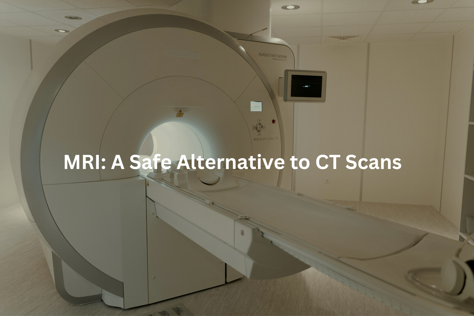
MRI scans offer deeper insights when ultrasounds can’t tell the whole story. These machines use magnets and radio waves (1.5 to 3 Tesla strength) to create detailed pictures without any radiation, making them safe for pregnant women and their babies.
Medical research supports MRI use during all pregnancy stages, including the first trimester(3). The process works by aligning hydrogen atoms in the body with magnetic fields, then measuring their movement with radio waves to build clear images.
Common reasons for pregnancy MRIs include:
- Checking placental health
- Investigating persistent pelvic pain
- Monitoring foetal development in high-risk cases
- Examining maternal tissue concerns
The procedure takes about 45 minutes in a large, tube-shaped machine that makes loud humming noises. Patients need to lie still for clear images. Most pregnancy check-ups won’t require an MRI, as ultrasounds typically provide enough information. But when needed, these scans offer valuable insights without added risks.
NIPT: A Different Kind of Testing
Non-Invasive Prenatal Testing (NIPT) stands out among medical screenings for its simplicity and safety(2). A single blood draw from the mother reveals crucial genetic information about the baby, without any risks to either of them.
The test screens for common chromosomal conditions:
• Down Syndrome
• Edwards Syndrome
• Patau Syndrome
By examining bits of fetal DNA in maternal blood (present from 10 weeks of pregnancy), NIPT provides early results with remarkable accuracy – over 99% for Down Syndrome detection. Many Australian clinics bundle NIPT with ultrasound services, creating a comprehensive screening package.
While NIPT excels at genetic screening, it doesn’t detect physical anomalies like heart defects. That’s where ultrasound scans become essential. Doctors recommend discussing NIPT options before the 10-week mark, as testing windows are specific. Medicare doesn’t cover NIPT costs (roughly $400-500), though some private health funds might offer partial rebates.
Considering the Risks with CT Scans
Sources: Radiology Video – Radiology Made Esay.
Medical imaging during pregnancy needs careful thought. Doctors must balance getting clear pictures with keeping mum and bub safe. CT scans and X-rays use ionising radiation, which might affect a developing baby.
CT scans create detailed body images that help spot serious problems:
• Lung blood clots
• Internal injuries
• Bad appendix cases
The radiation adds up differently for each scan type:
• Chest CT: 7 mSv (millisieverts)
• Head CT: 2 mSv
• Basic chest X-ray: 0.1 mSv
Safer options exist for most pregnancy checks. Ultrasound machines use sound waves instead of radiation, making them the go-to choice. MRI machines (which use magnets) work well too, though they take longer.
When CT scans can’t be avoided, special lead shields protect the baby area. Medical staff position these carefully to block scattered rays. Pregnant patients should ask their doctor about radiation-free alternatives first.
Making the Best Choice for Health Care

Pregnancy imaging tests need careful thought. Doctors match different scans to specific needs during pregnancy, making sure they’re both safe and useful.
The timing matters a lot. NIPT (Non-Invasive Prenatal Testing) works best around 10 weeks, while detailed anatomy scans happen at 20 weeks. Each test serves a different purpose – ultrasounds check the baby’s growth and organs, MRIs look at soft tissues, and NIPT screens for genetic conditions.
Safety comes first in pregnancy scans. Doctors avoid X-rays and CT scans when possible, since they use radiation. Instead, they pick safer options like ultrasound.
The cost of these tests varies (some reaching over $500), and not all insurance plans cover them. Patients should talk with their healthcare provider about:
- Medicare coverage options
- Which tests they actually need
- When to schedule them
- What each test checks for
FAQ
What is a CT scan and how does it work for pregnancy?
A CT (computed tomography) scan is an imaging test that uses X-rays to take detailed pictures of the body. For pregnant women, a CT scan can be used to examine the pelvic area and detect conditions like pelvic pain or soft tissue problems. The CT scan provides high-quality images, but it does expose the foetus to a low dose of radiation, so doctors will only recommend it when absolutely necessary.
What are the risks of radiation from imaging tests during pregnancy?
Radiation exposure from medical imaging tests like CT scans and nuclear medicine scans can pose a small risk to the developing foetus. The amount of radiation depends on the type of test, but in general, the risks are low, especially for tests done early in pregnancy. Doctors always aim to use the lowest dose of radiation possible and will only order these tests when the benefits outweigh the potential risks.
How do MRI scans compare to other imaging options for pregnant women?
MRI (magnetic resonance imaging) is often the preferred imaging method for pregnant women because it doesn’t use ionising radiation like X-rays or CT scans. MRI scans can provide detailed images of the foetus and placenta without exposing the baby to radiation. There is no evidence that MRI scans increase the risk of birth defects or other adverse effects, even when done in the first trimester.
When would a doctor recommend a nuclear medicine scan during pregnancy?
Nuclear medicine scans, like a perfusion scan, use small amounts of radioactive materials to create images of the body. These tests are generally avoided during pregnancy if possible, as they do expose the foetus to a small amount of radiation. However, in some high-risk cases, a nuclear medicine scan may be recommended to help diagnose a serious condition when the benefits outweigh the minimal radiation risks.
How can pregnant women reduce their health risks from imaging tests?
Pregnant women can take some steps to minimise the potential health risks from imaging tests. Choosing MRI over CT scans or nuclear medicine when possible can reduce foetal radiation exposure. Disclosing pregnancy to the healthcare team is also important, so they can tailor the test to be as safe as possible. And for any imaging test, it’s best to only undergo it when absolutely medically necessary.
Conclusion
Medical imaging during pregnancy needs careful thought. Ultrasounds and MRIs (magnetic resonance imaging) stand out as safe choices for both mother and baby. These methods let doctors check the baby’s growth without radiation risks.
Ultrasounds use sound waves to create pictures, while MRIs use magnetic fields and radio waves. Doctors pick the right scan based on what they need to see, and patients should ask questions about each option’s benefits. The right imaging helps track pregnancy progress safely.
References
- https://www.nps.org.au/news/ultrasound-during-pregnancy
- https://www.sonicgenetics.com.au/nipt/
- https://www.healthdirect.gov.au/magnetic-resonance-imaging-mri