This article gives you a clear view of imaging comparisons like CT and MRI scans, helping you understand their differences and uses.
When I first walked into a medical imaging centre in Palo Alto, I was amazed. The machines looked like something from a sci-fi movie. Big, shiny, and complex. I wanted to learn all about them. Imaging comparison is important because it helps doctors choose the right tests for their patients. You might have heard of CT scans and MRI scans, but do you know how they are different? Let’s explore this together.
Key Takeaway
- CT scans use X-rays to create detailed images of the body.
- MRI scans use strong magnets and radio waves, making them safer as they don’t use radiation.
- Both imaging types help doctors see inside our bodies to diagnose and treat conditions.
Understanding Imaging Comparisons
In a bright, bustling medical imaging centre in Palo Alto, one can see the hum of high-tech machines. They whir and beep, almost like they’re alive. CT scans and MRI scans stand out among the rest. CT, which stands for Computed Tomography, uses X-rays to create images of the inside of our bodies. Imagine a machine that takes lots of pictures of you from different angles, then a computer stitches them all together.
You end up with detailed images that can show bones, organs, and even the tiniest fractures or tumours that might be hiding inside. Doctors often rely on these scans for quick and accurate diagnoses.
MRI, short for Magnetic Resonance Imaging, takes a different route. Instead of X-rays, it uses magnets and radio waves. This means there’s no radiation involved, which is a huge plus for safety, especially for kids and pregnant women. MRI scans are like superheroes for looking at soft tissues.
They shine a light on muscles, the brain, and the spinal cord things that CT scans might miss. While some people might feel a little claustrophobic inside the big tube of the MRI machine, the images it produces are often worth it.
In Australia, many people are getting these scans, with numbers rising every year. This increase shows that imaging comparisons are becoming more important. Doctors want to choose the best test for each patient(1). They look at what’s needed based on the symptoms, the medical history, and even what part of the body needs checking. It’s like a puzzle, and each scan provides a piece of that puzzle.
How CT Scans Work
CT scans are like fancy X-rays, but they do so much more. Imagine lying on a bed that slides into a big round machine. The X-ray tube moves around you, sending out beams of radiation. As it spins, a computer captures all that data and puts it together to create detailed cross-sectional images of your insides. This process helps doctors see what’s happening in your body without needing to perform surgery.
CT scans shine in several key areas:
- Internal Injuries: They can quickly spot broken bones and internal bleeding.
- Disease Diagnosis: Doctors often use CT scans to check for diseases in the abdomen and pelvis.
- Emergency Situations: They provide fast results, which can be life-saving in urgent cases.
It’s true that CT scans involve some radiation, but it’s typically a low amount. Doctors carefully consider the risks and benefits before deciding to use this technology, ensuring patient safety at all times.
How MRI Scans Work
MRI scans are a different breed of imaging. They don’t use X-rays like CT scans. Instead, they rely on a powerful magnetic field and radio waves to create images of the body. Picture this: you lie down on a bed that slides into a large, tube-like machine. It might look a bit intimidating, but it’s actually quite safe. As the machine whirs and hums, it produces a magnetic field around you.
This field interacts with the water molecules in your body, and then radio waves are sent through you. Your body responds by sending signals back to the machine. Those signals are what create the detailed images that doctors need.
MRI scans are particularly good for looking inside the body. They excel at:
- Examining the brain and spinal cord. These scans can show if there’s swelling or other issues.
- Studying joints and soft tissues. MRI can help spot tears in muscles or ligaments.
- Diagnosing conditions like tumours or brain disorders. They provide a clearer picture compared to other imaging types(2).
Since MRI doesn’t use radiation, it’s often a safer choice for kids and pregnant women. It’s a bit like getting a sneak peek inside your body without any harmful side effects.
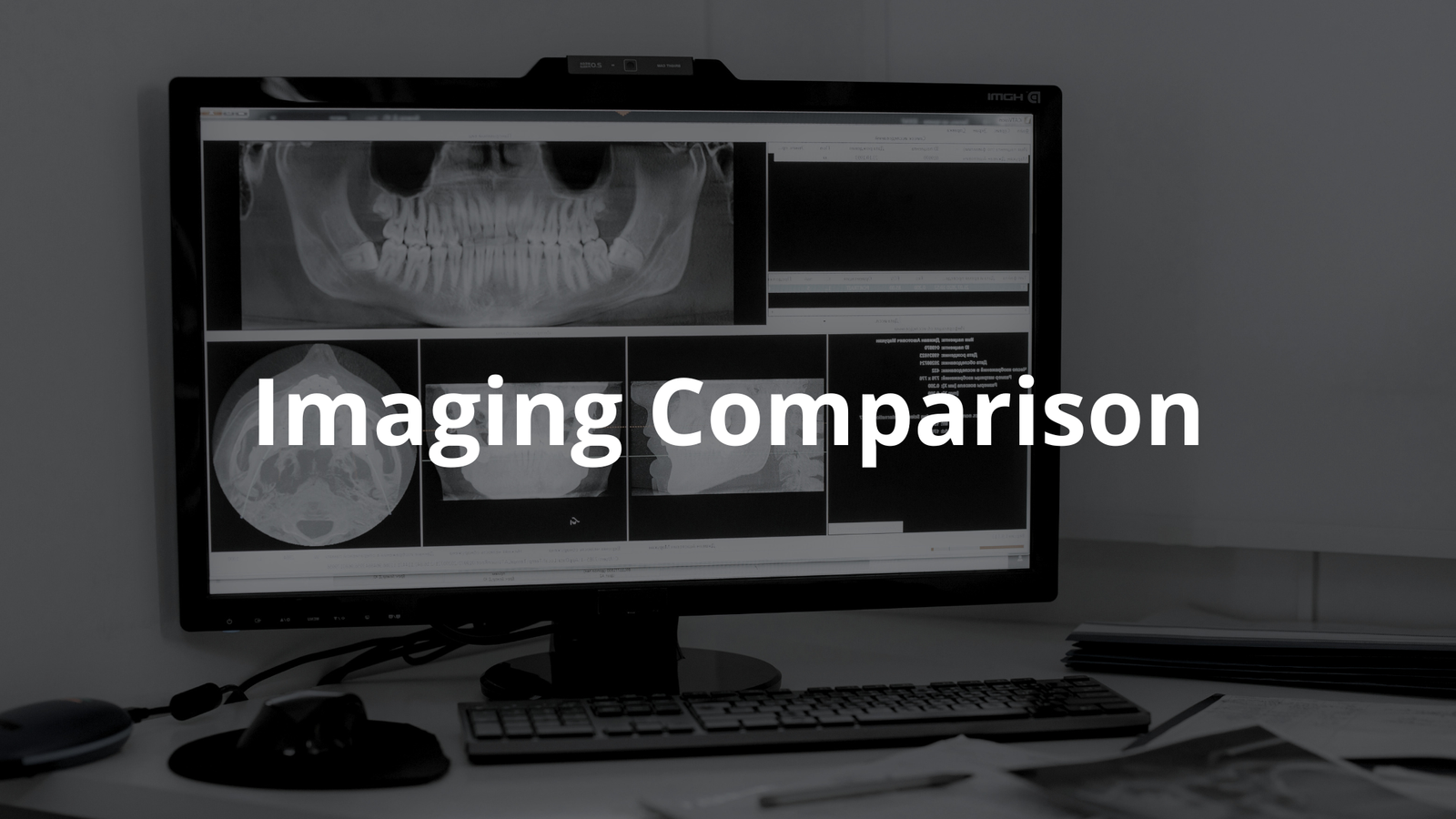
Comparing Image Quality
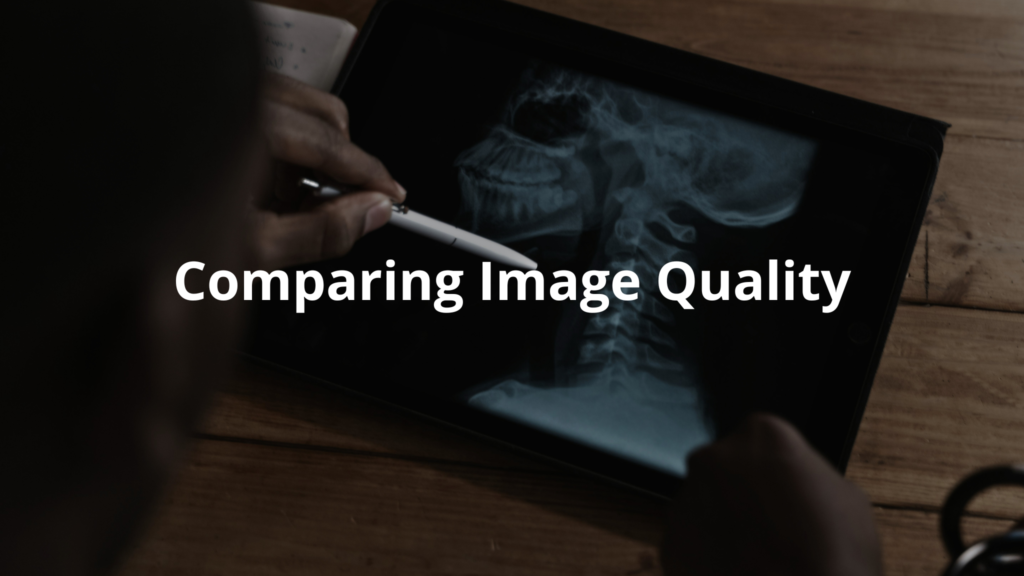
When comparing CT and MRI scans, it’s clear that both have their strengths. CT scans typically provide clearer images of bones and are quicker. This speed is why they’re often the go-to in emergency situations. If someone has a broken bone or internal bleeding, a CT scan can quickly give doctors the information they need.
On the flip side, MRI scans shine when it comes to soft tissues. They provide much more detailed images of the brain, muscles, and organs. If a doctor wants to see inside your brain or check for muscle tears, they might choose an MRI.
Here’s a quick look at what each imaging type does best:
- CT Scans: Great for bones, internal injuries, and quick diagnoses. They can show fractures and bleeding in a flash.
- MRI Scans: Best for soft tissues, brain scans, and detailed views of organs. They excel at revealing issues that CT scans might miss.
In this way, each type of scan plays a crucial role in medical imaging. It’s all about choosing the right tool for the job. Knowing what each scan can do helps patients understand what to expect and why their doctor might recommend one over the other.
The Importance of Safety
When thinking about imaging tests, patient safety is always top of mind. In the bustling atmosphere of a medical facility, the machines can look a bit daunting, but they’re built with care. CT scans use radiation, which can be harmful in high doses. That’s why doctors only order them when absolutely necessary. They consider each patient’s unique situation before deciding.
MRI scans provide a safer alternative since they don’t use radiation at all. Instead, they rely on powerful magnets and radio waves. However, some patients might not qualify for MRIs due to metal implants or pacemakers, which can interfere with the magnetic field.
Here are some key points to remember about safety:
- CT Scans: Used sparingly due to radiation risks.
- MRI Scans: Generally safer, but not for everyone.
- Doctor’s Decision: Always based on weighing risks and benefits.
This way, patients can feel informed and confident about their imaging choices.
The Cost of Imaging Tests
Costs for CT and MRI scans can vary greatly, which can be quite confusing for patients. Generally speaking, CT scans are more affordable than MRIs. This is partly because MRIs are more complex and take longer to perform. Imagine lying in a tube for about 30 minutes to an hour while the machine does its work. That extra time and complexity add to the cost.
In Australia, the Medicare system helps cover some of these costs, which is a relief for many. However, patients might still have to pay out-of-pocket depending on where they get the test done. Some hospitals charge more than clinics, and that can leave patients scratching their heads. It’s always wise to ask questions before scheduling a scan.
A practical step could be to call ahead and check the costs. “What will I owe after my Medicare coverage?” is a good question to ask. This way, patients can avoid any surprises when the bill comes. Knowing the cost upfront can help them plan their finances better.
Understanding the financial side of imaging tests is just as important as understanding the medical side. By being informed, patients can take charge of their health and make choices that work for them.
Conclusion
In wrap up, CT and MRI scans are both important tools for doctors. They help look inside our bodies to find out what’s wrong. CT scans are quick and great for bones, while MRIs are safer and better for soft tissues. Understanding these differences helps patients and doctors make the best choices. If you ever need an imaging test, remember to ask your doctor which one is right for you.
FAQ
How do CT scans and MRI scans differ in creating medical images of internal structures?
CT scans use ionising radiation and X-ray tubes to produce images of bones and internal organs, while MRI scans rely on magnetic fields and radio waves. MRI imaging excels at showing soft tissue detail, while CT imaging provides better images of bones and can be performed more quickly.
What makes magnetic resonance imaging particularly useful for diagnosing soft tissue conditions?
Magnetic resonance imaging creates detailed images using strong magnetic fields and radio waves. It shows excellent contrast between different soft tissues and can capture blood flow. MRI scanners produce high-quality images without ionising radiation, making them especially valuable for paediatric patients.
How do imaging techniques help healthcare providers diagnose and treat medical conditions?
Diagnostic imaging techniques like computed tomography, positron emission tomography, and magnetic resonance imaging provide detailed images of anatomical structures. These imaging modalities help in accurate diagnosis through high-resolution images that show internal organs, blood vessels, and the skeletal system.
What safety considerations should patients know about different imaging procedures?
Patient safety varies by imaging type. CT scans and PET CT involve radiation exposure, requiring careful attention to radiation dose. MRI scans are safe but require screening for implanted devices due to strong magnetic fields. Some procedures use contrast agents to enhance image quality.
How do advanced imaging systems achieve high-quality diagnostic results?
Modern imaging systems combine high-resolution capabilities with sophisticated image analysis tools. They offer improved spatial resolution, signal intensity, and noise ratio. These systems can perform quantitative analysis and create three-dimensional images, supporting a wide range of diagnostic imaging needs.
What role do imaging tests play in nuclear medicine and radiation therapy?
Nuclear medicine uses radioactive material in imaging tests like PET scans to produce images showing both structure and function. These imaging techniques help plan radiation therapy and monitor treatment effectiveness. Different imaging modalities can be combined, like PET CT, for more comprehensive diagnostic information.
References
- https://onlinelibrary.wiley.com/doi/abs/10.1111/1754-9485.13591
- https://pmc.ncbi.nlm.nih.gov/articles/PMC9024258/