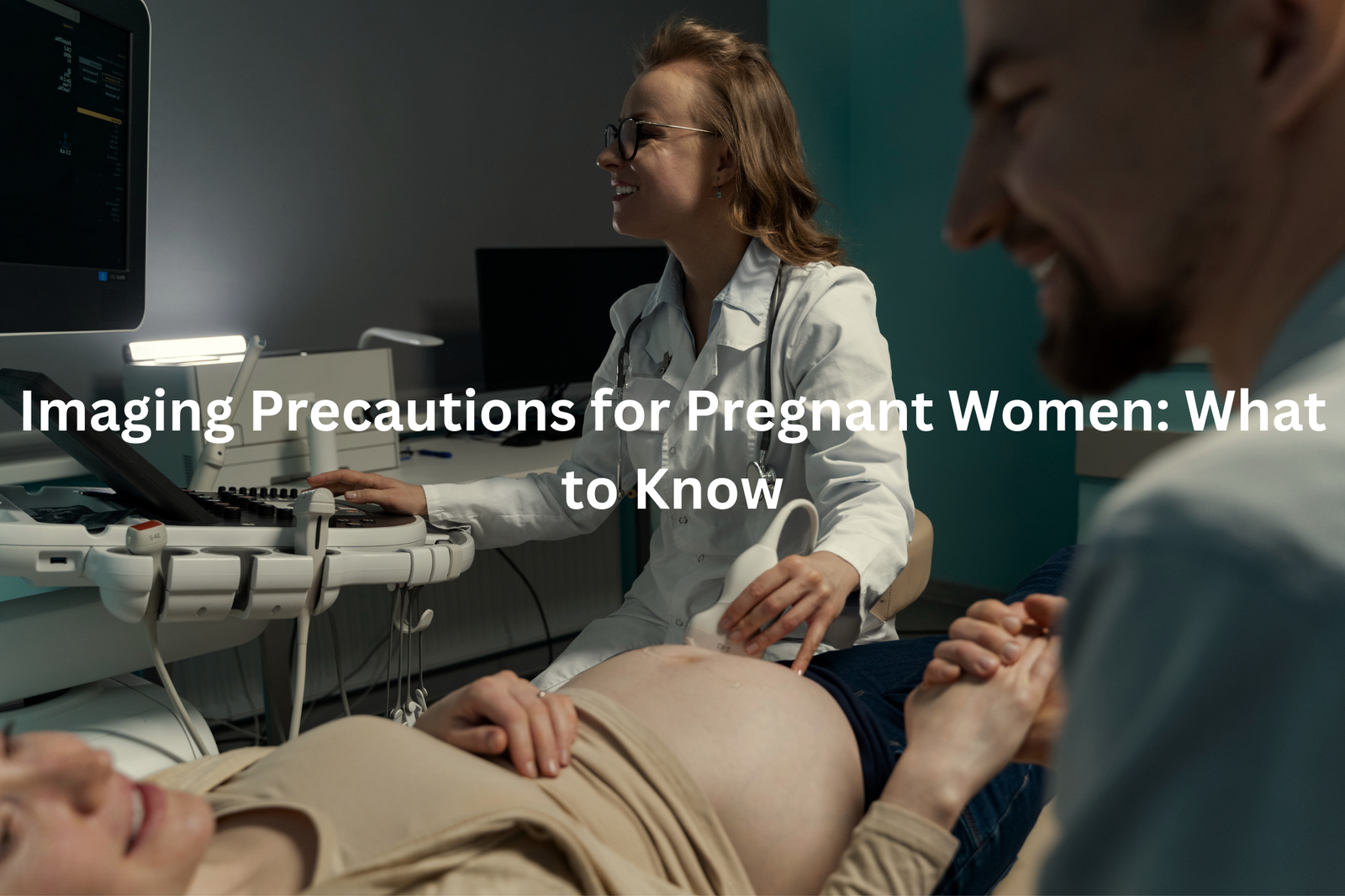
Imaging while pregnant? Know the precautions to keep your baby safe during X-rays, CT scans, and other tests. Here’s what you should consider.
When a pregnant woman needs medical scans, doctors must think about both mum and baby’s safety. Medical imaging brings up big questions during pregnancy (X-rays use small amounts of radiation, measured in millisieverts).
Ultrasound scans stand out as the first choice, since they don’t give off any radiation at all. For X-rays that can’t wait, lead aprons shield the growing baby, and doctors keep radiation doses as low as they can – usually under 5 millisieverts per scan.
Smart planning means talking to doctors about which tests make the most sense, weighing up what’s needed against any risks. Keep reading to learn more about keeping both mum and baby safe during these procedures!
Key Takeaway
- Non-radiation imaging methods like ultrasound and MRI are safest for pregnant women.
- If radiation is necessary, doctors use the lowest dose possible to protect the baby.
- Always confirm pregnancy status before any imaging that could involve radiation.
Understanding Imaging Precautions for Pregnant Women
Medical imaging helps doctors check inside the body, but pregnancy needs extra care when it comes to scans(1). Different types of imaging have different safety levels for mums and bubs.
Ultrasound scans use sound waves, making them the safest choice during pregnancy. They don’t use any radiation, which is why doctors use them to check baby growth.
MRI scans work with magnets and are generally safe, though they’re not the first choice unless needed. X-rays use small amounts of radiation (about 0.01 mGy), while CT scans use more (2-25 mGy depending on the area).
Doctors choose imaging based on what’s needed versus what’s safest. They might:
- Use protective shields over the belly
- Adjust machine settings
- Pick safer options when possible
For any scan during pregnancy, mums can ask about alternatives. Safety comes first when caring for two lives.
Non-Radiation Imaging Techniques
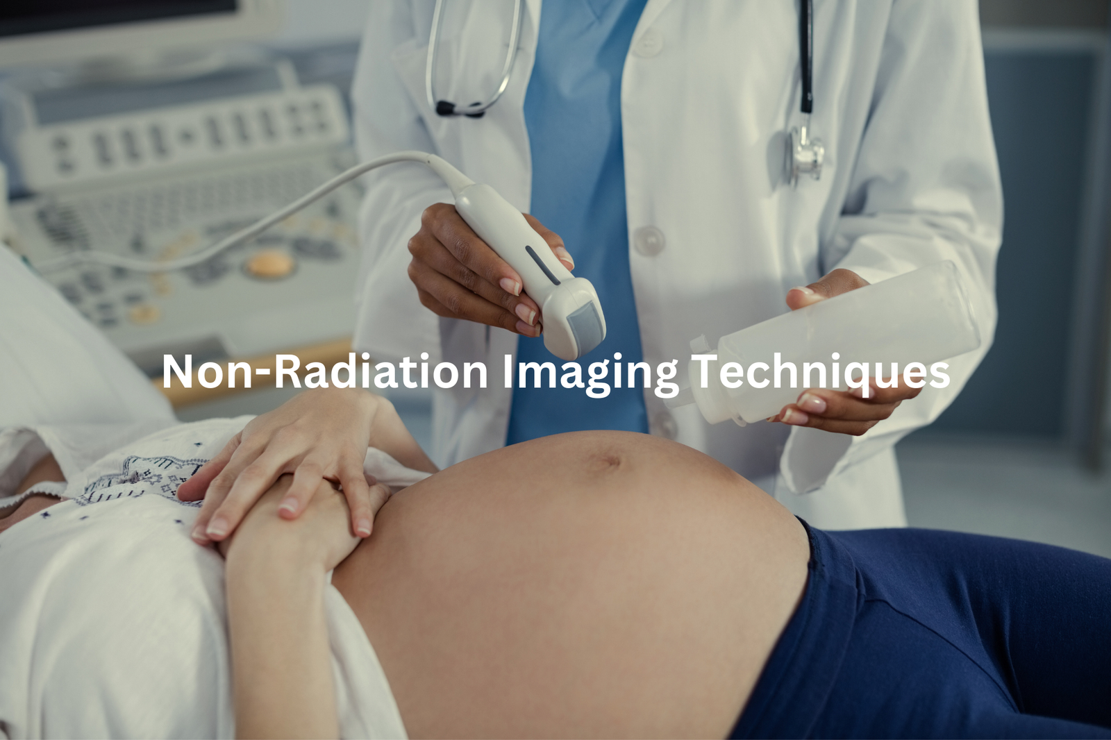
Doctors use different types of scans to check both mum and bub during pregnancy, but not all scans work the same way. Some are perfectly safe, others need more careful thinking.
Ultrasound scans lead the pack in pregnancy imaging. They use harmless sound waves to create pictures of the growing baby (showing everything from tiny fingers to heart chambers). Most mums get their first peek between 8-12 weeks.
MRI scans come second. These big machines use magnets and radio waves – no radiation at all. They’re brilliant for checking complicated things doctors can’t see well with ultrasound.
X-rays and CT scans are the last choice. While a single chest X-ray gives off just 0.01 mGy of radiation, CT scans can hit 25 mGy. That’s why doctors save these for emergencies only.
For safer scans, doctors use special shields and lowest possible settings. Smart mums always ask about alternatives – there’s usually a better way.
Radiation Risks and Safe Levels
Sources: Science With Chris.
Radiation during pregnancy needs careful handling in medical settings. The main focus stays on getting good images while keeping radiation levels safe for both mum and bub.
Medical professionals in Australia follow strict rules about radiation exposure (measured in millisieverts or mSv)(2). Different scans have different radiation levels:
• Chest X-rays give about 0.1 mSv – that’s less than a week of natural background radiation
• Dental X-rays are even lower at 0.01 mSv
• CT scans vary from 2 mSv for the head to 25 mSv for the belly area
The safe limit during pregnancy sits at 1 mGy (milligray), and doctors work to stay well below this number. They use special shields and adjust machine settings to protect the growing baby.
MRI and ultrasound scans don’t use radiation at all, making them safer choices when possible. Pregnant patients should ask their doctors about these options first. Most medical centres can offer alternatives that use less or no radiation.
Confirming Pregnancy Status
Medical scans use different amounts of radiation, making pregnancy checks essential before any imaging. Women aged 16 to 50 need to tell their doctors if there’s any chance they’re pregnant.
Different scans mean different radiation levels:
- X-rays give off small doses (0.1 mSv or less)
- CT scans can reach higher levels (1-25 mSv)
- Nuclear medicine tests might go over 10 mSv
Doctors set a safety limit of 1 mSv for unborn babies. When radiation doses might go higher, they look at other options like ultrasound or MRI scans, which don’t use any radiation at all.
Medical staff must ask about pregnancy before every scan. They use special shields to protect patients, and sometimes switch to different types of imaging that are safer for mums-to-be. The radiation from these scans can’t be reversed once it’s done, so checking first keeps everyone safe.
Careful Planning for Necessary Imaging
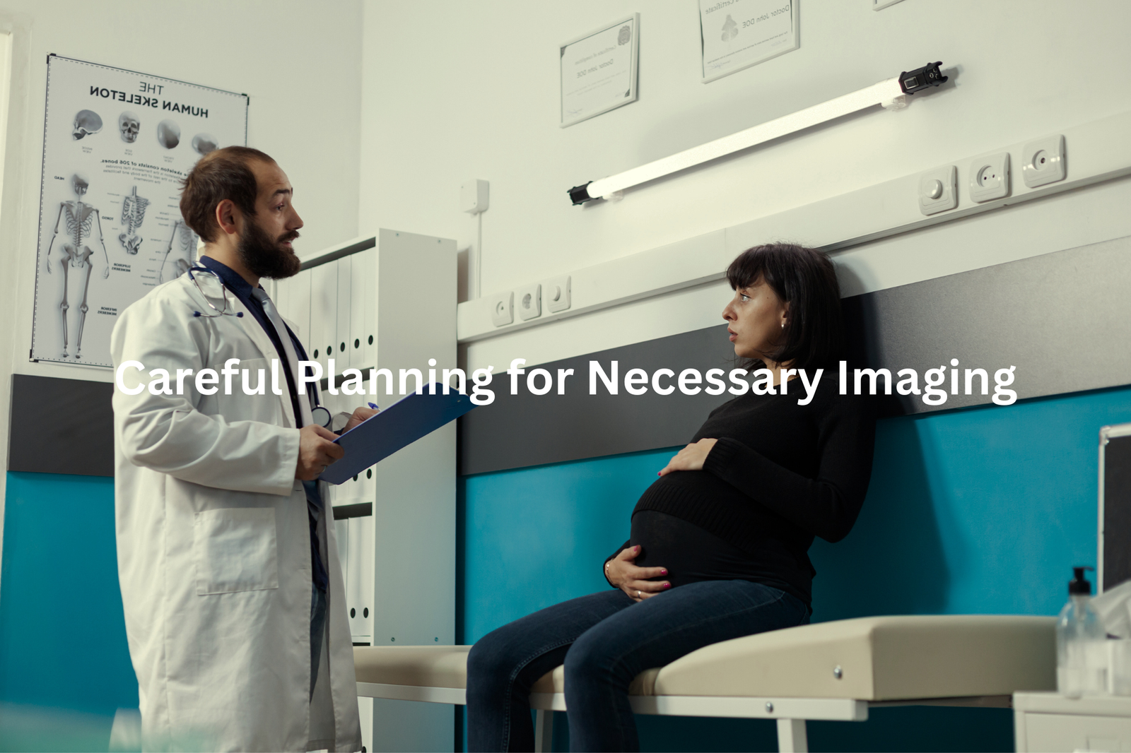
Medical imaging during pregnancy needs careful thought(3). The doctor’s choice between MRI and CT scans makes a big difference for both mum and baby.
MRI scans don’t use radiation, making them safer for pregnancy. But sometimes doctors need CT scans or X-rays to spot specific problems (like lung issues or broken bones). The radiation from these tests varies quite a bit – a chest X-ray gives about 0.1 mSv, while CT scans can reach up to 25 mSv.
Doctors should:
- Pick the lowest radiation dose possible
- Write down every scan’s details
- Use protective shields when needed
- Think about waiting if the scan isn’t urgent
The safe limit during pregnancy stays under 1 mSv. Anything more needs a good chat with the doctor. While radiation can’t be taken back, smart planning keeps risks low. Proper shielding and careful timing help protect the growing baby while still getting the needed medical info.
Special Considerations for CT Scans
CT scans offer detailed views inside the body that regular X-rays can’t match. These powerful machines capture images of organs, blood vessels, and tiny bone breaks in seconds (using radiation doses between 1-25 mSv).
During pregnancy, doctors face extra challenges when deciding about CT scans. The benefits must outweigh any risks to both mum and bub.
Medical teams check these points before ordering a scan:
- Could ultrasound or MRI work instead?
- Is the information needed right away?
- Can they use protective shields or lower doses?
Modern CT machines let doctors adjust radiation levels while keeping image quality clear enough for diagnosis. Half-dose scans often work just as well as full-strength ones, especially for basic checks.
When internal bleeding or dangerous clots might be present, CT scans become essential tools. The medical team picks the lowest dose that still shows what they need to see.
Follow-Up and Monitoring
Medical imaging during pregnancy needs careful tracking. The scan room’s machines (large white boxes with gentle whirring sounds) serve different purposes, each with specific safety levels.
Common prenatal scans include:
- Ultrasounds: No radiation, uses sound waves
- X-rays: Low radiation, about 0.01 mSv per scan
- CT scans: Higher radiation, up to 25 mSv
Pregnant patients should ask their doctors these questions after any scan:
- What type of imaging was done?
- Was radiation used? If yes, how much?
- Are there any follow-up scans needed?
- When will the results be ready?
Keeping records helps track exposure levels throughout pregnancy. A small notebook works well for writing down scan dates, types, and doses. Most imaging procedures pose minimal risks when properly managed, but documentation matters for future medical decisions.
Doctors recommend bringing scan records to all prenatal appointments, making sure nothing gets missed.
FAQ
What are the key radiation concerns for pregnant women during CT scans?
When considering CT imaging during pregnancy, healthcare providers carefully evaluate the radiation dose and potential risks to the unborn baby. Low-dose CT scanners and advanced techniques like half dose and tube current reduction help minimise fetal exposure. CT dose calculations using methods like Monte Carlo simulations help medical professionals assess potential radiation risks. While CT scans can be necessary in emergency care, physicians prioritise balancing diagnostic needs with protecting the mother and developing fetal thyroid and soft tissue from unnecessary radiation exposure.
How do medical professionals assess radiation risks during pregnancy?
Medical professionals evaluate radiation risks using comprehensive approaches including dose level assessments, fetal dose calculations, and understanding potential cancer risks. Experts in obstetrics and gynaecology fields recommend careful consideration of CT imaging methods, especially for pregnant women experiencing abdominal pain or pelvic pain. They analyse factors like field of view, image length, and absorbed dose to determine the lowest possible radiation exposure. Healthcare providers use animal studies and advanced techniques to estimate potential risks to the gravid uterus and unborn baby.
What imaging alternatives exist for pregnant patients?
Pregnant patients have several imaging alternatives that minimise radiation risks. MRI with contrast media offers a radiation-free option for soft tissue imaging, particularly useful for heart rate evaluations and nervous system assessments. Duplex Doppler and ultrasound techniques provide diagnostic information without radiation exposure. When CT scans are medically necessary, healthcare providers use low-dose protocols, reduced tube current, and precise field of view to minimise fetal exposure. Emergency care scenarios might require careful balancing of diagnostic needs and radiation protection.
What specific precautions are taken during chest CT and CT angiography?
During chest CT and CT angiography for pregnant women, medical professionals implement multiple radiation protection strategies. They use low-dose CT scanners, helical CT techniques, and precise X-ray beam configurations to reduce radiation exposure. Oral contrast and careful dose reduction methods help minimise risks to breast tissue, breast milk production, and fetal development. Physicians carefully evaluate the necessity of the scan, considering alternative imaging methods and potential health risks to both mother and unborn baby.
How do medical experts evaluate potential long-term radiation risks?
Medical experts assess long-term radiation risks through comprehensive studies like PIOPED II, examining potential impacts on the female breast, fetal thyroid, and overall cancer risk. They consider Australian radiation protection guidelines, analyse threshold dose levels, and use advanced computational methods like Monte Carlo simulations. Researchers study increased risk factors, comparing fetal doses across different imaging methods. By understanding amniotic fluid interactions and tissue heating potential, healthcare providers make informed decisions about radiation exposure during pregnancy.
Conclusion
Medical imaging during pregnancy demands careful thought. Ultrasound scans and MRI (magnetic resonance imaging) stand out as the safest options, since they don’t use any radiation. The medical team must check for pregnancy before any scan, and if X-rays become necessary, they’ll use the smallest possible dose. A chat between doctors and patients helps create a solid plan that keeps both mum and baby safe throughout the process.
References
- https://aci.health.nsw.gov.au/networks/eci/clinical/ed-factsheets/medical-imaging-in-pregnancy
- https://www.pregnancybirthbaby.org.au/radiation-exposure-during-pregnancy
- https://www.wacountry.health.wa.gov.au/~/media/WACHS/Documents/About-us/Policies/Radiology—Imaging-Pregnant-Patients-Procedure.pdf?thn=0

