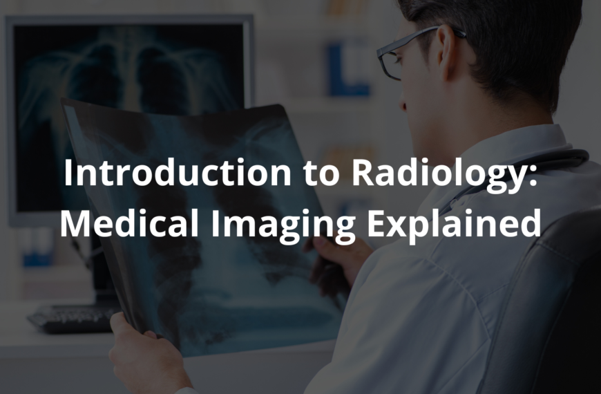
Learn about the invention of radiology, its impact on healthcare, and how it changed the way we see inside the human body.
Radiology is a special field that helps doctors look inside the human body without needing to cut it open. It all started with the invention of X-rays by Wilhelm Conrad Röentgen way back in 1895. Can you imagine how exciting it must have been to see the first X-ray images? Keep reading to find out how this amazing technology has changed healthcare forever.
Key Takeaway
- Wilhelm Conrad Röentgen discovered X-rays in 1895, starting the radiology revolution.
- Early radiologists in Australia quickly embraced this new tech for patient care.
- Radiology techniques, like CT scans and PET scans, help doctors diagnose diseases better.
The Beginning of Radiology
A long time ago, in the 19th century, life was very different. Doctors were like detectives, relying mostly on their eyes and touch to figure out what was wrong with patients. They had no fancy machines or gadgets to help them. Then, in 1895, a man named Wilhelm Conrad Röentgen discovered something amazing X-rays! This new technology could create images of the inside of the body, showing bones and soft tissues without surgery. Imagine taking a picture of your insides(1)!
When Röentgen made his discovery, it didn’t take long for the news to travel across the world. By early 1896, people in Australia were starting to experiment with X-rays. It was a thrilling time for science and medicine. One of the first to do this was Thomas Rankin Lyle, a professor at the University of Melbourne. He showed everyone how to use X-rays to look at a person’s foot, which was a big deal back then.
The excitement in the room must’ve been electric. Lyle demonstrated the simple yet revolutionary process of using X-rays to reveal bones and fractures that were hidden beneath the skin.
This was just the beginning. This discovery opened a door to a whole new world of medical imaging. The possibilities seemed endless. For the first time, doctors had a way to see inside their patients without needing to cut them open. It was a change that would shape the future of medicine, making it safer and more effective for everyone.
Early Radiologists in Australia
Three brave pioneers were among the first to create images in Australia:
- Thomas Rankin Lyle: He led the way in using X-rays for medical purposes. His demonstrations were captivating.
- Walter Filmer: An electrician who used X-rays to find a needle before surgery. He showed that X-rays could save lives.
- Father Joseph Slattery: He used X-rays to locate gunshot pellets in a student’s hand. This quick thinking probably saved that student’s hand from amputation.
These early adventures in radiology were thrilling for everyone involved. They helped doctors figure out what was wrong without needing to perform risky operations. Imagine using a ray machine to see inside someone’s body! The excitement of seeing an image appear on the screen must’ve felt like magic.
As these pioneers made their discoveries, X-ray technology quickly spread across Australia. Clinics and hospitals began to adopt this new method, and with it, the practice of radiology began to grow. Each new image revealed more about the human body, and doctors became better at diagnosing and treating illnesses.
These early efforts set the stage for what radiology is today. The ability to see inside the human body without surgery has saved countless lives and improved patient outcomes. The future was bright for this new field of medicine. The possibilities seemed limitless, and the impact on healthcare was just beginning to unfold.
How Radiology Changed Medicine
The new X-ray technology transformed medicine dramatically. Before this, doctors were like detectives without clues, relying on guesswork to determine what was wrong with their patients. But with X-rays, they could see bones, soft tissues, and blood vessels clearly. It was like gaining superpowers!
By the late 19th century, X-rays became instrumental in diagnosing various medical conditions, including:
- Broken bones: X-rays allowed doctors to see fractures instantly.
- Cancer detection: The ability to visualize tumours changed the game for early diagnosis.
This leap in technology gave rise to advanced imaging techniques like CT scans and PET scans. Doctors could now observe real-time images of the human body, enhancing their diagnostic accuracy and treatment planning.
CT scans provided cross-sectional views of the body, while PET scans illuminated metabolic activity in tissues a crucial factor in cancer diagnosis. This combination opened new doors for patient care and revolutionised the way medicine approached health issues.
The Role of Institutions
As radiology grew, schools and hospitals began to invest in this technology. The Ballarat School of Mines, for example, was one of the first institutions in Australia to conduct systematic experiments with X-rays. Frederick Martell and John Sutherland were key figures in these early experiments. They successfully produced radiographs of hands and wrists, which showed the practical uses of X-rays in diagnosing fractures.
These early radiologists probably felt a sense of excitement and responsibility. They were pioneers in a field that was still in its infancy. By sharing their findings in local newspapers, they helped spread the word about how valuable this technology could be. Their stories inspired others to explore the potential of radiology.
This was just the beginning of a long road that led to amazing advancements in medical imaging. As more institutions adopted X-ray technology, the quality of images improved. Schools began teaching future doctors about the importance of imaging tests. This education helped ensure that doctors could make accurate diagnoses and provide effective treatments. The collaboration between schools, hospitals, and medical professionals has been crucial in making radiology a cornerstone of modern medicine. It’s a reminder of how teamwork can lead to significant progress in healthcare.
Modern Radiology Techniques
Today, radiologists use many different technologies to help patients. Some popular methods include:
- CT Scans: These create detailed images of cross-sections of the body, making it easier to see inside.
- PET Scans: They use a special kind of radiation to show how organs and tissues are working, which is super helpful for detecting cancer.
- MRI Technology: This uses magnets and radio waves to create clear images of soft tissues, like the brain and muscles.
With these advanced imaging techniques, doctors can diagnose and treat diseases better than ever before. It’s incredible how far we’ve come since Röentgen first discovered X-rays!
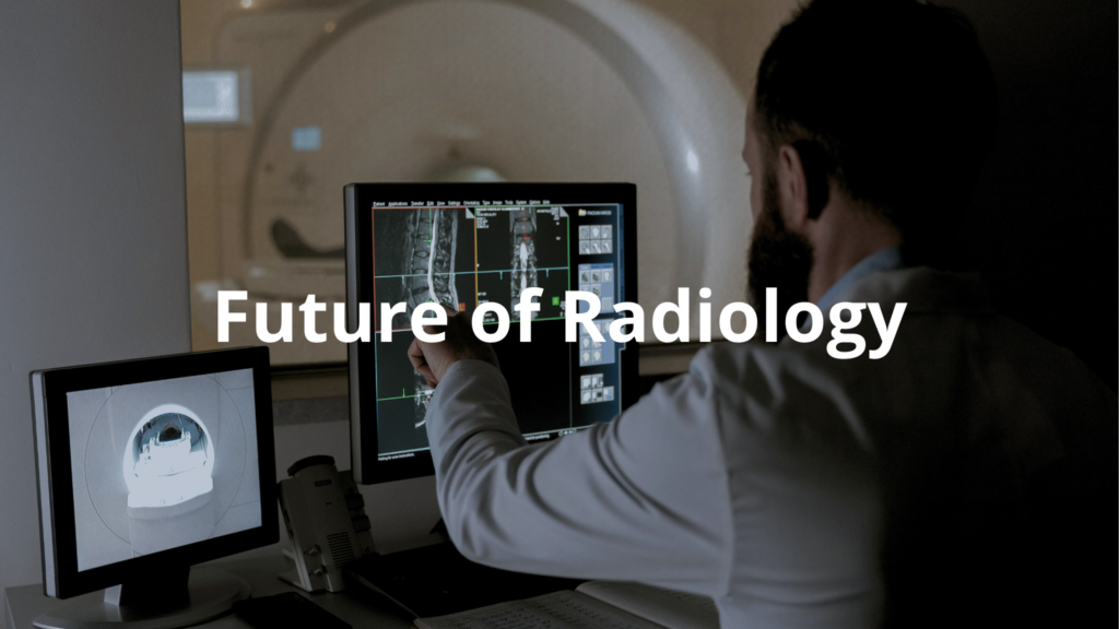
The Future of Radiology

The future of radiology shines bright like a clear blue sky. With the help of artificial intelligence (AI) and machine learning, doctors can analyse images more accurately and quickly. Imagine a computer assisting a doctor in spotting cancer cells or detecting blood flow issues! This kind of assistance might just improve patient outcomes significantly(2).
AI can sort through images much faster than a human can. For instance, a system could examine thousands of X-ray images in mere minutes. This speed means doctors can get results quicker, which is crucial in emergency situations. And with machine learning, these systems learn from previous images, getting smarter over time.
As technology continues to evolve, doctors can expect more amazing developments in imaging systems. Some exciting innovations include:
- Hybrid imaging: This combines different types of scans, like PET and CT, allowing for more detailed views.
- 3D imaging: This creates three-dimensional representations of organs, helping to plan surgeries better.
These advancements mean doctors can see more details and make even more accurate diagnoses. The possibilities are endless, and I think the future of radiology will bring a lot of hope for patients everywhere. It’s a reminder that technology can truly change lives for the better.
Conclusion
In wrapping up, the invention of radiology has transformed healthcare in ways we still benefit from today. It started with Wilhelm Conrad Röentgen’s discovery of X-rays, which allowed doctors to see inside the human body without surgery.
Early pioneers in Australia quickly embraced this technology, leading to the development of CT scans, PET scans, and more. As we move forward, advanced imaging will continue to improve patient care and treatment planning. So, let’s keep our eyes open for what’s next in the world of medical imaging!
FAQ
How did Wilhelm Conrad transform medicine with his X-ray discovery?
Using a cathode ray tube and photographic plates, Wilhelm Conrad made history by discovering X-rays in 1895. He created the first radiographic images, including the famous ray image of his wife’s hand, earning him the Nobel Prize in Physics. This breakthrough launched diagnostic imaging and transformed healthcare forever.
What key developments followed the initial X-ray discovery?
Early pioneers like Thomas Edison improved ray equipment and fluorescent screens for better image quality. The introduction of photographic film enhanced ray radiography. Doctors in field hospitals quickly adopted this imaging technology for diagnosing bone fractures and locating foreign bodies.
How did medical imaging evolve into different modalities?
Beyond projectional radiography, new imaging modalities emerged: computed tomography by Sir Godfrey Hounsfield, magnetic resonance imaging pioneered by Paul Lauterbur, and nuclear magnetic resonance. Each technique uses different types of radiation or electromagnetic radiation to create images of the human body.
What are the main branches of modern radiology?
Today’s radiology encompasses diagnostic radiology, interventional radiology, and radiation oncology. Clinical radiology includes specialized areas like pediatric radiology and emergency medicine. Medical physics supports these specialties, ensuring radiation safety and protection in radiology departments.
How has education and training in radiology developed?
The American College of Radiology and various radiological societies established comprehensive training programs. Medical education now combines doctor of medicine studies with specialized medical imaging training. The field demands expertise in medical physics, radiation therapy, and the latest imaging technologies.
What technological advances revolutionized radiology practice?
Digital radiography replaced traditional photographic plates, while artificial intelligence enhances diagnostic capabilities. Modern imaging techniques include minimally invasive procedures and real-time imaging. Contrast agents help create detailed images of body parts and soft tissue.
How has radiology shaped modern medical practice?
Radiology has become essential to patient care and health care delivery. From diagnosing broken bones to guiding complex interventional procedures, imaging modalities provide crucial diagnostic information. The field continues to advance with new technologies and techniques in medical imaging.
References
- https://federation.edu.au/about-us/our-university/history/geoffrey-blainey-research-centre/honour-roll/x/x-ray-pioneers
- https://nova.newcastle.edu.au/vital/access/manager/Repository/uon:7225


