Discover low-dose imaging that cuts radiation exposure while keeping images clear.
Low-dose imaging is a big deal in the world of medical imaging. It means getting important pictures of our insides without using too much radiation. Radiation can be harmful, and doctors want to make sure they use only what’s necessary.
In Australia, low-dose imaging has become a way to help keep patients safe while still getting the details they need for diagnosis. Curious about how it works? Let’s find out!
Key Takeaway
- Low-dose imaging reduces the amount of radiation used during scans.
- New technology helps keep images clear while using less radiation.
- Safety guidelines ensure that patient exposure stays low.
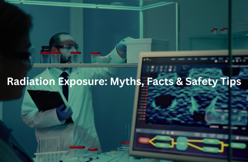
Understanding Low-Dose Imaging
It’s easy to take medical imaging for granted—until you’re the one lying on the table, watching the CT scanner whir to life. The hum of the machine, the faint warmth of the X-ray tube—it all makes you wonder just how much radiation is passing through your body. That’s where low-dose imaging comes in.
Low-dose imaging refers to medical scans that use significantly reduced levels of ionising radiation while still producing diagnostically useful images. It’s a balancing act—reducing radiation exposure while maintaining image quality. A standard chest CT scan, for instance, delivers about 6.7 millisieverts (mSv) of radiation. But a low-dose version? That can be as little as 0.2–0.3 mSv—nearly 95% less.
This isn’t just about cutting numbers. It’s about protecting patients. Radiation exposure accumulates over a lifetime, and unnecessary doses can increase long-term risks. By optimising scan protocols, radiologists ensure patients receive only the minimum radiation needed for an accurate diagnosis.
Radiation Exposure: What You Need to Know
Radiation isn’t just a product of medical imaging—it’s everywhere. Cosmic rays, radon gas, even the soil beneath our feet contribute to our annual exposure. In Australia, the average person absorbs around 2 mSv of background radiation each year. A cross-country flight adds another 0.03 mSv.
Now, throw in a medical scan, and that number can jump. A head CT? Around 2 mSv. A full-body PET-CT? Up to 25 mSv. That’s why dose optimisation is critical. The goal isn’t just to reduce exposure but to ensure that every scan is medically justified.
The Australian Radiation Protection and Nuclear Safety Agency (ARPANSA) sets strict radiation dose limits to protect both patients and medical staff. Their guidelines emphasise the justification principle—a fundamental concept in radiological protection. If a scan doesn’t provide clear clinical benefit, it shouldn’t be performed. (1)
The ALARA Principle: A Key to Safety
At the heart of radiation protection is the ALARA principle—“As Low As Reasonably Achievable.” It’s more than just a guideline; it’s a mindset. Radiologists, radiographers, and medical physicists all work within ALARA compliance frameworks to keep radiation doses as low as possible while maintaining diagnostic accuracy.
This involves:
- Time management in radiology: Reducing scan duration to limit exposure
- Distance from radiation source: Maximising patient-table positioning to reduce scatter radiation
- Shielding materials: Using lead shielding or mobile lead shields where appropriate
- Dose tracking systems: Monitoring cumulative radiation exposure over time
- Optimisation principle: Adjusting machine settings to the lowest effective dose
Every decision in radiology revolves around this principle. From tweaking CT scanner parameters to refining radiation monitoring protocols, the goal is simple—minimise exposure, maximise safety.
Technological Innovations in Low-Dose Imaging
Credit: Yale Radiology and Biomedical Imaging
Radiology isn’t what it used to be. A decade ago, a clear CT image required high radiation doses. Now? Advanced algorithms and smarter machines have changed the game.
Iterative reconstruction algorithms have been a breakthrough in dose reduction. These software techniques refine image quality without increasing radiation. Instead of raw X-ray data being processed just once, iterative methods cycle through corrections, reducing noise and enhancing detail. The result? High-quality images at lower doses.
Newer CT scanners also use automatic exposure control (AEC)—a system that adjusts radiation dose in real-time based on the patient’s size and the area being scanned. This ensures that smaller patients (like children) receive less radiation than larger ones.
Some hospitals now use ultra-low-dose CT scanners, particularly for lung imaging. These machines can produce clear images with radiation levels comparable to a few days’ worth of background exposure. For patients undergoing repeated scans—like those in lung cancer screening programs—this is a game changer.
Clinical Applications of Low-Dose Imaging
Low-dose imaging isn’t just a theoretical improvement—it’s already making a difference in clinical practice.
- Lung cancer screening: Australia’s National Lung Cancer Screening Program (2025) will use low-dose CT scans to detect cancer early in high-risk patients.
- Pediatric imaging: Children are more radiosensitive than adults, making dose reduction crucial. Studies like Aust-PERC guide best practices for paediatric scans.
- Cardiac CT angiography: Low-dose techniques allow detailed imaging of coronary arteries with minimal radiation.
- Orthopaedic imaging: Weight-bearing CT scans now use lower doses, helping assess joint conditions with reduced risk.
Safety Guidelines and Patient Protection
Following proper radiation safety audits ensures that imaging centres comply with regulatory standards. Hospitals must track radiation dose limits, use radiation monitoring devices, and regularly update their radiology best practices. (2)
Key patient protection measures include:
- Personal protective equipment (PPE): Lead aprons, thyroid shields
- Patient shielding techniques: Lead shielding for sensitive areas
- Radiological protection programs: Continuous education for radiology staff
- Radiological incident reporting: Ensuring errors in dose calculation are addressed
Risk-Benefit Analysis in Imaging Procedures
Not every scan is necessary. The risk-benefit analysis in radiology weighs the potential harm of radiation exposure against the diagnostic value of the test. In most cases, the benefit of detecting a serious condition far outweighs the minimal risk from a single scan.
But that doesn’t mean risks are ignored. Doctors take extra precautions when imaging pregnant patients, children, and individuals who require repeated scans. In these cases, alternative imaging techniques (like ultrasound or MRI) may be considered to avoid unnecessary radiation.
The Future of Low-Dose Imaging
Looking ahead, the future of low-dose imaging is promising. New AI-powered software continues to refine image quality at lower radiation doses. Continuous education on radiation safety ensures that radiologists stay up to date with best practices. Environmental impact considerations push the industry toward more sustainable radiation use, including radioactive waste management improvements.
As research advances, expect even safer, smarter imaging techniques. And for patients, that means one thing—better care with less radiation risk. Because in radiology, clarity should never come at the cost of safety. (3)
FAQ
How does low-dose imaging help reduce radiation exposure?
Low-dose imaging is designed to lower radiation exposure while maintaining image quality. Medical imaging techniques like CT scans and fluoroscopy use ionizing radiation, which carries some health risks. By optimising imaging protocols and using radiation dose reduction strategies, healthcare providers follow the ALARA principle (As Low As Reasonably Achievable) to keep exposure levels safe.
Flat-panel detectors and other imaging advancements also improve efficiency, reducing the cumulative radiation dose patients receive over time.
What is the cancer risk associated with low-dose imaging?
Low-dose imaging minimises the risk of radiation-induced effects, but even small doses of ionizing radiation can, in theory, contribute to cancer risk. The linear no-threshold model suggests that any exposure has the potential to cause DNA damage, chromosomal aberrations, and micronuclei formation, which are linked to long-term effects of radiation.
However, epidemiological studies show that the benefits of diagnostic imaging often outweigh these risks when imaging procedures are justified.
How do imaging protocols ensure patient safety?
Imaging protocols are designed to balance patient safety with diagnostic needs. Healthcare providers use effective dose measurement to assess exposure levels, while safety guidelines for imaging help reduce unnecessary scans. Low-dose protocols in medical radiology adjust scan parameters based on patient demographics and exposure history.
Regular radiation monitoring ensures compliance with radiological protection standards to limit the health risks of radiation.
How does the risk-benefit analysis apply to low-dose imaging?
A risk-benefit analysis helps determine whether an imaging procedure is necessary. The justification for imaging studies considers the patient’s medical condition, imaging frequency and risk, and potential radiation-induced effects. Research on low-dose effects continues to refine exposure assessment methodologies, ensuring that imaging technology evolves to reduce unnecessary radiation exposure while providing accurate diagnostic results.
What are the biological effects of radiation in low-dose imaging?
Even at low doses, ionizing radiation can cause biological effects of radiation, including DNA damage and chromosomal aberrations. These effects may contribute to the risk of cancer development and other long-term effects of radiation.
However, background radiation comparison shows that low-dose imaging contributes only a small fraction of overall radiation exposure, and healthcare provider education helps ensure appropriate use of imaging advancements to limit unnecessary scans.
How do technological innovations in imaging improve safety?
Technological innovations in imaging, such as flat-panel detectors and improved radiographic techniques, enhance image quality assessment while reducing radiation exposure. PET/CT scans, mammography radiation dose adjustments, and fluoroscopy techniques have all benefited from dose-response relationship studies that help optimise imaging techniques.
These advances help minimise the cardiovascular effects of radiation, reduce cataracts from radiation exposure, and improve patient safety.
What is the difference between ionizing and non-ionizing radiation in imaging?
Ionizing vs. non-ionizing radiation is a key distinction in imaging procedures. CT scans, fluoroscopy, and radiographic techniques use ionizing radiation, which carries some exposure risks. MRI safety is different because MRI uses non-ionizing radiation (radio waves and magnetic fields), posing no radiation-induced effects.
Understanding exposure assessment methodologies helps guide the safe use of medical imaging for different diagnostic needs.
What are the public health implications of increased imaging use?
Public health implications of imaging include concerns about cumulative radiation dose from frequent imaging procedures. Patient exposure statistics help track trends, and ethical considerations in imaging ensure that scans are only performed when necessary. Radiological protection standards aim to minimise unnecessary radiation exposure while maintaining high diagnostic accuracy in outpatient imaging procedures and other settings.
Conclusion
In conclusion, low-dose imaging is a vital part of modern medicine, helping to balance the need for clear diagnostic images with the importance of keeping patients safe from radiation exposure. With guidelines from ARPANSA and advancements in technology, Australia is leading the way in making sure that patients receive safe and effective imaging procedures. Continuous improvements will help maintain patient safety while providing the best care.
References
- https://pmc.ncbi.nlm.nih.gov/articles/PMC6849684/
- https://www.nature.com/articles/s41377-020-00353-0
- https://www.melbourneradiology.com.au/diagnostic-imaging/what-is-a-low-dose-ct-scan/