MRI: Detailed images using magnets & radio waves, a safe, radiation-free alternative to X-rays & CT scans. Discover MRI without radiation.
MRI, or magnetic resonance imaging, uses magnets and radio waves instead of radiation to see inside the body. It’s quite something, really. Doctors can get detailed pictures (using magnets about 30,000 times stronger than Earth) without harmful rays.
This means safer scans for patients. Less worry about radiation exposure. For many, MRI could be a smarter choice for imaging, probably. It offers a way to diagnose illnesses without the concerns of radiation. A rather clever method. There’s a lot to learn about how it works, so keep reading.
Key Takeaway
- MRI uses magnets and radio waves, making it radiation-free and safe for everyone.
- It helps doctors see soft tissues and internal structures in great detail.
- MRI is useful for detecting tumours and other health issues without using X-rays.
What is MRI?
An MRI, or Magnetic Resonance Imaging, it’s like looking under the bonnet without actually lifting it. Doctors reckon they can see all sorts of things, soft tissues, bones, organs, using this fancy technique. No X-rays either, which is probably a good thing, nobody wants too much of that.
Here’s the basic idea:
- Strong magnets are used. They line up tiny hydrogen atoms in the body (mostly water, after all).
- Radio waves are sent. These waves make the hydrogen atoms wobble about a bit.
- The wobbling atoms send signals back. The machine picks up these signals and, well, it gets clever.
The machine (a very expensive one at that) turns those signals into pictures. Pretty clever, that. They show the doctor muscles, bones and things like the heart and brain. It’s all about magnets and radio waves, and a bit of computing wizardry to make sense of it all. It might seem like magic; it’s actually science.
An MRI machine can cost somewhere around $1 million to $3 million dollars, depending on the versitility of the machine (a rather substantial investment indeed). Using the data, doctors can get an overall look at the human body to identify various injuries and ailments. This can help them create the right course of treatment for each patient.
How Does MRI Work?
Lying inside a metal tube isn’t exactly anyone’s idea of a relaxing afternoon. But it’s the magnetic field in an MRI that does the heavy lifting, mind you. These fields are strong, 1.5 to 3 Tesla, they reckon, (Tesla being the unit, in case you’re wondering). This strong field lines up the hydrogen atoms, like soldiers on parade, in the body. After all the human body is mostly water, so thats where these atoms reside.
How this works:
- A strong magnetic field is created. This makes hydrogen atoms line up.
- Radio frequency pulses are sent out. These pulses make the atoms wiggle about.
- Atoms return to their original position. Then send signals back.
The computer then gets to work processing these signals, turning them into images, this is where it gets interesting. Like a camera, but using magnets, not light. This gives doctors a much clearer picture, especially of soft tissues, you know, muscles and such. They can see things that might be missed by X-rays or other scans. If your doctor suggests one, its probably for good reason; they are looking for something.
Why is MRI Safe?
Radiation, nobody wants to be glowing in the dark; probably a good idea to avoid it. Many other scans, like X-rays, use radiation to create pictures; this probably isn’t the best thing for you. But MRI? No radiation at all; they use non-ionising technology, so there’s no risk of, you know, turning into a superhero (or something less desirable).
Reasons the MRI doesn’t use radiation:
- Non-ionising technology. This means no harmful radiation exposure.
- Safer for frequent scans. Good for kids or those needing regular check-ups.
- Good for pregnant women. Avoiding radiation is always a plus during pregnancy. [1]
This makes it a good option for kids, or expectant mothers who might need regular scans. The versatility of MRI is a real benefit, there’s not much to worry about with radiation exposure. If the doctor orders more then one, at least you will feel better knowing there are no harmful side effects.
What Can MRI Do?
Credits: Siemens Healthineers
Soft tissues, they’re often the tricky ones to get a good look at. Doctors use MRI for this sort of thing, that’s for sure. X-rays do not always work well, so MRI is a great second choice.
MRI Scans are used to look for these conditions:
- Tumours: MRI can help identify if lumps could be cancerous.
- Brain conditions: it can help see injuries and disease in the brain.
- Joint problems: good for looking at shoulders and knees.
Heart scans are also a big one. Doctors can understand how well your heart is functioning. It can check organs like the liver or kidneys, these are often hard to see otherwise. MRI is so detailed that it can even help with brain mapping, to understand how our brains work. Being able to watch blood flow throughout the body is also possible with the versitility of the MRI machine.
What’s the MRI Experience Like?
Now, getting an MRI, it takes a bit of time. Twenty to thirty minutes, lying still on a table. Not exactly a picnic. The table slides into the machine, and it can feel a bit snug, this is where some folks might feel a bit nervous or claustrophobic.
If you get the jitters in small spaces ask your doctor about an open MRI machine. These machines are less enclosed, so you might not feel so boxed in. The length of time in the machine can also make people anxious so take your time and relax.
During your MRI:
- A table will slide into the MRI machine.
- The process takes about 20-30 minutes.
- Claustrophobia is common; ask about an open MRI if needed. [2]
Sometimes doctors use a special dye, gadolinium, for what they call a contrast-enhanced MRI. This probably helps show certain areas more clearly on the scan. It’s like putting on special glasses to see better detail. Ask about options if you’re worried, the versitility of the machine is there for a reason.
How is MRI Used in Kids and Pregnant Women?
Kids, you know, they are often needing check-ups and scans; it’s just a part of growing up. Because an MRI doesn’t use radiation it’s a safe choice for young ones. Doctors can do repeated scans without any major worries, which is a good thing. The versitility of the machine can show lots of changes in a child’s body.
Safety factors include:
- No radiation exposure. Safer for repeated scans, unlike X-rays.
- Tracking growth: Helpful in monitoring health changes over time.
- Safe for Pregnant women. Doctors may check the baby without harmful radiation.
It’s also a safe way for pregnant women. Doctors might want to check the baby’s health, but want to avoid harmful radiation; that’s completely reasonable. So, if you’re a parent or expecting, MRI is likely a safe and versitile option for getting those important pictures.
Accessibility of MRI in Australia
Getting an MRI in Australia, it’s not always a walk in the park. You can’t just waltz in and demand one. The machines need to be licensed and a doctor has to give you a referral. It’s to stop people from just getting scans for no reason, and probably to make sure the right people are using them.
Things to Consider in Australia:
- Need a referral: A doctor must refer you for the scan.
- Licensed machines: Machines must be licensed to operate.
- RANZCR Guidelines: These guidelines ensure safety and proper usage.
The Royal Australian and New Zealand College of Radiologists (RANZCR), they have guidelines to keep things safe. You can probably assume, that they make sure the machines are used properly, you know, to avoid any risks. At least people can feel safer, knowing that they are in safe hands. MRI usage in Australia is a high priority for RANZCR and they take many precautions to ensure quality results.
MRI Safety Guidelines
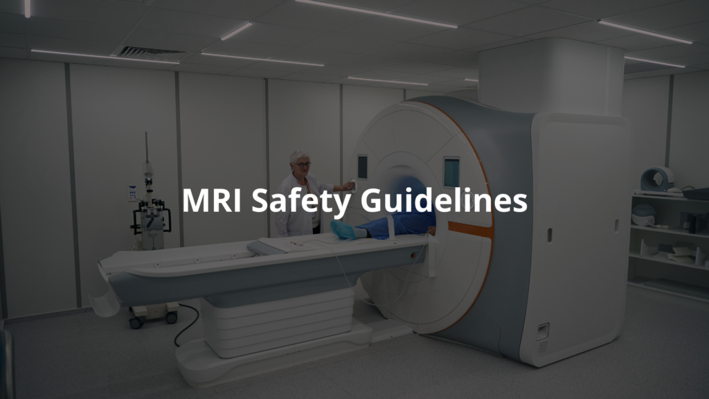
Keeping everyone safe is paramount. The RANZCR, (Royal Australian and New Zealand College of Radiologists), they’ve put together some guidelines to do just that during MRI scans. They think of everything, those folks. There is no room for errors, when working with such complicated equipment.
Some safety points:
- Zoning: MRI rooms are built to keep people safe from the magnets.
- Absorption rates: They keep an eye on how much energy the body absorbs during a scan.
- Screening: Before the scan, the staff checks for anything unsafe, like metal implants.
There are emergency plans: Each MRI facility has procedures to handle anything that may occur. By following these guidelines healthcare providers throughout Australia can offer services that provide high-quality diagnostic information, with less use of radiation. The versatility of the MRI helps maximise patient safety and comfort.
FAQ
How does Magnetic Resonance Imaging work without using radiation?
Magnetic Resonance Imaging uses magnetic fields and radio waves instead of harmful radiation. Unlike X-rays, MRI employs non-ionising energy that doesn’t damage cells. The machine creates a strong magnetic field that temporarily aligns hydrogen atoms in your body.
Then, radiofrequency pulses knock these atoms out of alignment. As they realign, they emit signals that are captured and turned into detailed pictures of your internal structures through image reconstruction.
Why is MRI considered safe medical imaging, especially for pregnancy-safe scans?
MRI is considered safe medical imaging because it’s radiation-free, using no X-rays or other harmful radiation. This makes it pregnancy-safe and appropriate for paediatric imaging when needed. The non-invasive nature of MRI means it doesn’t require cutting or inserting instruments into the body. Since MRI doesn’t use ionising radiation, doctors can order repeated scans without the radiation risks associated with other imaging techniques.
What makes MRI excellent for soft tissue imaging and detailed internal structures?
MRI excels at creating high-resolution scans with superior tissue contrast. It’s particularly effective for visualising soft tissue imaging of organs, muscles, and nerves that don’t show up well on X-rays. The technology can produce multiplanar imaging (views from different angles) and can be fine-tuned to highlight specific detailed internal structures. This makes MRI invaluable for examining the brain, spinal cord, joints, and other complex body parts.
What’s the difference between T1-weighted, T2-weighted, and diffusion-weighted imaging in MRI?
These are different MRI sequences that highlight various aspects of tissues. T1-weighted images show anatomy clearly, with fluid appearing dark and fat bright—great for seeing structural details. T2-weighted images make fluid appear bright, highlighting inflammation or edema—useful for finding abnormalities.
Diffusion-weighted imaging tracks water molecule movement, which is crucial for detecting early strokes and assessing tumours. Each sequence provides complementary information for comprehensive diagnosis.
How does functional MRI help with brain mapping and neuroimaging?
Functional MRI (fMRI) measures brain activity by detecting changes in blood flow. When a brain area is active, it uses more oxygen, changing the magnetic properties of blood that fMRI can detect. This allows doctors to create detailed brain mapping showing which parts activate during specific tasks.
Neuroimaging with fMRI helps identify critical brain regions before surgery, study brain disorders, and understand how different areas communicate through functional connectivity measurements.
What role do superconducting magnets and tesla strength play in MRI quality?
Superconducting magnets are the heart of MRI systems, generating the powerful magnetic field needed for imaging. Tesla strength (the unit measuring magnetic field strength) directly affects image quality—higher tesla strength (like 3T vs 1.5T) produces clearer, more detailed images in less time.
These powerful magnets work with gradient coils to precisely locate signals in the body. The magnetic field strength determines what subtle features can be detected, especially important for small structures or early disease signs.
How has MRI technology evolved to address claustrophobia and noise concerns?
MRI technology has adapted to improve patient comfort. Open MRI designs help people with claustrophobia by providing more space instead of the traditional “tunnel” design. Silent MRI techniques significantly reduce the loud banging noises traditional machines make, creating a more pleasant experience.
Manufacturers have also developed wider bores (openings), improved ventilation, and added features like lighting and even virtual reality to distract patients during scans. These innovations make this important radiation-free imaging option accessible to more people.
Conclusion
Right, so MRI, is a ripper tool for doctors. Without all that radiation, MRI helps get clear pictures inside your body. It’s safer for kids and expecting mothers, and it helps find health problems early on. With this technology getting better all the time, MRI will probably be a big part of healthcare. It’s important to remember, if you ever need one, that MRI can provide the information that’s needed and it is quite safe!
References
- https://mriquestions.com/uploads/3/4/5/7/34572113/safety_asnz_mri_safety_guidelines_v2.pdf
- https://www.melbourneradiology.com.au/guides/magnetic-resonance-imaging-mri-adult-patient-guide/