Learn about radiation-free imaging techniques that keep you safe while providing crucial health information.
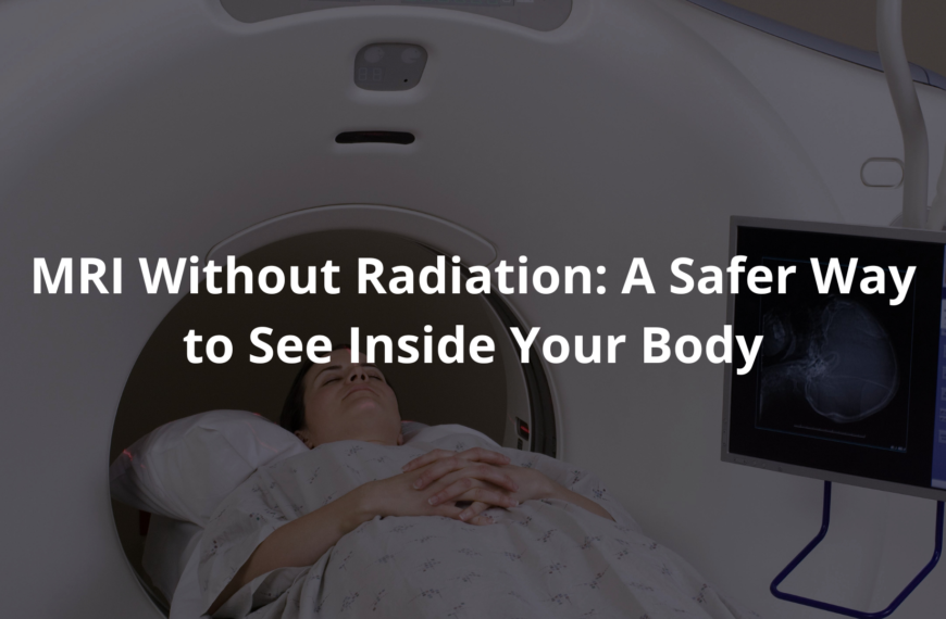
Seeing inside the body without radiation? That’s the promise of radiation-free imaging. In Australia, Magnetic Resonance Imaging (MRI) and Ultrasound are shining examples of this.
MRI uses strong magnets and radio waves (it’s quite fascinating, actually) to create detailed pictures. Ultrasound uses sound waves; think of it like sonar for the body, reflecting images of organs and such.
These methods are valuable; they assist doctors to diagnose medical conditions without the risk of radiation exposure. It’s clever stuff. They aren’t perfect of course; each has its limitations. But both keep patients safer. Keep reading to learn more about these amazing technologies and their clinical significance.
Key Takeaway
- MRI and Ultrasound are common radiation-free imaging techniques.
- These methods are safer for patients, especially kids.
- Understanding your imaging options can lead to better health decisions.
What is Radiation-Free Imaging?
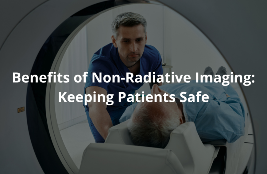
Radiation-free imaging; it refers to methods that show what’s going on inside a person without using the harmful ionising radiation of X-rays, so it’s safer. Doctors often need to see inside us, to assist us to get back to feeling grouse and these techniques do just that.
MRI, or Magnetic Resonance Imaging, is one such method, a rather clever way to peek inside. It uses strong magnets and radio waves to produce detailed images. Some advantages include:
- No Radiation: Since there’s no ionising radiation, it doesn’t damage cells, which makes it much safer.
- Soft Tissue: It creates very clear pictures of soft tissues like the brain, muscles, and joints.
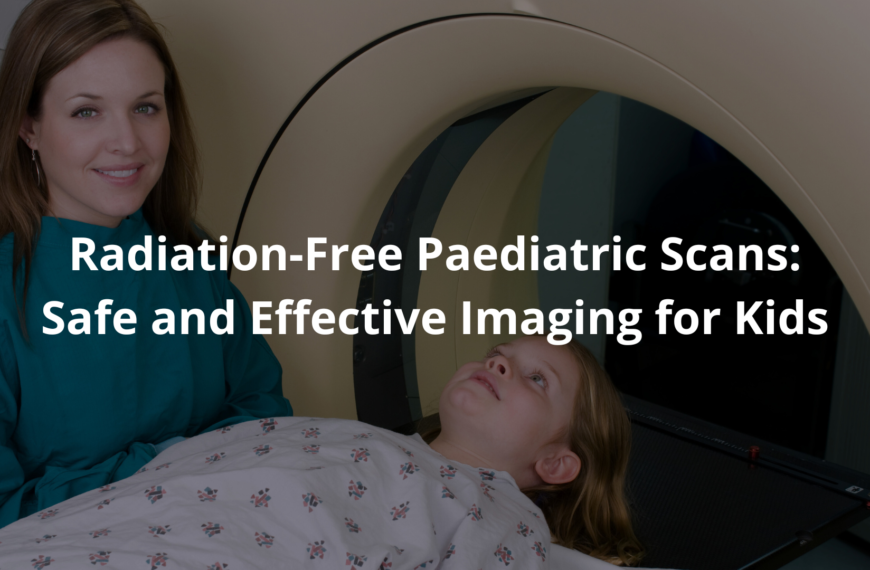
- Good for Kids: Doctors often use it on children (when necessary, mind you) because there’s no radiation risk.
- Slower, Though: It can take longer than other scans, but the image quality is usually worth the extra time. [1]
MRI machines can appear intimidating; they are large and cylindrical. A patient lies still inside, as the machine does its work. It’s quite remarkable how these pictures can be taken without any harm.
Ultrasound: Sound Waves in Action
Ultrasound is yet another type of radiation-free imaging; it’s quite common here in Australia. It bounces sound waves off internal structures to create real-time images. Here’s what you ought to know:
- Safe and relatively quick: Ultrasound is fast, and because it avoids radiation it’s safe for repeated use.
- Expecting mums: Doctors can use ultrasound to monitor babies during pregnancy.
- Image versitility: It can produce images in two dimensions, and three, giving doctors various viewing options (a neat trick, that).
- Detail limitations: It doesn’t always show deeper tissues or bones as clearly as MRI. But it still offers a good look around. [2]
Ultrasound is used in all sorts of situations. Doctors use it to check the heart, kidneys, liver, and gall bladder to check for abnormalities; it’s a true marvel.
The Importance of Safety in Australia
Credits: Brown’s Medical Imaging
Patient safety is paramount in Australia’s healthcare system. The Royal Australian and New Zealand College of Radiologists (RANZCR) sets standards to ensure best practices in all imaging techniques. Their goal; to provide patients with safe, high-quality care.
The Australian Radiation Protection and Nuclear Safety Agency (ARPANSA), meanwhile, works to educate the public about radiation risks. They aim to increase understanding about when radiation is necessary, and when it can be avoided.
“Reduction in Radiation Exposure to Children and Young People from CT Scans” is one program, reducing unnecessary radiation exposure in young patients.
Doctors here understand these concerns. They strive to use the most appropriate, and safest, imaging methods for each individual patient. It’s all part of providing thorough care.
Why Choose Radiation-Free Imaging?
Opting for radiation-free imaging, such as MRI and ultrasound, brings many advantages. Here’s a look at why these methods benefit patients:
- Generally safer: Because they avoid radiation, there’s no risk of radiation-induced damage.
- Repeated scans are less worrying: If a patient requires several scans, doctors can monitor their condition without the worry of cumulative radiation exposure.
- Better for children: Since children are more sensitive to radiation, radiation-free methods keep them safer.
It’s brilliant how technology is allowing us to see inside the human body, with minimal risk. When doctors have safe, effective options, they’re able to make better, more informed decisions. It’s progress; real progress.
When is Radiation-Free Imaging Used?
Radiation-free imaging finds its applications in all sorts of situations. Here are some common uses:
- Tumour detection: MRI can assist in locating tumours in soft tissues, which can be easily assessed using the technology.
- Organ assessments: Ultrasound is often used to look at organs, like the heart, liver, and kidneys.
- Monitoring foetal growth: Ultrasound is essential for monitoring foetal development throughout pregnancies.
- Guiding procedures: Ultrasound is used to assist doctors during certain procedures, such as placing tubes and needles (it’s rather precise, actually).
These examples show the importance of radiation-free imaging in modern medicine. It’s not just about producing pretty pictures; it’s about ensuring patients receive the highest standard of care without unnecessary risks.
New Technologies in Radiation-Free Imaging
New and exciting technologies in radiation-free imaging continue to emerge. Here are a few worth keeping an eye on:
- Magnetic Particle Imaging (MPI): This uses tiny magnetic particles to create images. It’s experimental, yet shows great promise.
- Contrast-Enhanced Ultrasound (CEUS): This method uses microbubbles to enhance the clarity of ultrasound images.
- Spatial Frequency Domain Imaging (SFDI): This looks at how light travels through tissue to create images.
- Diffuse Optical Spectroscopy: This can monitor tissue changes (useful for spotting diseases).
These advancements illustrate the ongoing efforts to develop better, and safer, ways to care for patients. Innovation is key.
Real-Time Visualisation

A standout feature of radiation-free imaging is its capacity for real-time visualisation. This allows doctors to see what’s happening inside the body as it happens.
- Quick decision-making: With real-time information, doctors can make rapid decisions during procedures.
- Enhanced guidance: For complex operations, imaging can guide surgeons to ensure accuracy.
- Reduced risk: Real-time imaging can reduce the need for invasive procedures, which are generally safer for the patient.
These technologies offer tangible benefits. When used correctly they can lead to improved patient outcomes. That’s what matters, doesn’t it?
FAQ
What is radiation-free imaging and how does it differ from traditional imaging?
Radiation-free imaging uses safer methods like ultrasound, MRI, and light-based imaging to see inside your body. Unlike X-rays, these non-ionising imaging methods don’t use harmful radiation. They’re better for people who need many scans, pregnant women, and kids.
These techniques use sound waves, magnetic fields, or light to make pictures of your insides. Doctors use these pictures to find problems without using radiation. As technology gets better, more doctors are using these safer methods.
How does Magnetic Resonance Imaging (MRI) work and what can it show?
MRI uses strong magnets and radio waves to make detailed pictures of your organs and tissues. It lines up tiny particles in your body and measures them as they move back to normal. MRI is a non-ionising imaging technique, which means no radiation touches you.
It shows soft tissues really well, like your brain, spine, and joints. It can also find tumours. MRI gives high-resolution medical imaging that shows lots of detail, though it takes longer than other tests and makes loud noises. People with certain metal in their bodies can’t have MRIs.
What is Ultrasound and Contrast-Enhanced Ultrasound (CEUS) and how do doctors use them?
Ultrasound uses sound waves to make pictures of what’s inside your body. The sound bounces off different parts of your body to create images right away. It’s quick, doesn’t hurt, and uses no radiation. Contrast-Enhanced Ultrasound (CEUS) goes further by adding tiny bubbles to your blood that make the pictures clearer.
Doctors use these for checking babies before they’re born, looking at organs like your liver and kidneys, and helping with certain procedures. These methods give real-time visualisation without any radiation exposure. CEUS is especially good at showing blood flow and finding small spots that regular ultrasound might miss.
How do doctors use Spatial Frequency Domain Imaging (SFDI)?
Spatial Frequency Domain Imaging shines striped light patterns (sinusoidal fringe patterns) on your skin and watches how the light bounces back. This wide-field structured light imaging checks optical properties like how much light your tissues soak up (absorption coefficient). SFDI helps find special substances (chromophore concentrations) in your tissues without using any radiation.
During surgery, it helps with intraoperative margin assessment—checking if all the bad tissue was removed. Using special math (demodulation techniques) and light models (diffuse light transport model), it gives useful pictures of tissue characteristics. Doctors use wavelength-dependent imaging with SFDI to tell different types of tissue apart by analysing backscattered light.
What is Fiber Optic RealShape technology and how is it helping with blood vessel surgery?
Fiber Optic RealShape technology uses tiny light fibers (optical fiber imaging) to make 3D colour imaging of your blood vessels during procedures. This light-based endovascular navigation system lets doctors see guidewires and catheters moving through your blood vessels right as it happens.
During tricky vascular procedures like aortic aneurysm repair, it shows many angles without using radiation. Surgeons can move through hard spots like the femoral artery better. The technology works with regular catheters and offers radiation-free technology for blood vessel navigation. It’s really helpful in complex aortic procedures where seeing clearly is super important.
What’s new with photoacoustic imaging and optoacoustic imaging?
Photoacoustic and optoacoustic imaging use both light and sound to see inside you. They work by sending light into your body that makes sound waves. The sound waves create pictures. New improvements have made spatial resolution imaging better, so doctors can see more details of your tissues.
These methods are great at finding blood vessels and checking oxygen in your tissues without radiation. Doctors use them more and more for finding breast cancer, looking at brains, and checking skin problems. These non-invasive imaging techniques show things about how your body works that old-fashioned pictures can’t show.
How do special light-based methods like fluorescence and bioluminescence imaging work?
Optical molecular imaging uses light to see tiny processes in your body. Fluorescence imaging adds special stuff that glows under certain light, making some parts of your body easier to see. Bioluminescence imaging catches light naturally made by your body (like fireflies make).
Near-infrared spectroscopy and Raman spectroscopy look at how your tissues interact with light to figure out what they’re made of. These techniques give quantitative optical imaging without any radiation. They help researchers, drug makers, and sometimes surgeons. These approaches let doctors and scientists watch diseases or treatments working in real time.
Conclusion
Radiation-free imaging techniques, like MRI and ultrasound, are essential. They keep patients safe, while providing valuable health insights. By avoiding the risks of radiation, these methods offer a variety of benefits. From monitoring pregnancies to guiding surgeries, radiation-free imaging plays a crucial role in modern healthcare. Remembering these technologies is important for you and your family’s health.
References
- https://pubmed.ncbi.nlm.nih.gov/24268967/
- https://www.arpansa.gov.au/sites/default/files/documents/2024-05/patienthandout.pdf