Understand the critical role of radiology for diagnosis, key imaging methods, and improving patient outcomes.
Radiology for diagnosis is a truly amazing field. Picture someone standing before a huge puzzle that reveals hidden truths about their health. It helps doctors peek inside without any invasive procedures. Just like discovering treasures inside a mysterious chest, radiology unlocks secrets.
By using special tools to create images, doctors gain insights into health issues, guiding them to make better decisions. This is a world filled with wonders, and it’s something everyone should learn more about.
Keep reading to uncover how radiology touches lives, making a real difference in healthcare and helping all of us stay healthy.
Key Takeaway
- Radiology helps doctors see inside the body using images.
- There are different types of imaging tests, like CT scans and MRIs.
- Radiology is important for diagnosing and treating diseases.
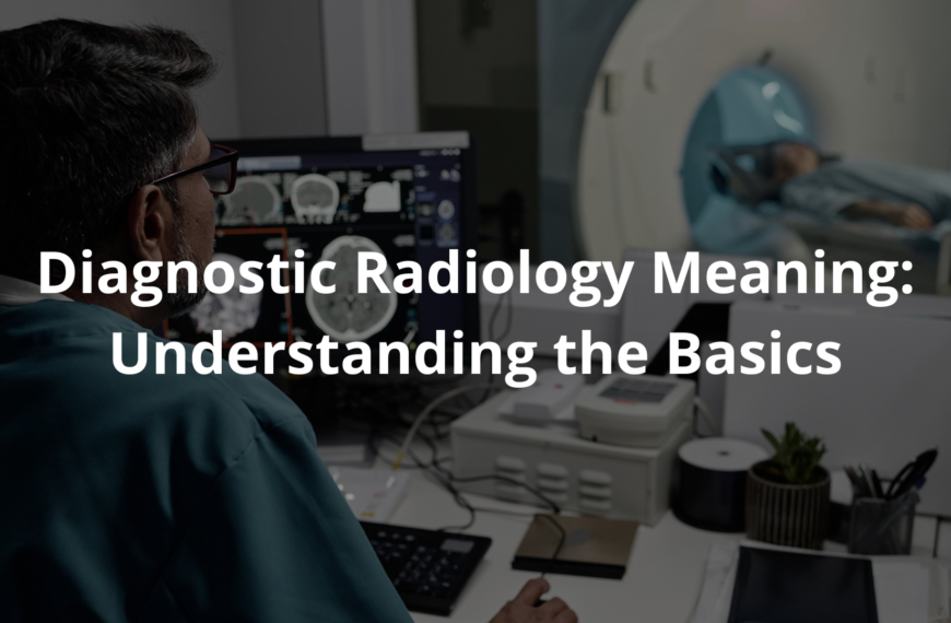
What is Radiology for Diagnosis?
Radiology for diagnosis is like having superhero glasses, allowing doctors to see inside our bodies without needing to make any cuts. This process uses special machines to create images of bones, organs, and tissues. A CT scan (computed tomography) is one of those machines. It takes tons of pictures from different angles to create a detailed view of what’s happening inside. This helps doctors diagnose problems, like heart disease or even breast cancer.
When someone goes in for a CT scan, it’s usually a pretty quick process. Patients lie down on a table, and the machine does its thing. There’s often a soft whirring sound, and it can take several pictures in just a few minutes. It’s super important to remember that doctors use low-dose radiation to keep patients safe while getting these images. They always have the patient’s safety in mind!
Sometimes, the images can reveal things that are hard to find just by feeling around. For example, if someone has unexplained back pain, a CT scan might show whether there’s an issue with the spine that needs looking at. It’s just amazing to think how these machines provide so much information just through pictures.
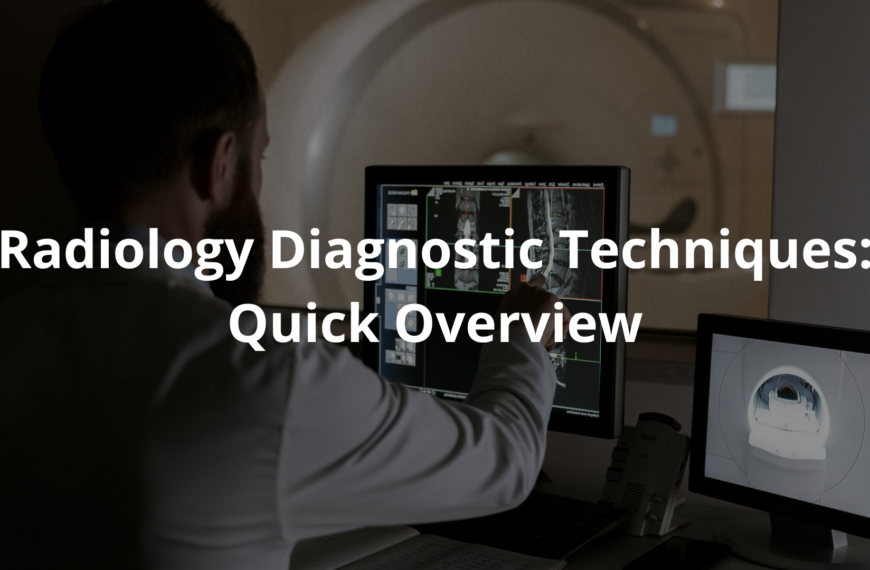
Types of Imaging Tests
There are many types of imaging tests that doctors can choose from. Each one has its special task. Here’s a quick list of some common ones:
- CT Scans: These scans give detailed pictures of inside the body. They can show soft tissues, blood flow, and even organs like the heart and lungs.
- X-rays: Often used to look for fractures or infections. They’re fast and easy, kinda like snapping a photo.
- MRIs (Magnetic Resonance Imaging): These tests use magnetic fields to create neat images of soft tissues. They’re great for looking at the brain, muscles, and joints.
- Ultrasounds: These use sound waves to create images, often used for checking on a baby during pregnancy and looking at soft tissues.
- Nuclear Medicine: This imaging uses small amounts of radioactive materials to see how organs are working. It helps doctors check out things like blood flow and organ function.
Each imaging test serves a unique purpose. Doctors choose the right one based on what the patient needs. For example, if a doctor suspects a heart issue, they might recommend a CT scan to get clear images of the heart. This way, they are better able to decide what treatment the patient might require.
The Role of Radiologists
Radiologists are the medical experts who read and make sense of these images. They spend many years getting trained—around four years of medical school and then a four-year residency in clinical radiology. That’s a lot to learn! Radiologists are like detectives of the body. They’re skilled at understanding what the images reveal and how to use that information to help treat diseases.
When radiologists look at images, they search for signs of problems like tumors, fractures, or other injuries. They work hand-in-hand with other doctors, like surgeons and general practitioners, to help make the best choices for their patients. This teamwork is super important in healthcare.
For example, if someone goes to the emergency room with chest pain, the doctor may order a chest X-ray or CT scan. The radiologist will analyze those images and report back on what they see. If there’s a spot on the image that looks worrying, the radiologist might suggest further tests or treatments. It’s all about working together to help patients get the care they need.
How Imaging Helps Diagnose Diseases
Imaging tests play a crucial role in detecting diseases early, as early detection can significantly improve outcomes. For example, identifying conditions like breast cancer or heart disease at an early stage can be life-saving. Imaging allows doctors to spot subtle changes in the body, which may indicate potential issues before symptoms become severe.
Key points:
- Early Detection Saves Lives: Timely imaging can help catch diseases such as cancer or heart conditions before they progress.
- Thyroid Scans: A thyroid scan helps evaluate how well the thyroid gland is functioning. If there are irregularities, treatment can be started promptly.
- Preventing Serious Conditions: Many diseases do not show symptoms until they are advanced. Imaging can detect these red flags early, reducing the risk of complications.
Imaging is a powerful tool that can make a significant difference in a person’s health by providing early insight into potential health concerns.
Patient Safety and Regulations
In Australia, radiology services are regulated so that patient safety is always a priority. The Australian Radiation Protection and Nuclear Safety Agency (ARPANSA) makes sure the radiation used during imaging tests is kept at safe levels. They set standards to protect patients from too much radiation, which is very important. [1]
Also, the Royal Australian and New Zealand College of Radiologists (RANZCR) helps keep high standards for radiology practices. They provide guidelines that ensure radiologists are well-trained and that imaging services are safe and effective. Thanks to these regulations, patients can feel confident that their safety is always a top priority during imaging tests.
Doctors always explain the procedures to patients beforehand. They tell patients what to expect from the imaging process. This helps patients feel more relaxed. If someone’s nervous about their scan, the doctor might say, “You might feel a little warm from the contrast agent, but it won’t hurt.” This kind of reassurance helps patients understand what’s about to happen.
Trends in Diagnostic Imaging Utilization

Between 2000 and 2021, Australia saw a significant rise in the use of diagnostic imaging, with over 436 million imaging studies conducted. [2] This sharp increase reflects advancements in technology and a growing reliance on imaging tests to support patient care. As people seek medical assistance more frequently, imaging has become a vital tool for doctors in diagnosing and monitoring health conditions.
Key Points:
- Increased Imaging Use: More than 436 million imaging studies were performed between 2000 and 2021, showcasing the rising demand for diagnostic imaging.
- Older Adults at the Forefront: Seniors are the largest group using imaging services, often requiring regular check-ups for conditions like arthritis or heart disease.
- Preventive Healthcare: Imaging plays a crucial role not just in treating illness, but in maintaining long-term health. Early detection through imaging can help identify potential problems before they become serious.
Encouraging regular check-ups with imaging ensures better health management and early intervention, particularly for older individuals.
FAQ
How do doctors use MRI and CT scans to look inside my body?
When doctors need to see inside your body, they use special cameras called MRI and CT scans to produce images. MRI uses magnetic fields – like super strong magnets – while CT scanning uses tiny bits of ionizing radiation. These help create detailed images that show what’s happening inside you.
What kinds of pictures can doctors take in the hospital’s radiology department?
The radiology department is like a photo studio for your body. They have different cameras that take medical images – from simple X-rays (plain radiography) to fancy magnetic resonance imaging machines. Each machine helps healthcare providers see different parts like soft tissues and check how your organs work (organ function).
How do special doctors use pictures to help fix health problems?
Some doctors, called interventional radiologists, use real time pictures to help fix problems inside your body. They can watch your blood flow on a screen and use sound waves to guide tiny tools through your body. It’s like using a video game to help make you better, and it doesn’t hurt as much as regular surgery because it’s minimally invasive.
How long do doctors study to learn about medical imaging?
Before doctors can take and read medical images, they need lots of training. They go to school to learn about medical physics and different imaging techniques. After medical school, they spend more years of training to become certified by the american board of radiology. They learn how all the imaging procedures work to help diagnose and treat patients.
How do X-rays help doctors see inside people?
Doctors use special types of light called ionising radiation (also spelled ionizing radiation in the united states and member states) to see inside you. They’re very careful and only use small amounts in medical diagnosis and radiation therapy. It’s like using a flashlight, but this special light can see through your skin!
How do doctors check breast tissue?
Doctors use special cameras to take detailed images of breast tissue. They have a range of imaging techniques that help them spot any problems early. Think of it like taking a super detailed photograph that helps medical doctors see if anything needs attention.
How do nuclear medicine and PET scans work?
These are like special cameras that use a tiny bit of radioactive material to show how your organs are working. It helps medical doctors see organ function – kind of like watching a movie of your organs at work. Positron emission tomography (PET scans) and nuclear medicine help doctors find problems they can’t see other ways.
What’s the difference between doctors who take pictures and doctors who use pictures to do treatments?
Some doctors (diagnostic radiologists) are like photographers – they take and study medical images to find problems. Others (interventional radiologists) are like mechanics who use these images to fix things. Both use imaging technology to help keep you healthy, but they do different jobs in health care.
What do doctors learn in medical specialisation that involves taking special pictures?
Doctors who work in diagnostic radiology learn about many different ways to take pictures of your body. They work in the radiology department using various imaging procedures to help other healthcare providers figure out what’s making you feel sick. It’s like being a detective who uses special cameras instead of a magnifying glass!
Conclusion
Radiology plays a huge part in healthcare. It helps doctors look inside our bodies and spot health issues. Different imaging tests like CT scans and MRIs guide doctors to make smart choices for treating patients. Radiologists work hard, ensuring we feel safe during these tests. So next time you hear about a CT scan or X-ray, think about how they help in finding and treating diseases. Understanding these tests is key; knowledge really is power when it comes to health!
References
- https://www.health.gov.au/topics/diagnostic-imaging/about
- https://onlinelibrary.wiley.com/doi/abs/10.1111/1754-9485.13591