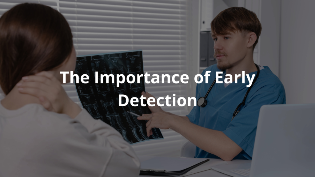
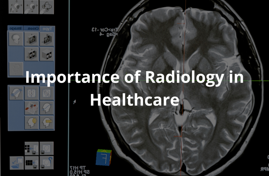
Radiology in cancer detection unveils life-saving insights, finding cancer early and transforming care for patients in Australia.
Radiology plays a crucial role in spotting cancer. It gives doctors a way to look inside the body, helping them find cancer early when treatment has the best chance to work. Across Australia, researchers and medical teams are pushing boundaries with advanced imaging technologies, aiming to make scans sharper, faster, and more precise.
It’s almost like a partnership between skilled hands and smart machines, all working to save lives. From CT scans to PET scans, these tools are changing how cancer is detected and treated. Keep reading to explore how these innovations are making a real difference for patients and families.
Key Takeaway
- Radiology uses special scans to find cancer early.
- New technologies are helping doctors get better images.
- Early detection means better chances for patients.
Understanding Radiology in Cancer Detection
Radiology is like a window into the body, letting doctors see what’s happening inside without needing surgery. It’s how they can find cancer and other problems early, often before symptoms even show up. These scans can truly make a difference.
CT Scans (Computed Tomography)
CT scans use X-rays to take detailed pictures of the body. They’re especially good for looking at the lungs, abdomen, and brain. What’s great is they can show both soft tissues and bones, which helps doctors spot things like tumours. The procedure itself is quick—usually just a few minutes.
A personal example comes to mind. My grandmother had a lung CT scan a few years ago. It found a tiny spot that turned out to be cancer, but because it was caught early, she got treatment and recovered. That scan might’ve saved her life.
MRI Scans (Magnetic Resonance Imaging)
MRIs work differently. Instead of X-rays, they use strong magnets and radio waves to create images. This means no radiation, which is a good thing. MRIs are great for showing soft tissues, like the brain, spine, and joints. They’re non-invasive and painless, though they can take longer—up to an hour sometimes.
Early detection matters so much. These scans can find problems before they grow, giving people a better chance at treatment and recovery.
The Importance of Early Detection

Catching cancer early can save lives. When tumours are small, they’re often easier to treat. Take lung cancer, for instance. It’s a serious disease, but with low-dose CT scans, doctors can screen high-risk patients and find it sooner. Early-stage lung cancer has a much better survival rate—up to 56% over five years. [1] Compare that to late-stage cancer, where survival can drop below 5%.
I once heard about a young woman who went for her regular breast MRI. She didn’t feel anything unusual, but the scan found a tiny tumour. Because it was caught so early, she only needed a small surgery and was able to move on with her life. Stories like hers show how important these scans are.
Screening programs for cancers like breast and colorectal cancer also help. They encourage regular scans, which can catch issues before they become serious. And for people with a family history of cancer, staying on top of screenings is even more critical. It could be lifesaving.
The Role of Radiologists
Radiologists are the doctors who read and interpret medical images. They’re trained to spot things others might miss, which makes them essential in diagnosing cancer and other conditions. Their work guides treatment decisions and helps patients get the care they need.
Here’s a quick breakdown of what they do:
- Interpreting Scans: Radiologists study images like X-rays, CT scans, and MRIs. They compare them to past scans to see if anything’s changed, like a tumour growing or shrinking.
- Using Technology: They use advanced software to make images clearer, which helps in spotting even tiny abnormalities.
- Collaborating with Specialists: Radiologists work closely with oncologists and other doctors to make sure patients get accurate diagnoses and effective treatment plans.
- Timely Intervention: By identifying issues quickly, they help the care team act fast, which can save lives.
- Ongoing Education: Radiologists keep learning through training programs, staying updated on the latest imaging techniques.
Their expertise is a big part of why cancer detection and treatment have improved so much.
How Technology is Changing Radiology
New tools and techniques are making cancer detection better and faster. Here are a few examples of what’s happening in Australia:
- Contrast Enhanced Mammography (CEM): This is for women with dense breast tissue. It uses a contrast agent to make small tumours easier to see, solving some of the problems with regular mammograms.
- In-line Phase-Contrast Computed Tomography (PCT): This improves imaging quality without needing to compress the breast, making it more comfortable for patients.
- Positron Emission Tomography (PET) Scans: These scans show how organs and tissues are working. By using a small amount of radioactive material, they can find cancers that might be hidden.
- Artificial Intelligence (AI): AI is helping radiologists by analysing images quickly and accurately. It can detect patterns that suggest cancer, allowing doctors to focus on the most urgent cases.
These advancements are making it easier to find cancer early, which means patients can start treatment sooner.
The Role of Organisations
Groups like the Royal Australian and New Zealand College of Radiologists (RANZCR) are working to improve cancer care. They set training standards for radiologists and make sure they’re skilled in using the latest imaging technology.
I once attended a talk where a radiologist explained how these organisations also educate the public about the importance of screenings. They run awareness campaigns to encourage people, especially those in high-risk groups, to get regular scans. This kind of outreach can lead to earlier detection and better outcomes. [2]
RANZCR also supports research to improve imaging techniques. They create guidelines that help doctors make better decisions based on scan results. Patients benefit from this because it means they’re getting the best possible care.
FAQ
How do pet and ct scans help find cancer in the human body?
PET and CT scanning technologies work together to create detailed pictures of what’s happening inside your body. When doctors combine these scans, they can spot cancer more accurately and see how far it has spread. These imaging tests help your care team make better decisions about your treatment.
What are the main differences between ct and mri when looking for tumors?
CT uses X-rays to create detailed cross-sectional images, while MRI uses magnetic fields to show soft tissue in great detail. MRI scans are especially good at showing the difference between normal cells and tumor cells. Your medical team might use both to get the most complete picture.
How does low dose ct imaging protect patients during scans?
Low dose CT uses less radiation while still providing quality images to help find cancer. This imaging test is particularly useful for high risk patients who need regular scans, like those being checked for cell lung cancer. The technology keeps radiation exposure as low as possible while maintaining a good noise ratio.
What role do blood flow and lymph nodes play in cancer detection?
Blood vessels and lymph nodes can show signs of cancer spread. Imaging tests can map out these systems to spot unusual patterns. When cancer cells travel through your body tissues, they often settle in lymph nodes first, making node mapping an important tool for diagnosis.
How do contrast dye and barium enema help in gi tract imaging?
These substances help create clearer pictures during upper gi and lower gi exams. A barium enema helps doctors see the gi series of your digestive system in real time imaging. Contrast dye is used in various scans to make certain body parts show up more clearly.
What is the importance of bone scans and bone marrow testing?
Bone scans use gamma rays to check if cancer has spread to your bones. In some cases, doctors might also need to check your bone marrow using fine needle sampling. These tests help determine if solid tumors have spread beyond their original location.
What are the side effects of different imaging tests?
Most imaging tests are safe with minimal side effects. Some patients might react to contrast dye, but this is rare. High energy ray imaging and ray machine exposure is carefully controlled. Your health care team monitors any potential risks during the procedure.
How do high field MRI and dual energy scans improve cancer detection?
These advanced technologies offer better image quality and can often detect early stage cancers. High field MRI provides detailed soft tissue images, while dual energy scanning can help spot subtle changes that might indicate cancer. Pet tracers are sometimes used alongside these methods for better accuracy.
What happens during image guided procedures?
Image guided procedures use sound waves or other imaging methods to help doctors perform precise procedures. This might include mri guided biopsies or treatments. In rare cases, special techniques might be needed to reach difficult areas.
How do stem cells relate to cancer treatment and imaging?
Stem cells play a crucial role in both cancer development and treatment. Special imaging techniques can track stem cell movement and effectiveness during treatment. This information helps your medical team monitor how well treatments are working.
Conclusion
Radiology’s pretty key in spotting cancer early here in Australia. Tools like CT scans, PET scans, and MRIs help doctors catch it when it’s easier to treat. Groups like RANZCR make sure radiologists get top-notch training and equipment. There’s hope for even better imaging to help patients down the track. If you’re worried about your health, have a chat with your doctor—they can guide you on the best screening options. Always worth asking.
References
- https://www.cancer.org.au/cancer-information/what-is-cancer/facts-and-figures
- https://www.aph.gov.au/DocumentStore.ashx?id=85f72e12-aabd-4192-8dc3-6148092ae3cb&subId=659814


