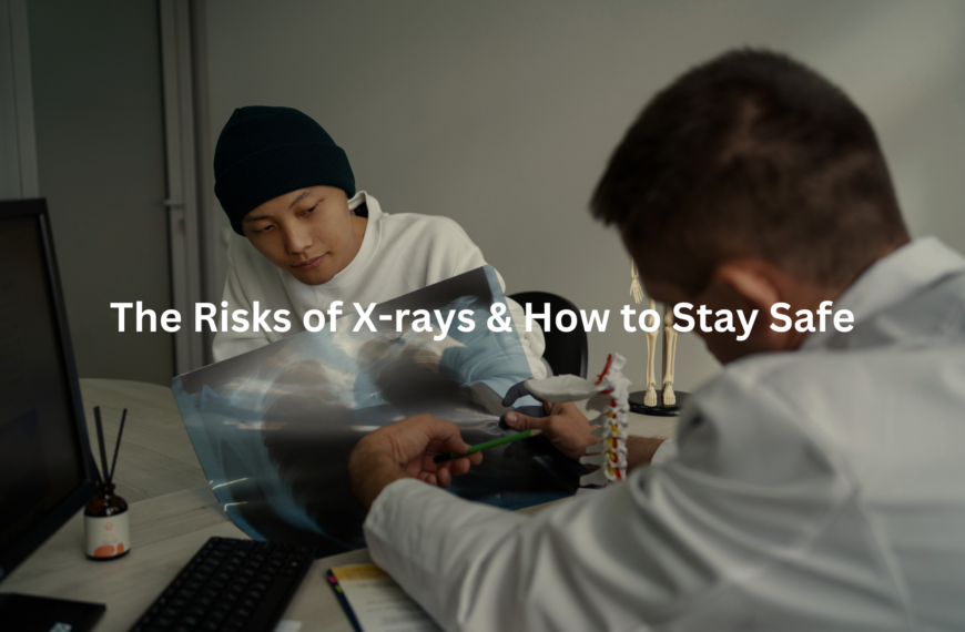
Learn how safe imaging practices protect patients by minimising radiation without losing image quality.
Safe imaging practices are essential for keeping patients healthy during medical tests. When doctors take pictures of our insides using machines like X-rays and CT scans, they have to be careful about radiation exposure. Too much radiation can be harmful, but with the right practices, we can get the information we need without putting patients at risk. Want to know how healthcare professionals do this? Let’s take a closer look!
Key Takeaway
- Safe imaging practices help reduce radiation exposure for patients.
- The ALARA principle guides healthcare providers to use the least amount of radiation necessary.
- Continuous education and training ensure that radiologists stay updated on the best safety practices.
Understanding Safe Imaging Practices
Radiology clinics are quiet places. The kind where time moves at its own pace, dictated by the soft hum of machines and the precise hands of radiographers adjusting controls. But behind every click, beep, and flicker of an image on the screen is a fundamental question—how do we get the clearest picture while exposing patients to the least amount of radiation?
Safe imaging practices are at the heart of medical radiology, ensuring that every X-ray, CT scan, or fluoroscopy procedure follows strict protocols to minimise unnecessary radiation exposure. This means careful calibration of machines, rigorous training for staff, and the application of imaging techniques that prioritise patient safety without compromising diagnostic accuracy. (1)
At the centre of these practices is the ALARA principle—”As Low As Reasonably Achievable.” It is the backbone of radiation safety, reminding radiologists and technicians that while ionising radiation is useful, it must be controlled. The goal is to deliver just enough exposure to capture high-quality images while avoiding excess that could contribute to cumulative radiation dose over time.
Radiation Exposure Reduction Techniques
Imaging tests are essential for diagnosing medical conditions, but they do come with radiation exposure. Some techniques help reduce that exposure while keeping image quality high.
- Justification for Imaging: Before any scan, doctors evaluate whether the benefits outweigh the risks. If an alternative like MRI (which uses non-ionising radiation) or ultrasound can provide the same diagnostic value, it may be chosen instead. This is especially important for young patients, who are more sensitive to radiation’s long-term effects.
- Dose Optimisation Techniques: Imaging protocols are adjusted based on a patient’s size, age, and the area being examined. For instance, paediatric imaging uses lower radiation doses than adult imaging. Low-dose protocols have been developed for common procedures like CT scans and fluoroscopy, ensuring necessary images are obtained with the least possible exposure.
- Effective Shielding Methods: Lead aprons, thyroid shields, and protective drapes are used to limit radiation to surrounding tissues. Although shielding is less commonly used in certain modern imaging techniques due to advancements in dose reduction, it remains an important tool in specific cases.
Patient Safety Protocols
Safety in radiology extends beyond just reducing dose—it includes informed decision-making, tracking exposure, and adapting imaging for vulnerable populations.
- Informed Consent: Patients need to understand the risks and benefits of imaging procedures before they agree to them. A doctor might explain that a PET/CT scan will involve exposure to ionising radiation, but the information it provides could be crucial in diagnosing or staging cancer.
- Monitoring Radiation Exposure: Some hospitals and radiology clinics track a patient’s cumulative radiation dose over time. If someone has had multiple CT scans or fluoroscopic procedures, doctors can factor in past exposure when deciding whether another scan is necessary.
- Paediatric Imaging Safety: Children are more sensitive to the biological effects of radiation, including DNA damage and chromosomal aberrations. Imaging guidelines for kids focus on reducing exposure by adjusting radiographic techniques and using alternative imaging methods whenever possible. (2)
Staff Training in Radiation Safety
Radiologists, radiographers, and medical physicists are the gatekeepers of imaging safety. Without ongoing training, even the best imaging technology can be misused.
- Continuous Education for Radiologists: Radiation safety isn’t a one-and-done lesson. Regular training sessions cover updates on radiological protection standards, dose-response relationships, and the latest research on long-term effects of radiation exposure.
- Quality Assurance in Radiology: Equipment doesn’t just need to work—it needs to work accurately. Flat-panel detectors, image quality assessment software, and radiation monitoring tools are routinely tested to ensure that each scan provides the best possible image at the lowest dose. (3)
Special Considerations and Technologies
Credit: MAHSA University
Advancements in imaging technology have transformed how radiation is used in diagnostics. Modern imaging machines are designed to optimise imaging techniques while keeping doses lower than older equipment.
- Digital Imaging Advancements: Computed radiography (CR) and digital radiography (DR) have largely replaced traditional film-based X-rays, providing better image quality with lower radiation doses. Technological innovations in imaging, such as automated dose modulation in CT scans, help adjust radiation levels based on patient anatomy in real time.
- Emergency Protocols for Radiation Incidents: While rare, unexpected radiation incidents can happen—equipment failures, miscalculated doses, or accidental repeat scans. Imaging facilities have established emergency protocols to manage and mitigate radiation-induced effects when necessary.
Collaborative Approaches to Radiation Safety
No single specialist owns radiation safety—it requires collaboration across medical fields, public health initiatives, and ongoing research.
- Interdisciplinary Communication: Radiologists work alongside referring physicians, medical physicists, and radiation safety officers to ensure imaging studies are justified. Clear communication helps prevent unnecessary imaging and ensures the best risk-benefit analysis for each patient.
- Public Health Initiatives on Imaging Safety: Government health agencies and professional radiology organisations run campaigns to inform patients and healthcare providers about radiation exposure risks. These efforts encourage ethical considerations in imaging, ensuring that patient safety always comes first.
Safe imaging practices are not just about reducing radiation—they’re about balance. Getting the clearest image while keeping exposure low is a science, an art, and a responsibility that radiologists take seriously every day.
FAQ
How do safe imaging practices help with radiation exposure reduction?
Safe imaging practices focus on reducing unnecessary radiation while maintaining image quality. The ALARA principle ensures that patients receive the lowest dose necessary for a diagnosis. Techniques like dose optimization techniques, effective shielding methods, and patient safety protocols help manage exposure. Imaging protocol standardisation also plays a role in making sure patients aren’t exposed to more radiation than needed.
What are the key radiation protection guidelines in medical imaging?
Guidelines focus on ionizing radiation management by setting dose limits, enforcing patient safety protocols, and using radiation dose recording systems. Use of lead aprons and shields, fluoroscopy best practices, and collaborative approaches to radiation safety help reduce unnecessary exposure. Regulatory bodies ensure compliance with these rules to improve patient and staff safety.
Why is justification for imaging procedures important in patient care?
Doctors must weigh the risks and benefits before ordering scans. Minimizing unnecessary imaging prevents excess exposure while maintaining diagnostic accuracy. Patient history evaluation for imaging helps determine if non-radiation alternatives like ultrasound or MRI are suitable. Risk-benefit analysis in diagnostics ensures imaging is only used when necessary.
How does patient communication strategies improve informed consent in radiology?
Patients need to understand the risks, benefits, and alternatives before undergoing imaging. Clear explanations of safe imaging practices, potential radiation effects, and long-term monitoring of exposure effects help patients make informed decisions. Awareness campaigns on radiation safety also play a role in public education.
What measures are in place for pediatric imaging safety?
Children are more sensitive to radiation, so dose optimization techniques and patient positioning techniques are used to lower their exposure. Minimizing repeat examinations and opting for non-ionizing imaging alternatives like ultrasound when possible reduce risks. Paediatric specialists ensure radiation exposure reduction without compromising diagnostic quality.
How do hospitals ensure imaging equipment safety and accuracy?
Regular maintenance and quality assurance in radiology prevent equipment malfunctions. Maintaining imaging equipment standards ensures accurate results and lower radiation doses. Technology integration for dose monitoring helps track and adjust radiation levels in real time.
What role does continuous education for radiologists play in safety?
Radiologists must stay up to date with radiation protection guidelines, staff training in radiation safety, and emergency protocols for radiation incidents. Ongoing education ensures professionals follow the latest fluoroscopy best practices and scatter radiation awareness strategies to keep both patients and staff safe.
How does monitoring radiation exposure protect patients?
Tracking exposure history helps prevent excessive cumulative doses. Radiation dose recording systems allow doctors to make informed decisions about further scans. Long-term monitoring of exposure effects helps researchers understand risks and improve safe imaging practices over time.
What are the best ways to minimize personnel exposure during exams?
Healthcare workers follow use of protective gear, use of collimation in X-rays, and reducing exposure time during procedures to limit exposure. Effective use of dosimeters helps track doses over time. Regulatory compliance in radiology ensures staff follow safety protocols.
How does patient positioning techniques affect safety?
Proper positioning improves image clarity, reducing the need for repeat scans. Optimizing imaging parameters and mechanical restraint techniques ensure accurate results while following radiation protection guidelines. This approach is especially important in paediatric and trauma imaging.
Conclusion
In conclusion, safe imaging practices are vital for protecting patients from radiation exposure while getting the necessary diagnostic information. By following the ALARA principle, continually educating healthcare providers, and utilizing advanced technologies, radiology is providing safe and effective imaging services. These practices not only help keep patients safe but also ensure that they receive the best possible care when it comes to their health.
References
- https://www.ncbi.nlm.nih.gov/books/NBK557499/
- https://www.ncbi.nlm.nih.gov/books/NBK585624/
- https://www.iaea.org/resources/rpop/health-professionals/radiology


