Radiology keeps healthcare running, but safety matters. Learn how to minimise risks and protect patients from radiation exposure.
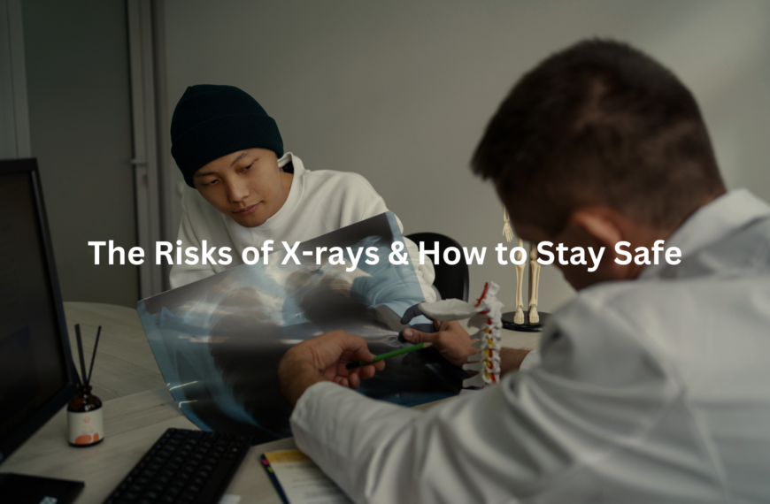
Radiology stands as a critical part of modern healthcare, letting medical teams see inside the body without surgery. Safety measures in radiology clinics across Australia protect both patients and staff from radiation exposure (measured in millisieverts, or mSv). Young patients need extra care, as their growing bodies show more sensitivity to radiation than adults.
Medical teams use lead aprons and strict protocols to shield patients during X-rays. Radiologists only order scans when the benefits outweigh any risks, following the ALARA principle – As Low As Reasonably Achievable. Keep reading to learn more about the smart ways we protect health while using these life-saving tools!
Key Takeaway
- Radiation exposure can be harmful, but there are ways to reduce it.
- Special care is needed for kids and pregnant women during imaging.
- Patient education helps everyone understand the risks and benefits of imaging.
Radiation Exposure
Radiation works like an invisible force in medical imaging(1). CT scanners, those big doughnut-shaped machines, use it to look inside the body. Each scan gives off different amounts of radiation, measured in millisieverts (mSv).
A single CT scan typically delivers 1-10 mSv, which is more than a chest X-ray (0.1 mSv) but less than a PET scan (25 mSv). The effects add up over time, and research shows higher doses might increase cancer risk.
Different body parts handle radiation differently. Some facts about radiation exposure:
- Each scan adds to lifetime exposure
- Some body parts are more sensitive than others
- Alternative tests like ultrasound or MRI use no radiation at all
Patients should ask their doctors these questions:
- What’s the radiation dose?
- Are there other options?
- Will this scan change the treatment plan?
Modern CT machines can adjust radiation based on body size, making scans safer than before. Still, every scan needs proper justification.
Pediatric Safety
Medical imaging with children requires special care. The CT scanner, a vital diagnostic tool, needs different settings for young patients due to their unique biology.
Children’s growing bodies react differently to radiation compared to adults(2). Their cells divide more quickly, making them more sensitive to radiation exposure. A CT scan that delivers 10 mSv to an adult can affect a child like 20 mSv (about 2,000 chest x-rays).
Medical centres follow these key practices for children’s CT scans:
- Reduced radiation doses (20% of adult settings)
- Quick scan times
- Protective lead shields
- Alternative imaging when possible
Parents should know their options. Some conditions might not need CT scans at all. MRI and ultrasound offer radiation-free alternatives for many cases. Modern hospitals with paediatric protocols use up to 50% less radiation than standard settings. Doctors balance diagnostic needs against radiation exposure, making sure each scan counts.
Pregnancy and Imaging

Medical imaging during pregnancy needs careful planning. Radiation affects unborn babies in different ways, based on the stage of growth and dose level.
High radiation doses (over 100 mSv) might cause birth defects in early pregnancy, while later exposure can affect the baby’s growth. Most medical scans use much lower doses – a chest X-ray gives about 0.1 mSv, and an abdominal CT scan around 10 mSv.
Medical staff follow these safe options for pregnant patients:
- MRI scans (no radiation)
- Ultrasound imaging
- Protective shields like lead aprons
- Lower-dose settings on X-ray machines
Sometimes X-rays can’t be avoided, like checking for pneumonia. But doctors can adjust their methods to keep radiation exposure low. They ask key questions before any scan:
- Is there a radiation-free choice?
- Can they lower the dose?
- Will the scan change the treatment plan?
The focus stays on getting needed information while keeping mum and bub safe.
Imaging Side Effects
Medical imaging often requires contrast agents – special liquids that make body parts show up better in scans. These agents come in two types: iodine-based for CT scans and X-rays, and gadolinium-based for MRIs.
The iodine-based liquid feels thick and chalky, with a metallic taste that lingers. Some patients notice a warm feeling spreading through their body during the scan (this is normal and goes away quickly). The gadolinium type tends to cause fewer issues.
Most common side effects:
- Feeling sick or dizzy
- Headaches
- Bloating
- Metallic taste
While these agents are safe for most people, patients should tell their doctors about:
- Any allergies, especially to iodine or shellfish
- Kidney problems
- Previous reactions to contrast
Before the scan, patients can ask about alternatives or whether contrast is absolutely needed. A simple blood test can check kidney function if there are concerns. The imaging staff monitors everyone closely during the procedure, ready to help if needed.
Radiation-Free Options
The medical imaging room stays quiet, save for the gentle whirring of machines. Non-radiation imaging offers safe ways to look inside the body, giving doctors clear pictures without X-ray exposure(3).
Two main types stand out in radiation-free imaging:
MRI (Magnetic Resonance Imaging):
- Uses strong magnets (1.5 to 3 Tesla strength) and radio waves
- Shows detailed views of soft tissues
- Takes 20-60 minutes per scan
Ultrasound:
- Creates images using high-frequency sound waves (2-18 MHz)
- Provides real-time movement views
- Typically takes 15-30 minutes
These methods work best for specific groups:
- Pregnant women
- Young children
- Patients needing regular scans
Each method has limits. MRIs can’t be used with metal implants, while ultrasound doesn’t work well for viewing bones. Patients should ask their doctors about:
- Scan duration
- Image quality needs
- Whether contrast agents are needed
Different conditions need different approaches, and choosing the right scan makes all the difference.
Contrast Material Safety

Contrast agents act like special dyes that make body parts show up better in medical scans. These clear liquids might look simple, but they do an important job helping doctors spot problems.
Most people don’t have trouble with contrast agents, though some reactions can occur:
Mild reactions (happen in about 3% of cases):
- Feeling warm or sick
- Metal taste in mouth
- Slight itching
More serious reactions (less than 1% of cases):
- Skin rashes
- Getting dizzy
- Trouble breathing
Medical staff watch patients closely during scans. They keep medicine ready, just in case. People with kidney problems or past allergic reactions need extra care, and doctors might choose different types of contrast.
Simple steps make scans safer:
- Drink 2-3 litres of water afterwards
- Tell staff about any allergies
- Watch for unusual feelings for 24 hours
Modern contrast agents (like LOCM) work better and cause fewer problems than older ones.
Ethical Practices
Medical imaging needs careful thought. Doctors face this question daily – which scan helps, and which might not?
The ACR (Australian College of Radiologists) sets clear rules for medical imaging. They rate different scans from 1 to 9, with 9 being most suitable. These guidelines help doctors pick the right test.
Key points to think about:
- Medical need (symptoms must match the test)
- Treatment impact (scan results should guide care)
- Safer choices (ultrasound before CT when possible)
Too many scans happen without good reason. Some patients ask for MRIs when they don’t need them, while others get CT scans “just to check.”
Smart steps for safer imaging:
- Ask about the scan’s purpose
- Look into gentler options first
- Tell doctors about previous scans
- Check if results will change treatment
The best scan isn’t always the most advanced – it’s the most appropriate one for each case.
Imaging for Elderly
Medical imaging needs careful thought for older patients. The ageing body processes radiation and contrast materials differently than younger ones.
Doctors consider several factors when scanning elderly patients:
Radiation exposure – Each scan adds to lifetime exposure (typical chest X-ray delivers 0.1 mSv)
Kidney function – Contrast dye clearance slows with age
Bone density – Positioning becomes more challenging on hard surfaces
Patient comfort – Longer scans can cause discomfort
Alternative options often work better for older patients. Ultrasound uses no radiation and costs less (around $100-300 AUD per scan). MRI, while expensive, avoids radiation exposure completely.
Key questions for elderly patients and doctors:
- Is the scan medically necessary?
- Could a lower-radiation option work?
- Does kidney function allow for contrast use?
- Can scan time be reduced?
Simple adjustments like lower radiation doses and shorter scan times help protect older patients while still getting needed diagnostic information.
Patient Education
Medical imaging tests often bring up worries for patients in waiting rooms across Australia. The machines look big, make strange noises, and sometimes need special dyes or preparations.
Common imaging tests include:
- X-rays (quick snapshots of bones and chest)
- CT scans (detailed 3D pictures using more radiation)
- MRI scans (magnetic fields for soft tissue views)
Each scan works differently. X-rays pass through the body in seconds, while CT scans rotate around it. MRI machines use powerful magnets and radio waves, creating detailed pictures without radiation.
Some facts about safety(4):
- X-rays use very low radiation (about the same as 2 days of normal background radiation)
- CT machines adjust radiation based on body size
- MRI doesn’t use radiation at all
Patients can feel better about these tests by asking their doctors basic questions about what to expect. The staff can explain the process, timing, and any special instructions needed before the scan.
Myths About Safety
Sources: Nabil Ebraheim.
Medical imaging brings up questions about radiation safety in healthcare settings. A basic chest X-ray delivers about 0.1 millisieverts (mSv) of radiation, which equals roughly seven days of natural background exposure that everyone gets from the environment.
Different scans have different radiation levels:
- Chest X-ray: 0.1 mSv
- Head CT scan: 2 mSv
- Dental X-ray: 0.005 mSv
MRI machines don’t use any radiation at all, they work with magnetic fields instead. The contrast dye used in some scans needs checking against kidney function first, but it’s safe for most patients.
Lead aprons provide protection during X-rays, though they aren’t always needed for every scan. Medical centres follow strict safety guidelines (set by the Australian Radiation Protection and Nuclear Safety Agency) to keep exposure levels low.
Patients should discuss any worries about medical imaging with their healthcare provider, who can explain the actual risks and benefits.
FAQ
What is CT dose and how can it be reduced?
CT scans use X-rays to create detailed images of the body. While CT is a valuable diagnostic tool, the radiation exposure from CT scans can increase the lifetime risk of cancer, especially in children. To reduce CT dose, it’s important to only order CT scans when medically necessary, optimise scan protocols, and use dose reduction techniques like automated exposure control and iterative reconstruction.
What is the BEIR VII report and how does it impact radiation risk?
The BEIR VII report is a comprehensive review of the health effects of exposure to low levels of ionising radiation. It provides updated estimates of the risks associated with radiation exposure, including the risk of cancer. The BEIR VII report is an important resource for understanding the potential health impacts of medical imaging procedures that use ionising radiation.
Who is considered high risk for radiation exposure?
Certain groups are at higher risk for the harmful effects of radiation exposure, including children, pregnant women, and individuals with certain genetic conditions or prior radiation exposure. It’s important for healthcare providers to be aware of these high-risk groups and take extra precautions to minimise their radiation exposure during imaging exams and procedures.
What are lead-free radiation shielding options?
While lead aprons and thyroid shields have traditionally been used to protect patients and healthcare workers from radiation exposure, lead-free options are now available. These include materials like bismuth and tungsten that can provide effective radiation shielding without the weight and potential toxicity concerns of lead. Exploring lead-free shielding options can help improve radiation safety in healthcare settings.
How can MRI safety be ensured?
MRI is a valuable imaging technique that does not use ionising radiation. However, there are still potential safety risks associated with MRI, such as projectile effects from ferromagnetic objects, burns from radiofrequency energy, and acoustic noise. Careful screening of patients and staff, proper equipment maintenance, and following safety protocols are crucial to ensuring the safe use of MRI in healthcare.
What is the importance of image quality in radiology?
Producing high-quality medical images is essential for accurate diagnosis and effective patient care. Factors like radiation dose, contrast agent use, and technical parameters can all impact image quality. Healthcare providers should strive to optimise imaging protocols to achieve the best possible image quality while minimising patient radiation exposure and other risks.
How can low back pain be evaluated using medical imaging?
Medical imaging can play an important role in diagnosing the underlying causes of low back pain, such as disc herniation, spinal stenosis, or vertebral fractures. However, imaging should be used judiciously, as unnecessary or excessive imaging can lead to increased radiation exposure and healthcare costs without improving outcomes. Providers should follow evidence-based guidelines to determine when imaging is appropriate for evaluating low back pain.
What are the key considerations for managing patient radiation dose?
Minimising patient radiation exposure is a critical priority in radiology. Key strategies include justifying the medical necessity of each imaging exam, optimising imaging protocols to reduce dose, and educating patients about the potential risks and benefits of radiation-based imaging. Healthcare providers should work closely with medical physicists to implement dose reduction techniques and monitor radiation exposure.
How can the safety of imaging exams be improved?
Enhancing the safety of imaging exams involves a multifaceted approach. This includes providing thorough training for healthcare staff on radiation safety, implementing robust quality control measures for imaging equipment, and using decision support tools to guide appropriate imaging utilisation. Engaging patients in shared decision-making about imaging exams and their potential risks is also important for improving overall imaging safety.
What are the health effects of radiation exposure from medical imaging?
Ionising radiation used in medical imaging, such as X-rays and CT scans, can increase the lifetime risk of developing cancer. The risk is generally low for individual imaging exams but can accumulate over a person’s lifetime, especially with repeated or extensive imaging. Healthcare providers should be aware of these potential health effects and work to minimise unnecessary radiation exposure.
Conclusion
Safety stands as a cornerstone in radiology departments across Australia. Medical teams watch radiation doses (measured in millisieverts) like hawks, making sure patients get the lowest amount needed. Proper shielding, clear instructions, and careful monitoring create a secure environment for everyone.
Patients should ask their radiologists questions about the process, risks, and safety measures. The medical staff tracks exposure through digital badges that measure accumulated radiation in real-time, keeping the numbers well below the yearly limit of 20 millisieverts for workers.
References
- https://www.ansto.gov.au/education/nuclear-facts/what-is-radiation
- https://www.safetyandquality.gov.au/sites/default/files/migrated/RIRECCTS-SUMMARY-REPORT-Web-Accessible-D15-45689.pdf
- https://www.skg.com.au/media/Multisite5525/radiation-in-medical-imaging-patient-guide_v6-0619-current.pdf
- https://www.racgp.org.au/afp/2013/june/radiation-safety