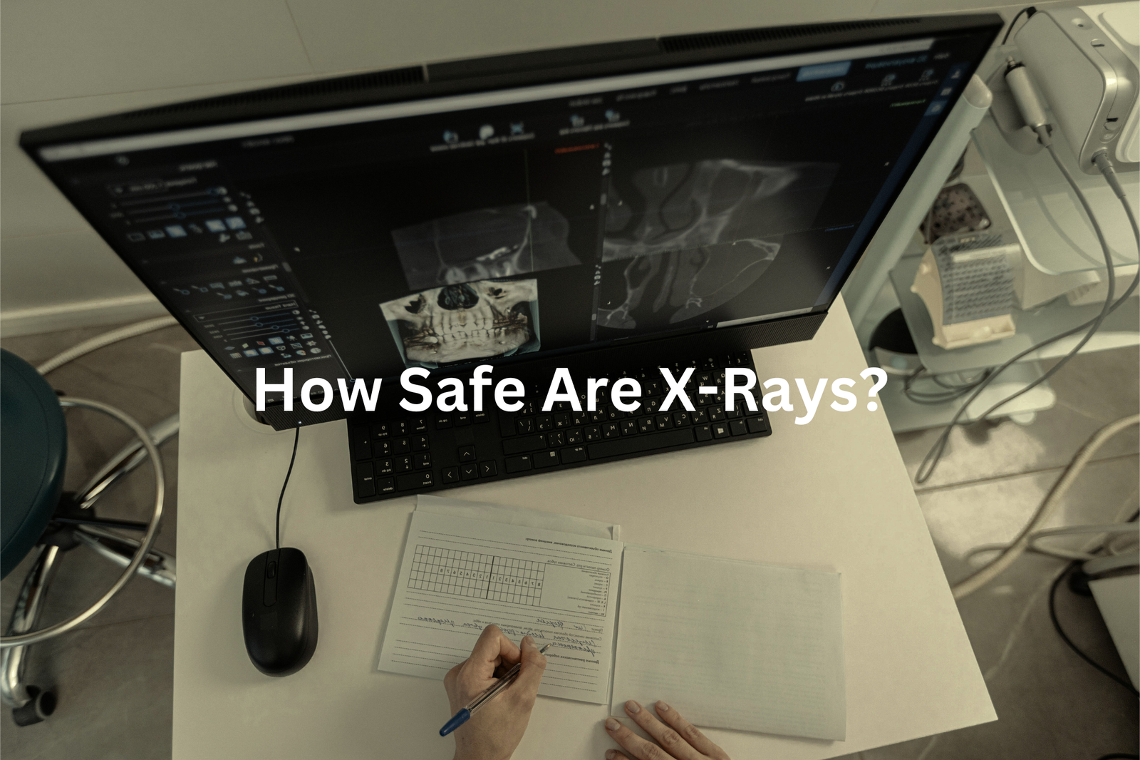
Discover how X-rays work! A special light reveals what’s inside your body, helping doctors diagnose injuries and illnesses safely and quickly.
X-rays are a fascinating type of light that allow doctors to see inside our bodies without any surgery. They take pictures of bones, lungs, and other organs, helping to find problems we can’t always feel. When we’re hurt or unwell, X-rays give a clear view of what’s happening inside us.
This makes it easier for doctors to diagnose and treat us. While X-rays are very helpful, there can be some risks like exposure to radiation, but they’re generally safe when done correctly. Keep reading to discover more about how X-rays work and their importance in medicine!
Key Takeaway
- X-rays help doctors see inside our bodies to find problems like broken bones or infections.
- The X-ray machine uses a small amount of radiation to create images.
- X-rays are safe when done correctly, with strict rules to keep patients safe.
What Are X-Rays?
X-rays are pretty common in Australia. There are over 8,000 accredited imaging facilities across the country (that’s a lot!). They’re often the first test doctors use to check for injuries or illnesses. If you’ve ever broken a bone or had a bad cough, you might’ve had one yourself.
The process is really quick—just a few minutes. And the radiation? It’s tiny. A standard chest X-ray gives you about the same radiation as you’d get naturally in a month. So, it’s safe for most people.
In 2022-23, nearly 39% of Australians used some kind of imaging service, like X-rays. Medicare covered over 27 million tests during that time. That’s huge. X-rays help doctors find things like broken bones, infections, or fluid in the lungs early, which means they can treat them faster.
Newer digital X-ray machines make the images clearer and use even less radiation. Plus, places like the Australian Radiation Protection and Nuclear Safety Agency (ARPANSA) make sure these facilities follow strict safety rules. So, whether you’ve had one or not, X-rays are a big part of keeping Aussies healthy. If you ever need one, don’t stress—they’re quick, safe, and super helpful.
How Do X-Rays Work?
Thinking about X-rays, it’s kind of incredible how they let us see inside the human body without cutting it open. When someone gets an X-ray, what’s really happening is a beam of energy (a type of electromagnetic radiation) is being sent through their body. This energy moves differently depending on what it passes through. Bones, for example, are dense and block most of the X-ray energy, which is why they show up white on the image. Softer tissues, like muscles or skin, let more of the energy pass through, so they appear darker.
The X-ray beams hit a special detector or film on the other side of the body, creating a detailed picture(1). It’s fast, usually just a few minutes, and doesn’t hurt. I remember when my little brother fell off his bike and hurt his arm. The doctor used an X-ray to check, and we could see his bone was broken—it was clear as day on the screen.
If you ever need an X-ray, don’t worry. It’s quick and safe. Just remember to stay still so the picture comes out clear.
Why Do We Use X-Rays?
Source: MonashUniMNHS.
X-rays are pretty amazing when you think about it. They let doctors see inside our bodies without needing to cut us open, which is kind of like having superpowers. One of the main things they’re used for is spotting broken bones. Say you fall off your bike or trip while playing footy—an X-ray can show if there’s a crack or break in the bone. It’s quick and doesn’t hurt, which is a relief, especially for kids who might already be scared.
But X-rays aren’t just for broken bones. They’re also used to check for infections, like in your lungs (pneumonia is a big one) or even in your bones. That’s pretty handy because catching infections early means doctors can start treatment faster. And then there’s the part about finding tumours—those weird lumps that shouldn’t be there. Early detection can make a huge difference.
Doctors also use X-rays to keep track of diseases over time(2). For example, if someone has asthma or arthritis, X-rays can show how things are changing inside. It’s like a snapshot of your health. So, next time you hear about X-rays, remember—they’re not just cool; they’re lifesavers.
How Safe Are X-Rays?

You might be wondering if X-rays are safe. Well, they’re generally safe when done the right way. An X-ray machine uses a very low dose of radiation to take pictures inside your body. Radiation might sound scary, but it’s just a type of energy. High levels of it can be harmful, sure, but X-rays are carefully controlled to use only a tiny amount. This keeps the risk really low.
In Australia, there are strict rules to make sure X-rays are done safely. The Australian Radiation Protection and Nuclear Safety Agency (ARPANSA) oversees this. They follow something called the “ALARA” principle, which stands for “As Low As Reasonably Achievable.” This means they always aim to use the smallest amount of radiation possible while still getting clear images.
Still, it’s worth knowing that having lots of X-rays over many years could slightly increase the risk of cancer. That’s why doctors only recommend X-rays when they’re truly needed. It’s all about balancing safety with the benefits of diagnosing or treating a health problem. If you’re ever unsure, just ask your doctor—they can explain why an X-ray is necessary and answer any questions you’ve got.
What Happens During an X-Ray?
Getting an X-ray is pretty straightforward. You go to a clinic or hospital with the right equipment, and they’ll guide you through it. Usually, you’ll need to remove any jewellery or clothing that might block the X-ray, sometimes wearing a gown instead. (It’s all about getting a clear image.)
The technician will ask you to sit, stand, or lie down depending on what part of your body needs the scan. Staying still is key—wiggling can make the image blurry. Once you’re in position, they’ll step behind a screen and press a button. The machine sends out X-ray beams (you won’t feel a thing), and you might hear a faint click.
Afterwards, a radiologist (a doctor who specialises in reading X-rays) will examine the images for anything unusual. They’ll share the results with your doctor. It’s usually quick—I remember my first X-ray only took about 10 minutes! No need to stress.
Other Types of Imaging Tests
X-rays are just one way doctors can see inside our bodies(3). They’re quick and useful for looking at bones, like checking for breaks or fractures. But sometimes, doctors need more detailed images.
CT scans (short for computed tomography) use lots of X-ray pictures taken from different angles to create a detailed image of soft tissues, organs, or even blood vessels. They’re especially helpful for looking at the brain, chest, or abdomen.
MRIs (magnetic resonance imaging) are different. Instead of X-rays, they use strong magnets and radio waves to create pictures. These are great for seeing soft tissues, like muscles, ligaments, or even the brain.
Ultrasound is another test. It uses sound waves to make images and is often used during pregnancy to check on babies. It’s also good for looking at organs like the liver or kidneys. Each test has its own job. Doctors choose the best one based on what they need to see.
Technology and Advances

X-ray technology has really come a long way. Back in the day, X-ray machines used film (kind of like old cameras), but now most places use digital X-rays. This switch has made a big difference. For starters, digital X-rays give much clearer images. The pictures are sharper, which helps doctors see what’s going on inside the body more easily.
It’s like upgrading from an old TV to a high-definition one. Another big thing is safety. Digital X-rays can use less radiation compared to the old film ones. This means patients are exposed to lower levels of radiation, which is definitely a good thing.
And here’s the kicker—speed. Digital X-rays are super fast. Doctors can see the images almost immediately, which means they can figure out what’s wrong and start treatment quicker. So, with clearer pictures, less radiation, and faster results, digital X-rays have made a huge difference in medical care.
FAQ
What is a CT scan?
A CT (computed tomography) scan is a type of X-ray examination that produces detailed images of the body’s internal structures. It uses a series of X-ray beams to create cross-sectional images, allowing doctors to see internal organs, bones, and soft tissues in great detail.
How do X-rays work?
X-rays are a type of high-energy radiation that can pass through the body and create images of internal structures. An X-ray machine sends a small amount of radiation through the body, and a detector on the other side captures the rays that make it through, producing an image.
Can X-rays cause hair loss?
In very high doses, X-ray exposure can potentially lead to temporary hair loss, but this is extremely rare and only occurs with very high levels of radiation. The small amounts used for most medical X-ray examinations do not cause hair loss or other side effects.
What is an X-ray image?
X-ray images, also called radiographs, are created when X-ray beams pass through the body and are captured by a detector. This allows doctors to see bones, organs, and other internal structures in great detail, which is helpful for detecting injuries, diseases, or foreign objects.
What are gamma rays?
Gamma rays are a type of high-energy electromagnetic radiation, similar to X-rays but with even higher energy levels. They are sometimes used in medical imaging and cancer treatments but are generally produced through natural radioactive decay rather than medical equipment.
Is there a lifetime risk from X-ray exposure?
While high levels of X-ray exposure can increase the lifetime risk of developing certain cancers, the small amounts used for most medical imaging examinations pose a very low risk. Doctors carefully monitor radiation doses and work to minimise exposure as much as possible.
What is a barium enema?
A barium enema is a type of X-ray examination that uses a contrast dye called barium to coat the colon and rectum. This allows doctors to obtain detailed images of the large intestine, which can help diagnose conditions such as colorectal cancer, polyps, or other digestive issues.
How are X-ray detectors used?
X-ray detectors capture the rays that pass through the body during an X-ray examination. These detectors convert the X-ray energy into electrical signals, which are then processed by a computer to generate the final image. Modern detectors can produce high-quality digital X-ray images.
What is a barium swallow?
A barium swallow is an X-ray examination that uses a contrast agent called barium to coat the oesophagus, stomach, and upper intestines. This allows doctors to obtain detailed images of these organs and look for any abnormalities, such as blockages or signs of cancer.
What is X-ray radiography?
X-ray radiography is the process of using X-rays to create images of the body’s internal structures. It is one of the most common and important medical imaging techniques, allowing doctors to diagnose a wide range of conditions by examining bones, organs, and other tissues.
Conclusion
X-rays are special pictures that help doctors look inside our bodies. They’re super helpful for finding things like broken bones or infections. Even though there’s a tiny bit of risk from the radiation, X-rays are safe when done properly. So, if you need one, don’t stress! Remember, it’s just a quick picture to help you feel better. X-rays make a big difference in health care, assisting many people every day with their health needs.
References
- https://www.arpansa.gov.au/understanding-radiation/what-is-radiation/ionising-radiation/x-ray
- https://www.healthywa.wa.gov.au/Articles/U_Z/X-ray
- https://www.adia.asn.au/about-diagnostic-imaging/types-of-imaging/


