
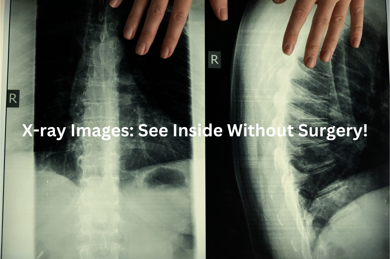
X-ray images let doctors see inside your body—bones, lungs, and more—without surgery. Learn how they work and why they’re so important for your health.
X-ray images are incredible tools that let doctors look inside our bodies without surgery. Think of them like peeking inside a locked treasure chest! X-rays produce pictures of bones, lungs, and other vital organs. They work by using a small amount of radiation that passes through our bodies, creating shadows on a film or digital sensor.
This helps doctors see broken bones, detect infections, or spot other health issues. X-rays are quick, safe, and very useful in diagnosing medical problems. So, if you’re curious about how these images help keep us healthy, keep reading!
Key Takeaway
- X-ray images help doctors see inside our bodies.
- They are used to find problems like broken bones or lung issues.
- X-ray procedures are generally safe with low radiation doses.
How Are X-Ray Images Made?
It’s kind of amazing how an X-ray machine works, showing us what’s hidden inside. The X-ray tube sends out a small amount of radiation, which passes through the body to create an image.
Here’s the process(1):
- X-ray beams travel through the body.
- A detector on the other side captures the image.
- Dense parts, like bones, absorb more radiation and show up white.
- Softer tissues let more radiation through, appearing darker.
It’s like a shadow photo, where each part of the body leaves its mark.
I remember when my mate broke his arm playing footy. The X-ray showed the fracture so clearly, like a crack in a piece of wood. The doctor used it to plan how to fix it. If you ever need an X-ray, just stay still. It’s quick and painless, and the clearer the image, the better it helps the doctors. Simple but brilliant.
What Are the Common Uses of X-Ray Images?
There’s something oddly captivating about how doctors read X-rays. It’s like they’re detectives, piecing together a story from shadows and light. These images are used in so many ways, each one telling a different tale.
- Broken bones: A fall off a skateboard, a sharp pain, and then the X-ray shows a crack—clear as a pencil line on paper.
- Chest checks: Lungs can be tricky. X-rays help spot pneumonia, fluid, or even how someone’s healing.
- Dental health: Ever bite down on that weird plastic thing at the dentist? They’re looking for cavities or sneaky wisdom teeth.
- Foreign objects: My cousin once swallowed a marble. The X-ray showed it sitting in his tummy, round and obvious.
- Surgery prep: Surgeons use X-rays like a guide, planning every move carefully.
X-rays are quick and painless, but they’re powerful. They let doctors see what’s hidden, helping them figure out what’s next.
Different Types of X-Ray Examinations
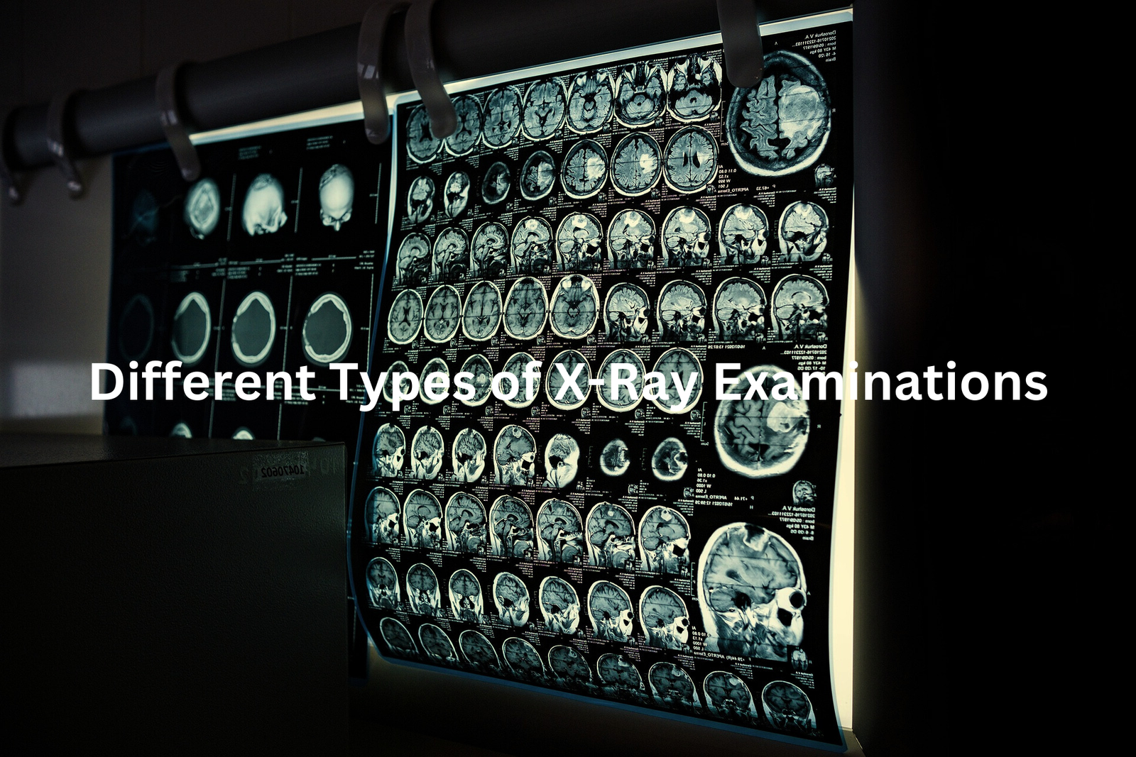
There’s something fascinating about how X-rays can see what we can’t. They’re like secret storytellers, each type revealing a different chapter of what’s going on inside us.
Plain X-rays are the simplest. They’re great for spotting fractures or infections—like a quick snapshot of your bones.
CT scans are more detailed. Think of slicing a loaf of bread and looking at each piece. They create 3D images, showing things hidden in the layers.
Fluoroscopy works in real-time, capturing moving organs, like watching your heart beat or food travel through your stomach.
Mammograms focus on breast tissue, helping detect cancer early(2).
Angiograms map blood vessels using dye, like tracing rivers on a map.
Bone density scans measure bone strength, checking for osteoporosis.
Each X-ray type has a purpose, helping doctors piece together the puzzle of your health. It’s science working quietly, but powerfully.
Safety and Risks of X-Ray Imaging
Sources: University of South Australia.
Radiation. Just saying it makes some folks nervous. But X-rays? They’re actually pretty safe. A chest X-ray, for example, gives about 0.1 millisievert (mSv) of radiation. That’s the same as what you’d naturally get from the environment in ten days. Things like flying or just being outside expose us too, but no one worries much about that.
Doctors always weigh the benefits of an X-ray against its risks, which are tiny. Modern machines use much less radiation than older ones. Still, there are times to be cautious. If you’re pregnant or think you might be, tell your doctor. Foetal tissue is more sensitive. And if you’ve had a lot of scans recently, it’s okay to ask questions.
When I sprained my ankle, I needed an X-ray. I hesitated but trusted the doctor. It wasn’t broken, just a sprain. That quick image? It saved me weeks of guessing.
Preparing for an X-Ray
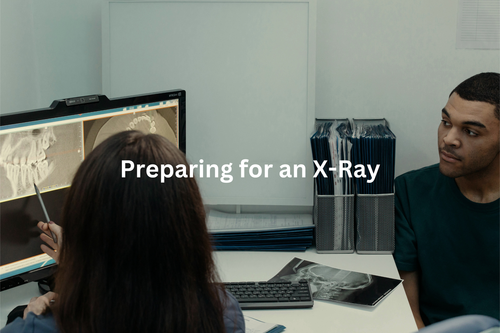
Getting ready for an X-ray is a bit like prepping for a snapshot, except this one looks inside you. Usually, it’s quick. You follow the radiographer’s instructions, hold still, and it’s done. But sometimes, like with barium studies, there’s extra prep. You might need to fast—no food or drinks for hours beforehand.
Metal is a no-go. Jewellery, belts, even underwire bras can mess with the image. A radiographer once told me it’s like trying to read a book with smudges on the page. So, off it all goes.
Here’s what to expect:
- Take off metal items.
- Change into a gown (it’s not stylish, but it’s practical).
- Follow any fasting or prep instructions.
- Stay still for a clear image.
When I had a chest X-ray last year, I forgot about my necklace. The tech kindly handed me a tray for it. That little gesture stuck with me—X-rays are about precision, but there’s care in the process too.
How Are X-Ray Images Read?
The X-ray machine hums softly, capturing the image in seconds. But the real work? That happens after. A radiologist—a medical detective, in a way—examines the image. They’re trained to spot things most of us wouldn’t notice. A faint shadow might mean a broken bone. A small blur could hint at fluid in the lungs.
Here’s the process(3):
- Radiologist reviews the images closely.
- They write a report, explaining their findings clearly.
- The report goes to your doctor, who decides the next steps.
- You get the results, usually within a day or two.
I remember waiting for my own chest X-ray results last year. It felt like holding my breath. When the doctor said it was all clear, I could finally exhale. If you’re waiting, ask how long it’ll take. Knowing the timeline helps. And remember, it’s all about getting answers to move forward.
FAQ
How can neural networks be used for chest X-ray images?
Neural networks have shown promising results in analysing medical imaging like chest X-rays. They can automatically detect patterns and abnormalities that may be difficult for the human eye to spot. This can help doctors make faster and more accurate diagnoses, especially for conditions like COVID-19 which have distinct X-ray signatures.
What is the role of medical imaging in the COVID-19 pandemic?
Medical imaging like chest X-rays has played a critical role in the COVID-19 pandemic. X-rays can reveal signs of COVID-19 infection in the lungs, such as patchy opacities or pleural effusion. This allows healthcare providers to quickly identify potential COVID-19 cases and guide treatment. Automated detection using deep learning on X-ray images has also been explored to enhance COVID-19 case detection.
How can artificial intelligence and deep learning aid in COVID-19 detection?
Researchers have developed deep learning models that can analyse chest X-ray and CT scan images to automatically detect signs of COVID-19 infection. These AI-powered approaches have shown promising results in identifying COVID-19 cases, often with high accuracy. By complementing human experts, these deep learning techniques can help improve COVID-19 detection, especially in areas with limited access to testing.
What are some key considerations for designing deep learning models for medical imaging?
Designing effective deep learning models for medical imaging tasks like COVID-19 detection requires careful consideration of factors such as the dataset size and quality, preprocessing techniques, neural network architecture, and training strategies. Approaches like self-supervised learning, transfer learning, and human-machine collaborative design can all play important roles in developing robust and reliable models for real-world deployment.
How can medical imaging data help advance COVID-19 research?
The COVID-19 pandemic has spurred the collection of large, diverse datasets of medical images like chest X-rays and CT scans. This data has enabled researchers to develop and refine deep learning models for automatic COVID-19 detection. Additionally, analysis of this imaging data can provide valuable insights into the progression of COVID-19 and help inform treatment strategies. Responsible sharing and use of this data is crucial to accelerate research and development for combating the pandemic.
Conclusion
X-ray images are amazing tools that let doctors look inside our bodies safely. They help find broken bones, check lung health, and more. Yes, there’s a tiny bit of radiation, but the good things they do for your health are much bigger. If you need an X-ray, don’t stress! It’s just a way for doctors to help you get better. If you’re unsure about anything, make sure to ask your doctor. They’re there to help!
References
- https://csiropedia.csiro.au/x-ray-phase-contrast-imaging/
- https://nbcf.org.au/about-breast-cancer/detection-and-awareness/mammograms/
- https://ris.com.au/x-ray-imaging-types-uses-and-revolutionary-technology/


