
Learn how X-ray radiation powers medical imaging and the safety protocols to protect patients.
X-ray radiation plays a crucial role in modern healthcare by enabling precise diagnostic imaging. It helps doctors detect fractures, infections, and even life-threatening conditions like cancer.
However, understanding the balance between effective use and radiation safety is essential for both healthcare providers and patients. This article covers the properties of X-rays, their medical applications, health effects, and safety protocols to ensure optimal usage.
Key Takeaways
- X-ray properties: X-rays are high-energy electromagnetic radiation that can penetrate tissues, aiding in diagnostic imaging.
- Medical applications: X-rays are widely used in medical imaging, including radiography, CT scans, and radiation treatment for cancer.
- Radiation safety: It’s crucial to manage X-ray exposure carefully, following safety guidelines to minimise health risks, especially for vulnerable groups like pregnant patients.
Properties of X-Ray Radiation
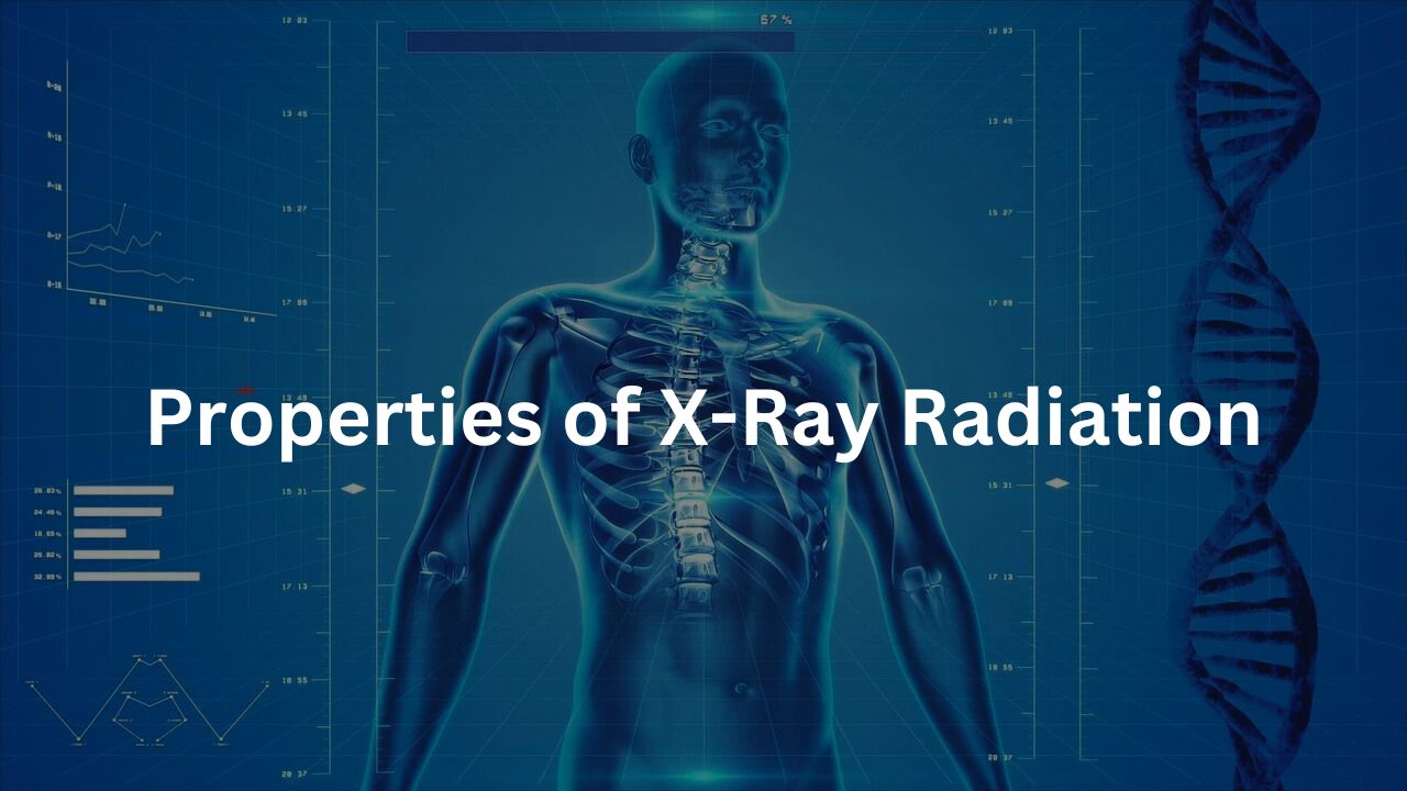
X-ray radiation sits comfortably between ultraviolet rays and gamma rays in the electromagnetic spectrum.
It’s a curious place to be—an invisible force that has the power to reveal our inner worlds, quite literally. X-rays are a form of electromagnetic radiation with wavelengths between approximately 10−810^{-8} and 10−1210^{-12} meters. This puts them in the category of high-energy radiation. (1)
ionising Radiation: Not Just Another Beam of Light
X-rays are categorised as ionising radiation, meaning that their energy is high enough to strip electrons from atoms. This ability allows them to alter the chemical structure of molecules, and in doing so, they can cause cell damage.
If cells are sufficiently damaged or altered, the risk of cancer can increase. That’s why safety measures are crucial in any environment where X-rays are in use.
Penetrating Power: Why X-Rays Can See Inside Us
The penetrating power of X-rays varies depending on the substance they encounter. Denser materials, like bones, absorb more X-rays and appear white on a radiograph.
Softer tissues, such as muscles or organs, absorb fewer X-rays, which makes them appear darker in the image. This is how doctors can distinguish between different types of tissues when diagnosing fractures, infections, or other medical conditions. (2)
Production of X-Rays
X-rays aren’t just something that exist in the air. They’re created in a very controlled environment, typically in an X-ray tube.
High-velocity electrons are accelerated toward a metal target, and when these electrons collide with the target, their energy is converted into X-ray photons. The magic happens in two distinct ways:
- Bremsstrahlung Radiation: When the high-energy electrons slow down near the nucleus of the target atoms, they release energy in the form of X-rays. This is a bit like a car braking hard—it sheds its excess energy.
- Characteristic Radiation: In this process, when an electron collides with an inner electron of the atom, it displaces it, creating a vacancy. As electrons from higher energy levels fill the vacancy, they release energy in the form of X-rays. The energy of these X-rays is characteristic to the material, like a fingerprint.
Medical Applications of X-Rays
The value of X-rays in medical diagnostics cannot be overstated. They are the go-to tool for detecting a wide array of conditions, from simple bone fractures to more complex diseases like cancer.
Diagnostic Imaging: A Snapshot of Your Health
- Radiography: The classic X-ray image. These still images provide a snapshot that doctors use to identify fractures, infections, or even early signs of disease.
- Fluoroscopy: This method is more dynamic. It provides real-time imaging, allowing doctors to observe how your internal structures move and function. It’s often used for things like swallowing studies or guiding catheters into specific areas of the body.
- CT Scans: Think of CT scans as a 3D version of the classic X-ray. They combine multiple X-ray images to create detailed cross-sectional images of the body. CT scans are especially useful for detecting conditions like tumours or infections in organs and soft tissues.
Special Cases: Protecting Vulnerable Populations
As much as X-rays are useful, there’s also the question of safety. When it comes to pediatric patients and pregnant women, extra care is taken to limit exposure to X-rays.
The risks of radiation are higher for children due to their developing tissues, and for pregnant women, especially in the first trimester, the potential effects on the fetus must be carefully considered.
Health Effects of X-Ray Exposure
While X-rays are undoubtedly beneficial, they also come with risks, particularly when used excessively.
Low-Dose Exposure: Generally Safe, But Be Cautious
Low-dose exposure from medical imaging procedures, like routine X-rays and CT scans, is usually considered safe. The key is keeping radiation exposure as low as reasonably possible (the ALARA principle—As Low As Reasonably Achievable). This is why healthcare professionals will often avoid repeating imaging exams unless absolutely necessary.
High-Dose Exposure: Not So Simple
High-dose radiation, on the other hand, can cause serious damage. Short-term exposure to large doses can result in burns or acute radiation sickness, while long-term exposure increases the risk of cancer. That’s why radiation safety protocols are carefully followed, especially in medical settings.
Protection Protocols: Shielding and Safety Standards
To mitigate these risks, various safety protocols are in place. For example, lead aprons are commonly used to protect parts of the body not being examined. X-ray rooms are designed with shielding to prevent radiation from escaping, and the exposure time is minimised.
Regulations and Certification for X-Ray Equipment
In Australia, safety standards for X-ray equipment are set by bodies like the Royal Australian and New Zealand College of Radiologists (RANZCR). They provide stringent guidelines to ensure the safe use of radiation in medical settings.
Safety Standards: RANZCR Guidelines
RANZCR has established clear standards regarding radiation exposure, ensuring that both practitioners and patients are protected. This includes regular checks on equipment calibration, safety certifications for operators, and radiation shielding in clinics and hospitals.
Certification Process: How Hospitals Get Their X-Ray Equipment Approved
Medical institutions must comply with registration requirements and go through a certification process to ensure their X-ray equipment meets safety standards. This includes a rigorous inspection of the machinery and the protocols used to minimise radiation exposure.
Radiation Protection Measures
Under RANZCR guidelines, healthcare providers must ensure that staff is trained in radiation protection and that patients receive the minimum amount of exposure necessary to obtain an accurate diagnosis.
Emerging Technologies in X-Ray Imaging
The field of X-ray imaging is far from stagnant. New technologies are constantly being developed to improve both the quality and safety of imaging procedures.
Advancements in X-Ray Technology: 3D Imaging and Beyond
Recent advancements include 3D X-ray imaging, which allows for more detailed images, especially useful in detecting small fractures or intricate structural abnormalities. Next-generation CT scans are now available with higher resolution, enabling doctors to make even more accurate diagnoses.
MRI vs. X-Ray: When to Use Which
While X-rays are fantastic for visualising bones and certain soft tissues, Magnetic Resonance Imaging (MRI) is often preferred for imaging soft tissues like muscles, brain, or spinal cord. X-rays have the advantage when it comes to quick diagnosis and cost-efficiency, but MRI excels in providing more detailed soft-tissue images without radiation.
X-Ray Use in Cancer Treatment
Beyond diagnostics, X-rays also play an integral role in cancer treatment. External beam radiation therapy uses high-energy X-rays to target cancer cells in the body. It can be used to treat a wide range of cancers, including lung cancer and bone fractures caused by tumours.
Benefits and Risks: A Delicate Balance
While X-rays are an effective treatment method, there is always a balance between treatment success and the potential risks of radiation exposure. The focus is always on using just enough radiation to target the cancer cells, while minimising damage to surrounding healthy tissues.
Common Concerns and FAQs
Credits: NIBIB
Pregnancy and X-rays: The main concern with X-rays during pregnancy is the potential risk to the fetus. In most cases, if X-rays are medically necessary, the benefits outweigh the risks, but precautions like shielding are taken to protect the abdomen.
Environmental Impact: Another growing concern is the environmental impact of radiation waste from medical imaging. The disposal of radioactive materials is tightly regulated, ensuring that there are no harmful environmental consequences.
Certification FAQs: The certification process for X-ray equipment is thorough and involves multiple stages, including inspection, operator training, and routine checks to ensure the equipment remains safe to use.
Conclusion
X-ray radiation is one of the most powerful tools in modern medicine. It has the ability to reveal what’s hidden beneath the surface, helping doctors diagnose and treat patients effectively.
But like any powerful tool, it comes with risks. Proper safety protocols, regulations, and advancements in technology ensure that the benefits of X-rays continue to outweigh the dangers.
FAQ
What are doses of radiation used in medical procedures?
In medical procedures, radiation is carefully controlled to ensure that doses of radiation are just enough to help doctors diagnose or treat a condition. The amount of radiation used depends on the type of procedure, such as Dental X-ray, X-ray imaging, or Computed Tomography (CT).
For example, a CT scan uses a higher dose of radiation compared to a regular X-ray because it takes detailed images of the body. However, it’s important to note that medical X-rays and other medical imaging procedures are designed to minimise radiation exposure while providing the necessary information for accurate diagnosis and treatment.
What is electromagnetic radiation, and how does it affect the human body?
Electromagnetic radiation is a form of radiation that includes visible light, radio waves, X-rays, and gamma rays. X-ray beams and other forms of electromagnetic radiation have enough energy to pass through the body and are often used in medical imaging to create images of bones, soft tissues, and other internal structures.
When exposure to radiation is too high, it can lead to harmful effects such as tissue damage or an increased risk of cancer. Radiation protection measures, such as shielding and careful management of radiation doses, are crucial to minimise the risks.
What are the risks of X-ray exposure for pregnant patients?
Pregnant patients should be cautious about X-ray exposure due to the potential risks to the unborn child. High levels of radiation exposure can increase the risk of birth defects or harm to the developing baby.
In cases where X-rays are needed for diagnosis or treatment, healthcare providers take extra care to limit radiation exposure. Special precautions are often taken, such as avoiding X-rays in the early stages of pregnancy or using techniques that minimise radiation exposure. For example, X-ray machines can be adjusted to target only the area of concern, reducing exposure to other parts of the body.
How do medical X-rays affect soft tissues in the body?
X-ray machines are designed to create detailed images of bones and other tissues inside the body. While bones absorb more X-rays, soft tissues allow more X-ray photons to pass through, which is why soft tissues may appear darker on an X-ray image.
Although soft tissues are less sensitive to X-rays than bones, exposure still needs to be managed carefully to avoid potential damage. With radiation safety protocols in place, the risk to soft tissues can be minimised, and the benefits of medical imaging often outweigh the potential risks.
How can radiation treatment be used in cancer treatment?
Radiation treatment, such as external beam radiation therapy, is often used to treat cancers like lung cancer or bone fractures caused by cancer. It works by using high-energy X-ray photons to target and destroy cancer cells.
The radiation dose must be carefully calculated to ensure that it effectively kills cancer cells while minimising damage to healthy tissues. Health care providers follow strict guidelines to ensure the safety and effectiveness of radiation treatment.
What is radiation protection, and why is it important?
Radiation protection refers to the methods and measures taken to reduce exposure to harmful radiation, ensuring the safety of both patients and healthcare providers.
In medical environments, radiation protection can include shielding (such as lead aprons or walls) to block radiation, limiting exposure time, and keeping a safe distance from radiation sources. These measures are especially important in procedures like Diagnostic and Interventional X-ray Procedures and Computed Tomography, where X-ray equipment is used.
What is the effective dose of radiation from medical X-rays?
The effective dose of radiation refers to the amount of radiation absorbed by the body during a medical X-ray procedure. It takes into account not only the energy of the radiation but also how different tissues and organs respond to it.
For example, doses to patients’ bones may differ from those to soft tissues, and this can impact the overall health risks. Medical X-rays are generally considered safe when performed by trained healthcare providers, but the dose should always be kept as low as possible while still achieving the necessary diagnostic result.
References
- https://www.arpansa.gov.au/understanding-radiation/what-is-radiation/ionising-radiation/x-ray
- https://www.racgp.org.au/afp/2013/june/radiation-safety


