
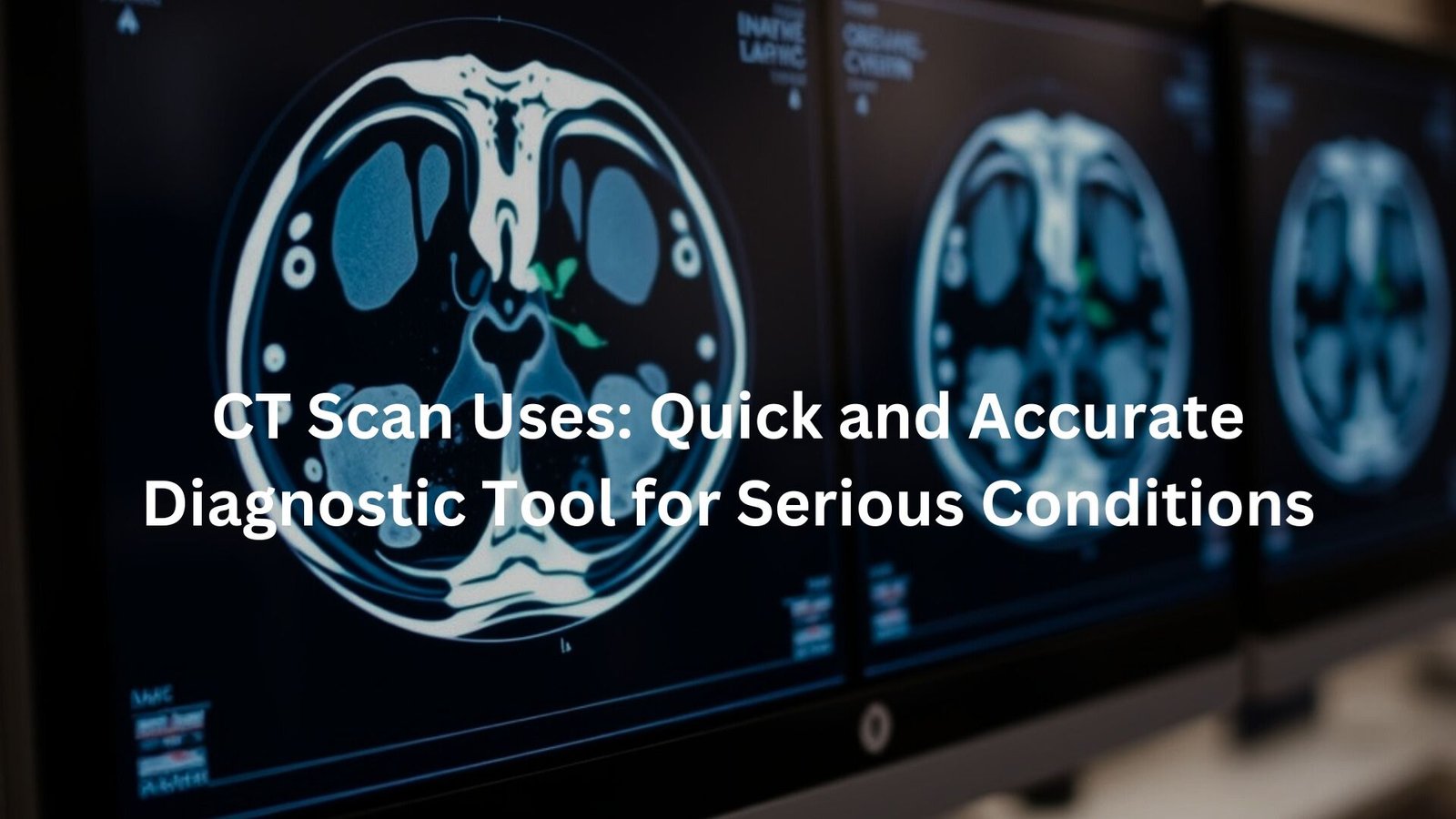
Learn how CT scans help diagnose heart disease, cancer, and trauma quickly with high-resolution imaging.
CT scans are essential diagnostic tools that use advanced X-ray technology to create detailed cross-sectional images of the body. Unlike traditional X-rays, they offer a clearer view of internal structures like bones, organs, and blood vessels, making them crucial for diagnosing various medical conditions such as heart disease, cancer, and traumatic injuries.
This article explores the common uses of CT scans and how they benefit patients in both emergency and routine medical care.
Key Takeaway
- CT scans provide high-resolution, 3D images of bones, tissues, and blood vessels for accurate diagnoses.
- They are vital for diagnosing critical conditions like heart disease, bone fractures, and pulmonary embolism.
- Radiation exposure is minimised with Clinical Decision Rules (CDRs) to ensure only necessary scans are performed.
What is a CT Scan?
A CT scan, or Computed Tomography scan, is a medical imaging technique that uses X-rays and computer processing to create detailed images of the inside of the body. Unlike traditional X-rays, which capture flat, two-dimensional images, CT scans take a series of detailed cross-sectional “slices” of the body.
These images are then combined to create a 3D representation of the area being scanned. CT scans help doctors identify a variety of health problems that can’t be seen with a regular X-ray.
The main difference between a CT scan and a traditional X-ray is the amount of detail. A CT scan provides high-resolution images, which means it’s much more useful for diagnosing complex conditions.
While regular X-rays are great for spotting broken bones or lung conditions, CT scans can give a clearer picture of soft tissue injuries, internal bleeding, and diseases like cancer. (1)
When and Why is a CT Scan Used?
CT scans are used for diagnosing serious conditions that require more detailed imaging. Some of the conditions commonly detected through CT scans include:
- Heart disease: CT scans can identify issues like blockages in arteries, which may lead to heart attacks.
- Bone fractures: In cases of serious trauma, CT scans can help spot fractures that might not be visible on a traditional X-ray.
- Kidney disease: CT imaging can spot kidney stones or abnormalities in the kidneys.
- Pulmonary embolism: CT scans can detect blood clots in the lungs, which could lead to serious complications.
- Cancer: CT scans are used to find and monitor various types of cancer, helping doctors determine the size, shape, and location of tumours.
Emergency Applications
In an emergency, time is critical. CT scans are incredibly useful for detecting issues like internal bleeding, trauma from accidents, and other acute conditions. For example, if a patient is involved in a car accident, doctors may use a CT scan to quickly check for injuries to internal organs or brain damage, which would be hard to detect without detailed imaging. (2)
How CT Scans Work
CT scans work through a combination of X-ray tubes and electronic detectors. When you’re in the scanner, the machine rotates around you, sending X-rays through your body. The detectors capture these X-rays, which are then processed by a computer to create detailed “slices” of the area being scanned.
Unlike MRI (Magnetic Resonance Imaging), which is better suited for examining soft tissues like the brain and spinal cord, CT scans excel at visualising bones, soft tissues, and blood vessels. This makes them incredibly versatile, especially when looking for conditions that affect multiple types of tissue at once.
The resulting images give doctors a comprehensive 3D view of the scanned area, which helps them make more accurate diagnoses. This ability to view the body in slices makes CT scans especially effective in complex cases.
Radiation Exposure and Safety Considerations
Like all forms of X-ray imaging, CT scans do involve some radiation exposure. However, the amount of radiation you’re exposed to during a CT scan is relatively low, and the benefits typically outweigh the risks.
The radiation levels in a CT scan are far higher than a standard X-ray, but the images produced can help detect life-threatening conditions early, which could prevent further complications.
Clinical Decision Rules (CDRs)
To help reduce unnecessary exposure to radiation, Clinical Decision Rules (CDRs) are often used. These guidelines help doctors determine when a CT scan is necessary, ensuring that scans are only performed when they’re likely to provide useful information.
For example, in cases of suspected pulmonary embolism (blood clots in the lungs), specific criteria will be followed to determine whether a CT scan should be done, based on the patient’s symptoms and risk factors.
Though the risk is low, doctors aim to limit CT scans to situations where the potential benefit of accurate diagnosis outweighs the small risk of radiation.
CT Scan Benefits
There’s no denying the significant advantages of CT scans in diagnosing medical conditions. Some of the key benefits include:
- High-resolution images: CT scans provide clearer, more detailed images than regular X-rays, which can make a huge difference when diagnosing complex issues.
- Quick process: CT scans are fast, which is essential when it comes to diagnosing and treating emergencies. In a busy hospital setting, doctors need results quickly, and CT scans are one of the fastest imaging tools available.
- Comprehensive imaging: Unlike traditional X-rays that can only show bones, CT scans are capable of visualising bones, soft tissues, and blood vessels at the same time. This is especially useful for detecting injuries that affect multiple types of tissue.
CT Scan vs. Other Imaging Techniques
Credits: Med today
There are a few other imaging techniques that doctors use, but each has its strengths and weaknesses. Let’s compare CT scans to X-rays and MRI to see where they differ:
CT vs. X-ray
While both use X-ray technology, the major difference is in the type of images they produce. X-rays provide flat, two-dimensional images, while CT scans offer detailed, three-dimensional cross-sections. This means that CT scans can give a far more accurate picture of internal structures and conditions.
CT vs. MRI
MRI scans are better for imaging soft tissues like muscles, the brain, and spinal cord, whereas CT scans excel at visualising bones and conditions affecting both soft tissue and bone. So, while MRI is often used for neurological or musculoskeletal problems, CT is preferred for trauma, cancer, and other acute conditions.
Types of CT Scans
There are several types of CT scans, each designed to focus on a specific part of the body or type of condition. Some of the common types include:
- Abdominal CT: This type of CT scan is used to check for conditions that affect the abdomen, such as abdominal pain, fluid buildup, and other internal problems.
- CT Angiography: This scan uses a contrast dye to highlight the blood vessels in the body. It’s typically used to spot blockages, blood clots, or vascular conditions.
Preparing for a CT Scan
If you’re getting a CT scan, the preparation is usually straightforward. Here’s what you can expect:
- Lying on the table: You’ll be asked to lie on a narrow table that slides into the CT scanner. The scan is typically painless, but you might need to hold your breath for short periods during the process.
- Removing metallic objects: Before the scan, you’ll need to remove any metallic objects such as jewellery, watches, or glasses, as these can interfere with the imaging.
- Contrast scans: In some cases, you may be given a contrast dye (usually injected into your veins or swallowed) to help improve the clarity of the images. If this is the case, make sure to tell the technician if you have any allergies or kidney problems, as these may affect your ability to safely receive the contrast dye.
Conclusion
CT scans are an essential diagnostic tool, providing fast, high-resolution images that help doctors make accurate diagnoses. From detecting heart disease and cancer to identifying injuries from trauma, CT scans play a key role in diagnosing serious medical conditions.
While the radiation exposure is a concern, the benefits typically outweigh the risks, especially in emergency situations. With the help of Clinical Decision Rules and advanced imaging technology, doctors can ensure that CT scans are used effectively and safely.
If you’re preparing for a CT scan, don’t stress. It’s a quick, non-invasive procedure that could make a big difference in your treatment and recovery. Just follow the preparation guidelines, and trust that the scan will provide the doctor with the detailed information they need to provide the best care.
FAQ
What is the purpose of a CT scan?
A CT scan is used to create detailed images of the inside of your body. It helps doctors see what’s going on with internal organs like the brain, heart, or kidneys, and it’s especially useful for diagnosing bone fractures, cancer, heart disease, and even checking for blood clots in your blood vessels. Unlike a traditional X-ray, it provides cross-sectional images, which gives a much clearer picture of what’s happening inside.
How does a CT scan work?
A CT scan works by using an X-ray tube to send X-rays through your body while you’re lying on a narrow table. These X-rays are then captured by electronic X-ray detectors that send the information to a computer. The computer creates image slices, which are stacked to form a 3-D image. This technique allows healthcare professionals to view your body from many angles and helps them diagnose and monitor conditions more accurately.
Is a CT scan safe?
A CT scan is generally safe, but like any medical procedure that involves radiation exposure, there are some risks. Radiation dose from a CT scan is higher than a regular X-ray, but doctors make sure to only recommend the test when it’s needed. While the risk of cancer from a CT scan is small, healthcare professionals aim to minimise radiation exposure by using the lowest doses possible and avoiding unnecessary scans. It’s always a good idea to ask your doctor about the risks if you have concerns.
What should I do before a CT scan?
Before your CT scan, you might be asked to remove any metal objects such as jewellery, glasses, or other items that can interfere with the scan. Depending on the type of CT scan, you may also need to drink a special liquid (called contrast) or get an injection to help make certain parts of your body show up more clearly. If you’re getting an abdominal CT, for example, you may need to fast for a few hours beforehand.
Can a CT scan detect everything?
While a CT scan can provide a lot of information about your body, it can’t detect everything. It’s great for spotting things like bone fractures, kidney disease, pulmonary embolism, cancer, and abdominal pain, but it might not always show soft tissues as clearly as an MRI. Your healthcare professional will decide the best imaging method based on your symptoms and what needs to be looked at.
How long does a CT scan take?
The actual scanning process is quick, usually only lasting a few minutes. However, depending on the type of scan you’re getting, you may need to spend some time getting ready, like removing your clothes or having contrast injected. It’s important to stay still during the scan, as movement can blur the images.
How is a CT scan different from an X-ray?
Both CT imaging and conventional X-rays are types of medical imaging, but a CT scan is much more detailed. An X-ray provides a flat, 2D image, whereas a CT scan creates detailed cross-sectional images of the body, giving doctors a much clearer picture of what’s going on inside. It’s like looking inside a loaf of bread with an X-ray versus cutting it into slices and looking at each slice – the CT scan gives you more information.
References
- https://brightonradiology.com.au/?gad_source=1&gclid=Cj0KCQiA4rK8BhD7ARIsAFe5LXInQx59sDxV_Qy6dSl3wzdypYrHhxQ7jJk-6bAeh8kfwvl6AHdndcsaAsGzEALw_wcB
- https://i-med.com.au/procedures/ct-scan#gsc.tab=0


