
Discover how PET/CT hybrid imaging combines two powerful scans to detect cancer and help doctors understand your health better. Find out more!
PET/CT hybrid imaging is a unique scan that helps doctors see inside our bodies. It uses two types of imaging: PET (which shows how our tissues are working) and CT (which shows what they look like). This combination is really helpful, especially for finding and understanding diseases like cancer.
With PET/CT, doctors can make better decisions about treatment and health. It’s like looking at a picture and understanding its story. If you want to learn more about how this technology works and its benefits, keep reading!
Key Takeaway
- PET/CT scans combine two imaging techniques to give detailed pictures of the body.
- These scans help doctors detect cancer and see how well treatments are working.
- They can lower radiation exposure and speed up diagnosis.
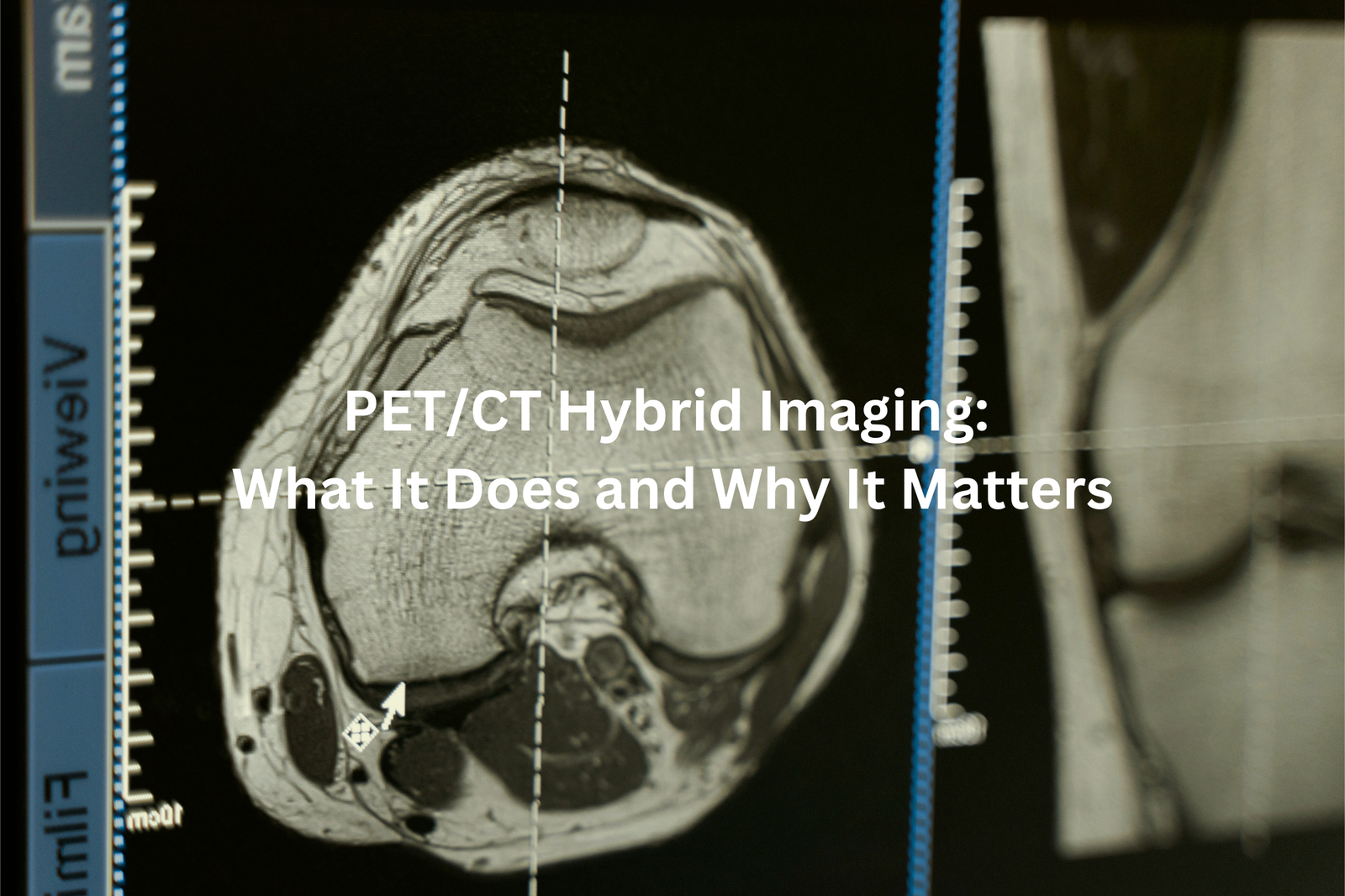
What is PET/CT Hybrid Imaging?
PET/CT scans create a powerful medical duo, combining two different types of imaging into one detailed picture. The process starts with a small injection of a radioactive tracer (usually a form of glucose) that travels through the body.
The PET scanner detects areas where cells are more active, showing up as bright spots on the scan. These active spots might point to cancer cells, which use more energy than normal cells. Meanwhile, the CT scanner takes detailed X-ray pictures of bones, organs, and tissues.
When these scans join forces, they produce:
- Clear anatomical maps
- Spots of unusual cell activity
- Precise locations of potential problems
The combined scan helps doctors find issues that might not appear on standard tests. While no medical test is 100% accurate, PET/CT scans spot problems early, making treatment planning more effective.
Patients should discuss with their healthcare provider about scan preparation, which typically includes fasting for 4-6 hours beforehand.
How Does PET/CT Work?
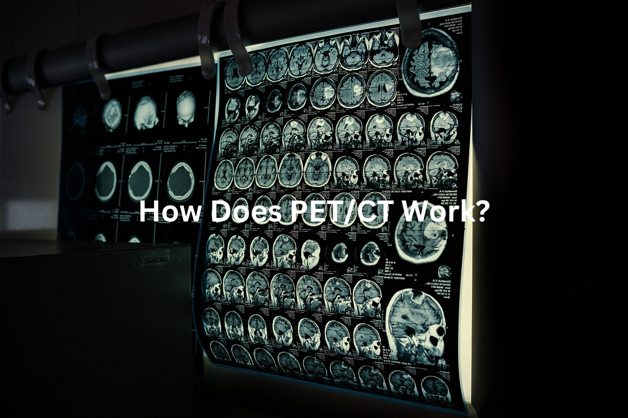
A PET scan at Royal North Shore Hospital follows a simple process, though the medical terms might sound tricky at first.
The procedure starts with a small injection of FDG (fluorodeoxyglucose), a radioactive sugar that helps doctors see active cells. Patients rest quietly for 60 minutes after the injection, giving the FDG time to move through their body.
The scanning machine looks like a large white donut, about 70 centimetres wide – much roomier than an MRI scanner. During the 30-minute scan, patients lie on a sliding bed while the machine takes two different types of images(1):
• PET images show which cells are using lots of energy
• CT images create a map of the body’s structure
Patients should wear warm, comfortable clothes without metal parts. The scan doesn’t cause pain, but staying still is essential for clear images. Most medical centres suggest bringing a book for the waiting period.
Common Uses of PET/CT Scans
Sources: Queensland X-Ray.
PET/CT scans combine two types of medical imaging into one powerful diagnostic tool (Positron Emission Tomography and Computed Tomography). The large white machine captures detailed pictures of what’s happening inside the body, making it easier for doctors to spot problems.
During the scan, patients get an injection of radioactive sugar solution that makes cancer cells glow on the images. Cancer cells use more sugar than normal cells, so they show up brighter on the scan. The whole process takes about 30 minutes.
These scans help doctors(2):
- Find where cancer is located
- Check if cancer has spread
- Monitor treatment progress
- Watch for cancer coming back
Before having a PET/CT scan, patients need to:
- Fast for 6 hours
- Wear loose clothing without metal parts
- Tell staff about any medications
- Bring previous scan results
The procedure is painless, though patients must lie still on a narrow table while the scanner moves around them.
Advantages of PET/CT Hybrid Imaging
PET/CT scans combine two powerful medical imaging tools into one streamlined process. The CT scanner captures detailed pictures of the body’s structures, while PET technology shows how cells use energy – creating a complete picture of what’s happening inside.
These dual-imaging machines (developed in the early 2000s) work like two cameras taking photos at the same time. The process takes about 30 minutes, making it faster than separate scans.
Benefits of combined PET/CT scans:
- 30% less radiation exposure compared to separate scans
- Quicker results for doctors to start treatment
- More accurate diagnosis with merged images
- One appointment instead of two
The merged images help doctors spot problems that might not show up on a single scan. For example, the CT might show an odd-shaped area, while the PET reveals if it’s using energy differently than normal tissue. This technology makes a big difference in finding and treating illnesses early.
Safety Considerations
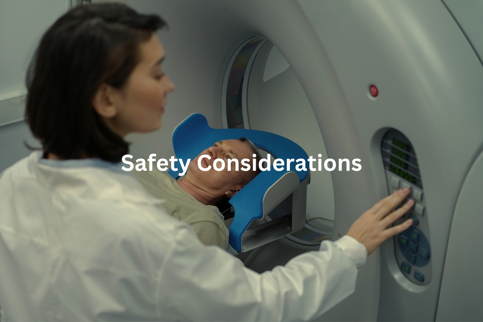
PET/CT scans combine two powerful imaging tools into one machine that looks like a large white donut(3). The scanner (a 64-slice CT model) captures detailed pictures of what’s happening inside the body.
The radiation dose from these modern scanners equals roughly the same amount received during a return flight from Sydney to Perth. Medical teams use the lowest possible dose needed for clear images.
Before the scan, patients should:
- Tell medical staff about allergies
- Report past reactions to contrast dye
- Bring their Medicare card for billing
Most scans take about 30 minutes to complete. The newer machines produce up to 40% less radiation compared to older models, making them safer for patients. Medicare covers a large portion of the scan costs at Australian hospitals. The process runs smoothly when patients discuss any concerns with their doctors beforehand and follow the preparation guidelines provided by the imaging department.
FAQ
What is PET/CT hybrid imaging?
PET/CT hybrid imaging combines positron emission tomography (PET) and computed tomography (CT) scans to provide detailed information about the body’s structure and function. This technique allows doctors to see both the anatomical details from the CT scan and the metabolic activity from the PET scan, providing a comprehensive view for diagnosis and treatment planning.
How does PET/CT imaging work?
In a PET/CT scan, the patient is first injected with a small amount of a radioactive tracer, usually FDG (fluorodeoxyglucose). This tracer is then detected by the PET scanner, which creates images showing where the tracer has accumulated in the body. The CT scanner then provides detailed 3D images of the body’s anatomy. The PET and CT data are combined to produce a single, fused image.
What are the benefits of PET/CT imaging?
PET/CT imaging offers several advantages over traditional imaging methods. It can provide more accurate detection and staging of certain cancers, as well as information about blood flow, metabolism, and other physiological processes. PET/CT is also useful for evaluating treatment response, detecting recurrent disease, and guiding biopsy or surgical planning.
How is PET/CT imaging used in cancer care?
PET/CT is widely used in the management of many types of cancer, including lung cancer, lymphoma, and certain solid tumours. It can help determine the stage of cancer, identify the best treatment approach, monitor response to therapy, and detect recurrence. PET/CT is particularly helpful in identifying small or subtle lesions that may be missed on other imaging tests.
What are the safety considerations for PET/CT imaging?
PET/CT scans involve exposure to low doses of ionising radiation from the CT and PET components. However, the benefits of the information obtained from the scan typically outweigh the small risks. Patients with certain medical conditions, such as pregnancy or kidney disease, may require additional precautions. Overall, PET/CT is considered a safe and well-tolerated imaging technique when performed by qualified healthcare professionals.
Conclusion
PET/CT scans merge two powerful medical tests into one clear picture. These scans show doctors both what tissues look like and how they work (using special tracers that light up active cells). The combined images make it easier to spot cancer and track treatment progress. Medical teams use these scans to check tumours as small as 1 centimetre. Patients can ask their healthcare provider about scan details, preparation steps, and what to expect during the 30-minute procedure.
References
- https://www.sydney.edu.au/courses/subject-areas/spec/hybrid-imaging0.html
- https://www.insideradiology.com.au/pet-scan/
- https://www.msac.gov.au/sites/default/files/documents/1632%2520Redacted%2520Application%2520Form.pdf


