Discover how radioactive tracers in PET scans help doctors spot diseases like cancer early and understand what’s happening inside your body.
Radioactive tracers play a key role in medical tests like PET scans. These special substances allow doctors to see inside our bodies, helping them locate issues such as diseases or cancer. When a tracer is injected, it travels through the body and highlights areas that need attention (like tumours).
This process lets doctors understand what’s happening inside, making it easier to diagnose conditions and decide on treatments. If you’re curious about how these tracers are made and why they’re so important, keep reading! There’s so much more to discover about these amazing tools in medicine!
Key Takeaway
- Radioactive tracers help doctors find diseases by showing where things are happening in our bodies.
- They are usually safe, with very low radiation doses.
- They are especially useful for spotting cancer and watching how well treatments are working.
What Are Radioactive Tracers?
Nuclear medicine scans use special radioactive substances to see inside the body. These substances, called tracers or radiopharmaceuticals, work like tiny torches lighting up different parts inside patients.
The process starts when a patient receives a small amount of tracer through a needle – about the size of a few water drops. As it moves through the bloodstream, the tracer gets picked up by certain cells, including cancer cells that tend to absorb more of it than normal cells.
The scanning process follows three main steps:
- Injection of the tracer into the patient’s body
- Waiting period while the tracer travels around
- Taking pictures with special gamma cameras
The amount of radiation used in these scans is quite low, similar to what someone might get from a regular X-ray. Australian medical guidelines make sure these tests are safe for patients who need them. Doctors use these scans to find problems early, which helps them plan better treatments for their patients.
How Do They Work?

PET scans stand as one of modern medicine’s most advanced tools for seeing inside the body. The process begins with a radioactive tracer, injected through a small needle into a patient’s bloodstream.
This tracer (usually a form of glucose mixed with radioactive material) travels through blood vessels, seeking areas of high cellular activity. Cancer cells and infection sites often show increased energy use, making them visible targets for the tracer.
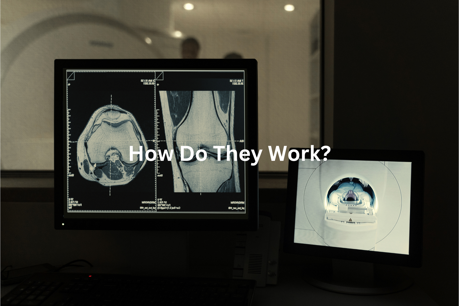
When the tracer reaches these active spots, it releases gamma rays that special cameras detect. The large, donut-shaped PET scanner captures these signals, creating detailed images on computer screens. Different colours on these images show varying levels of activity – brighter areas typically indicate more intense cellular processes.
Medical teams use PET scans to:
- Detect cancer locations
- Monitor treatment effectiveness
- Find infection sites
- Identify brain and heart conditions
Modern PET scanners can spot areas as small as 4 millimetres, making them crucial for early detection and treatment planning(1).
Commonly Used Radioactive Tracers
PET scans use a special sugar called FDG (Fluoro-deoxyglucose) to find cancer cells in the body. These cells act like hungry shoppers at a sale, grabbing more sugar than normal cells do.
When patients get FDG through an IV, it moves through their body for about an hour. Cancer cells take in this special sugar and start to glow on the scan images, making them easy for doctors to find.
Common PET scan tracers include:
• FDG – used for most cancer types
• Gallium-68 – works well for prostate cancer
• Carbon-11 – helps with brain studies
The scanner takes pictures from different angles (usually 20-30 minutes), showing where the tracer collects in the body. Doctors use these images, which look a bit like a heat map, to spot unusual cell activity. Modern PET scanners can find tumours as small as 8mm, making them quite good at catching cancer early.
Safety and Side Effects
PET scans might sound scary, but the radiation levels aren’t as high as many think(2). The process uses a special sugar called FDG (Fluoro-deoxyglucose) that gives off about 7 millisieverts of radiation, similar to what someone gets naturally over two years.
Before getting a PET scan, patients should tell their doctors about:
- Past reactions to radioactive materials
- Any allergies, mainly to medical substances
- Problems with previous imaging tests
The scan helps doctors find serious health problems like cancer. Most patients find the process quick and simple, taking about 30 minutes for the actual scan. During this time, they lie still on a table while the machine takes pictures.
Doctors use PET scans because they show how organs work, not just how they look. This makes them great for finding diseases early, when treatment works best. The benefits of finding health problems early make the small radiation risk worth it.
Preparing for a PET Scan
Sources: NIBIB.
A PET scan reveals crucial details about the body’s inner workings, much like a detective searching for hidden clues. The process starts well before anyone steps into the scanning room.
Patients must skip meals for 4-6 hours prior to the scan (drinking water remains essential). Once at the clinic, a radioactive tracer gets injected into their arm, marking the beginning of a 60-minute waiting period in a quiet room.
The process includes these steps(3):
• Fasting 4-6 hours before scan
• Drinking plenty of water
• Getting the tracer injection
• Resting 60 minutes
• Completing the scan (about 30 minutes)
During the scan, the machine detects areas where cells use more energy than others. These spots often need closer examination, as they might signal health concerns. The entire procedure typically takes about 2 hours from start to finish. Bringing entertainment like books or music helps pass time during the waiting period – just no electronic devices during the actual scan.
Why Are They Important?

PET scans at Australian hospitals use radioactive tracers that shine like tiny lights on medical screens. These special substances help doctors find cancer in ways regular X-rays can’t.
The tracers work by:
• Attaching to cancer cells, making them show up clearly
• Measuring tumour sizes (often down to 2-3 millimetres)
• Showing if treatments are successful
Cancer cells consume the tracer mixture (radioactive material combined with glucose) roughly 70% more quickly than healthy cells. This difference makes spotting cancer much easier for medical teams.
The process takes about an hour at most Australian hospitals. A healthcare worker injects the tracer through a small needle, then patients rest on the scanning bed while the machine captures detailed images. The procedure doesn’t hurt, and patients can usually go home the same day.
Most Medicare-covered PET scans happen at major public hospitals across Sydney, Melbourne, Brisbane and other capital cities.
FAQ
What is a VQ scan?
A VQ scan, also called a lung scan, uses a small amount of a radioactive tracer and special cameras to take images of your lungs. It can help doctors see how well air and blood are flowing through your lungs, which is important for diagnosing lung conditions.
How does a CT scan work?
A CT scan, also called a computed tomography scan, uses X-rays and a computer to take detailed images of the inside of your body. It can provide information about your organs, bones, and other structures that a regular X-ray can’t. The CT scan takes pictures from many angles to create a 3D image.
What is the difference between a CT scan and a VQ scan?
A CT scan and a VQ scan are both imaging tests that use radiation, but they provide different information. A CT scan uses X-rays to create detailed images of the inside of your body, while a VQ scan uses a small amount of a radioactive tracer to look at the airflow and blood flow in your lungs.
What is a bone scan?
A bone scan is an imaging test that uses a small amount of a radioactive tracer to look at the bones in your body. The tracer is injected into a vein and then travels to the bones, where it shows up on special cameras. This can help doctors identify bone problems or injuries.
How does a gamma ray work in medical imaging?
Gamma rays are a type of high-energy radiation that can pass through the body and be detected by special cameras. In medical imaging, a small amount of a radioactive tracer is injected into the body. As the tracer moves through the body, it releases gamma rays that are picked up by the camera to create an image.
What is the purpose of a blood flow test?
A blood flow test, also called a perfusion scan, uses a small amount of a radioactive tracer to look at the blood flow in different parts of the body. This can help diagnose conditions like blood clots, heart disease, or lung problems by showing where the blood is flowing well and where it may be blocked or reduced.
How long does a radioactive tracer stay in the body?
Radioactive tracers used in medical imaging tests are designed to have a short lifespan in the body. They are usually eliminated from the body within a few hours or days, depending on the specific tracer used. This allows doctors to get the information they need without exposing the patient to high levels of radiation for a long time.
What is a PET imaging scan?
A PET scan, or positron emission tomography scan, is an imaging test that uses a small amount of a radioactive tracer to look at how the organs and tissues in the body are functioning. The tracer is injected into a vein, and then a special camera detects the radiation given off by the tracer as it moves through the body.
What are the side effects of a radioactive tracer?
The amount of radiation used in medical imaging tests with radioactive tracers is very small and considered safe for most people. Some people may experience minor side effects like nausea, headache, or redness at the injection site, but these are usually mild and go away quickly. More serious side effects are very rare. People with certain conditions may need to take extra precautions.
How is a radioactive tracer administered?
Radioactive tracers used in medical imaging are usually injected into a vein, but they can also be swallowed or inhaled in some cases. The tracer is designed to travel to the part of the body being imaged and emit radiation that can be detected by special cameras. This allows doctors to see how your body is working in that area.
Conclusion
Radioactive tracers work like tiny beacons in medical tests, such as PET scans (Positron Emission Tomography). These special substances travel through a patient’s body, lighting up areas where cells are most active. Doctors use these glowing signals to spot problems like cancer or check if treatments are doing their job. The tracers contain very small amounts of radiation (less than a chest X-ray), making them safe for medical use. Healthcare providers can explain more about these helpful diagnostic tools.
References
- https://www.healthywa.wa.gov.au/Articles/S_T/Technetium-scans
- https://www.arpansa.gov.au/sites/default/files/legacy/pubs/rps/rps14_2.pdf
- https://www.svhs.org.au/ArticleDocuments/3973/Patient%20Preperation%20for%20FDG%20PET%20CT%20-%20Jan%202020.pdf.aspx?embed=y


