Learn how PET scans are revolutionising cancer diagnosis and treatment monitoring with precision and functional insights.
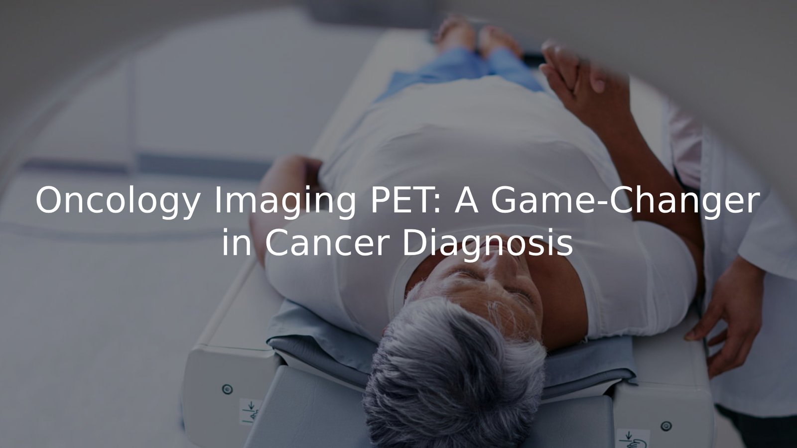
Positron Emission Tomography (PET) is changing the way cancer is diagnosed and treated by providing key insights into the metabolic activity of tumours. This technique allows doctors to not only identify cancer but also monitor the effectiveness of treatments and detect any recurrence.
By combining functional and anatomical data, PET offers a more comprehensive view of cancer behaviour compared to traditional imaging methods.
Key Takeaway
- Precision Diagnosis: PET scans are vital in distinguishing between benign and malignant lesions.
- Treatment Monitoring: PET plays a crucial role in evaluating therapy response and adjusting treatment plans.
- Early Detection: PET scans can identify cancer at earlier stages, improving patient outcomes.
What is Positron Emission Tomography (PET)?
Positron Emission Tomography (PET) is a crucial tool in oncology, allowing doctors to better understand how cancer cells behave in the body. This is particularly important, as cancer cells typically have higher metabolic activity than normal tissue. PET scans help detect cancer, monitor treatment effectiveness, and identify recurrence.
What sets PET apart is its focus on cellular function, not just structure. Unlike traditional imaging methods like CT scans and MRIs, which show detailed structural images, PET highlights how tissues are metabolising nutrients. Since cancer cells consume more energy, PET can pinpoint active areas that may be missed by other imaging techniques.
Key points:
- PET scans provide functional insights into how cells metabolise nutrients
- Detects cancer earlier by identifying high metabolic activity in tumours
- Often combined with CT (PET/CT) for a comprehensive view, merging structural and functional data
How PET Imaging Works
The Role of the Radioactive Tracer (18F-FDG) in Detecting Cancer
PET scans use a radioactive substance called a tracer, with 18F-fluorodeoxyglucose (18F-FDG) being the most commonly used in oncology. This tracer mimics glucose, which cancer cells absorb more of compared to normal cells. When injected into the bloodstream, the tracer is absorbed by metabolically active tissues, like tumours.
Once the 18F-FDG reaches the tumour, it emits radioactive particles detected by the PET scanner. The scanner then generates detailed images, highlighting areas with higher metabolic activity, providing doctors with a clear view of the cancer’s location.
Key points:
- 18F-FDG mimics glucose, targeting metabolically active tissues
- Tracer accumulates in tumours, which consume more glucose than normal cells
- PET scanner detects radioactive particles to create detailed images
- Helps pinpoint the exact location of cancer by highlighting areas of high activity
Scanning Process
The PET scanning process begins with the injection of a radioactive tracer into the patient’s bloodstream. This tracer targets areas with high metabolic activity, such as cancer cells. After the injection, the patient typically waits for a short period, allowing the tracer to accumulate in the tissues of interest. This waiting time is essential to ensure accurate results.
Once the tracer has been absorbed, the patient is positioned in the PET scanner. The machine detects the gamma rays emitted by the decaying tracer, and this data is used to create detailed, three-dimensional images of the body. These images help doctors assess the cancer’s size, location, and activity, allowing for precise treatment planning. (1)
- Injection of radioactive tracer into bloodstream
- Short waiting period for absorption
- PET scanner detects gamma rays
- Three-dimensional images to assess cancer size and activity
Clinical Applications of PET in Oncology
Diagnosis
PET imaging plays a crucial role in differentiating between benign and malignant lesions. While CT and MRI offer structural images, PET reveals whether a lesion is actively metabolising glucose. Malignant lesions generally exhibit higher metabolic activity than benign ones, assisting in more accurate diagnoses.
- Differentiates benign from malignant lesions
- Highlights metabolic activity of lesions
- Improves diagnostic precision
Staging
PET scans play a vital role in staging cancer, allowing doctors to assess how far the cancer has spread. By detecting areas of high metabolic activity, PET can reveal whether the cancer has reached critical organs like the liver, lungs, or bones.
- Identifies spread to lymph nodes and distant organs
- Enhances staging accuracy for treatment decisions
- Supports better prognosis and treatment planning
Monitoring Treatment
PET imaging is not only crucial for diagnosis and staging, but it also plays a vital role in tracking treatment effectiveness. After chemotherapy or radiation, PET scans help doctors evaluate the tumour’s response by detecting changes in metabolic activity.
- Decreased metabolic activity signals effective treatment
- Persistent high activity may indicate treatment resistance
- Provides real-time insight into treatment progress
Advantages of PET Imaging
Early Detection of Cancer with High Sensitivity
PET’s sensitivity is one of its greatest strengths, as it detects metabolic changes that can reveal cancer early on. Traditional imaging methods like CT or MRI often miss small tumours, but PET can identify them before they become detectable by touch.
- Detects cancer earlier than CT/MRI
- Identifies even the smallest tumours
- Early detection leads to better outcomes and survival rates
Providing Functional Insights Into Tumour Metabolism
PET scans offer a unique advantage by providing functional information, showing how a tumour metabolises nutrients. This insight is crucial in determining the tumour’s aggressiveness and shaping treatment strategies.
- PET highlights tumour metabolism
- Increased metabolic activity signals the need for more aggressive treatment
- Slower-growing tumours may be treated with a more conservative approach
Improved Diagnostic Accuracy by Combining PET with CT
Combining PET with CT imaging into a PET/CT scan significantly boosts diagnostic accuracy. The CT scan offers detailed structural information, while the PET scan highlights areas of increased metabolic activity, giving a more comprehensive view of the tumour’s size, location, and activity. (2)
- CT provides structural details
- PET identifies metabolic activity
- Combining both improves cancer diagnosis and assessment
Limitations of PET Imaging
False Positives/Negatives Due to Inflammatory Conditions
While PET imaging offers many benefits, it does have some limitations, such as the potential for false positives and false negatives. Inflammation or infection can sometimes mimic cancer, leading to false positives. Additionally, small or low-grade tumours may not be clearly visible, resulting in false negatives.
- False positives from inflammation or infection
- False negatives for small or low-grade tumours
- PET should be used alongside other diagnostic tools
Radiation Exposure Risks and Efforts to Minimise Patient Dose
Radiation exposure is a consideration with PET scans, though the dose is typically low and safe. For patients needing multiple scans, medical professionals carefully plan each procedure to minimise exposure.
- Radiation dose is low but monitored
- ALARA principle ensures safety
- Careful planning for repeated scans
Guidelines for PET Imaging in Oncology
RANZCR Guidelines
In Australia, the Royal Australian and New Zealand College of Radiologists (RANZCR) ensures PET imaging accuracy through comprehensive guidelines. These guidelines address everything from patient prep to quality control, emphasising the importance of standardised protocols for consistent results across institutions.
- RANZCR guidelines ensure imaging accuracy
- Covers patient preparation and quality control
- Standardised protocols for consistent results
Credentialing and Expertise Required for Accurate Imaging
PET scans are complex procedures requiring skilled professionals to operate the equipment and interpret the results. As per RANZCR guidelines, radiologists and nuclear medicine specialists must have proper credentials and advanced training. This ensures that PET scans provide precise, reliable information for accurate diagnosis and treatment planning.
- Credentialing of radiologists and nuclear medicine specialists
- Advanced training in technical and clinical aspects
- Ensures meaningful and accurate PET scan results
PET Imaging in Specific Cancers
Credits: Art of Healing Cancer
Non-Small Cell Lung Cancer (NSCLC)
PET is essential in detecting and staging non-small cell lung cancer (NSCLC), the most common lung cancer type. It helps assess whether the cancer has spread to lymph nodes or other organs, enabling precise staging. Additionally, PET scans are key in monitoring treatment response, ensuring treatment plans are effective.
- Detects spread to lymph nodes and organs
- Provides precise staging of NSCLC
- Monitors treatment response to guide adjustments
Prostate Cancer and Metastatic Breast Cancer
In prostate cancer, PET imaging with tracers like 11C-choline can identify spread to bones or lymph nodes, crucial for treatment decisions. For metastatic breast cancer, PET helps pinpoint the spread’s location, guiding tailored treatment options. Early assessment of cancer spread can significantly influence the treatment plan.
- Detects prostate cancer spread to bones or lymph nodes
- Identifies metastatic breast cancer locations
- Enables early treatment decision-making
Applications in Gastric Cancer and Neuroendocrine Tumours
PET imaging is also valuable in diagnosing and staging gastric cancer and neuroendocrine tumours. In gastric cancer, it helps assess tumour spread and monitor treatment response. For neuroendocrine tumours, PET detects metastases that may be missed by other imaging methods.
- Assesses tumour spread in gastric cancer
- Monitors treatment response in gastric cancer
- Detects metastases in neuroendocrine tumours
PET Imaging in Clinical Studies and Prognosis
The Role of PET in Prospective Studies and Multicentre Trials
PET imaging plays a significant role in clinical research, especially in multicentre trials and prospective studies. It’s used to assess how tumours respond to new treatments, providing crucial data to help researchers refine and develop more effective therapies.
- Used in multicentre trials and prospective studies
- Provides data on tumour response to treatments
- Aids in developing more effective cancer therapies
PET as a Prognostic Tool for Treatment Response and Survival Outcomes
PET imaging is becoming a crucial prognostic tool in cancer treatment. It offers early indications of how well a patient’s tumour is responding to therapy. A significant decrease in metabolic activity signals a positive response, while persistent or increased activity could suggest resistance to the treatment.
- Early signs of treatment response
- Decrease in metabolic activity suggests a positive response
- Increased activity may indicate treatment resistance
Conclusion
Positron Emission Tomography (PET) is a crucial tool in cancer care, offering early detection and detailed insights into tumour metabolism. Despite limitations like false positives and radiation exposure, its ability to guide diagnosis, staging, and treatment monitoring makes it invaluable.
Adhering to guidelines like those from RANZCR ensures accuracy and safety. Ultimately, PET imaging plays a significant role in improving cancer treatment and patient outcomes, making it a vital asset in oncology.
FAQ
What is Positron Emission Tomography (PET) and how does it help in cancer diagnosis?
Positron emission tomography (PET) is an advanced imaging technique that assists doctors in observing how cancer cells behave within the body. PET scans detect areas with high metabolic activity, which is often present in cancer cells compared to normal tissue. This method helps identify and locate cancer by observing how cells metabolise nutrients.
How do PET scans work and why are they important for detecting cancer?
PET scans use a radioactive drug to highlight areas of the body with increased metabolic activity, often caused by cancer cells. The most common tracer is 18F-FDG, which mimics glucose, as cancer cells tend to consume more glucose than normal tissue. PET scans are vital for detecting cancer early, sometimes even before the tumour is visible with conventional imaging methods.
What is the role of PET scans in staging cancer?
PET scans play an important role in the staging of patients with cancer. They help determine how far cancer has spread, especially in cases of metastatic disease. PET scans provide key insights into the metabolic tumour volume and can guide decisions on treatments like surgical resection or chemotherapy.
References
- https://guoncologynow.com/post/psma-pet-imaging-modalities-a-game-changer-in-prostate-cancer
- https://www.lifehealthcare.co.za/news-and-info-hub/cancer/life-healthcare-spotlights-game-changing-pet-ct-technology/


