
Nuclear imaging uses tiny amounts of radiation to help doctors view organs and bones. Explore its uses and how it aids in accurate diagnosis.
Nuclear imaging is a cool way for doctors to see inside our bodies. It helps them understand what’s happening with our organs and bones. By using tiny bits of radioactive material, they can take pictures that show how well everything is working. Some common types of nuclear imaging are CT scans and PET scans.
These tools help doctors find problems early and decide the best treatment. Understanding nuclear imaging is important for better health. If you want to learn more about how this important medical tool works and its benefits, keep reading!
Key Takeaway
- Nuclear imaging helps doctors see how our organs work.
- It uses low doses of radioactive materials.
- It can find problems early, helping with better treatments.
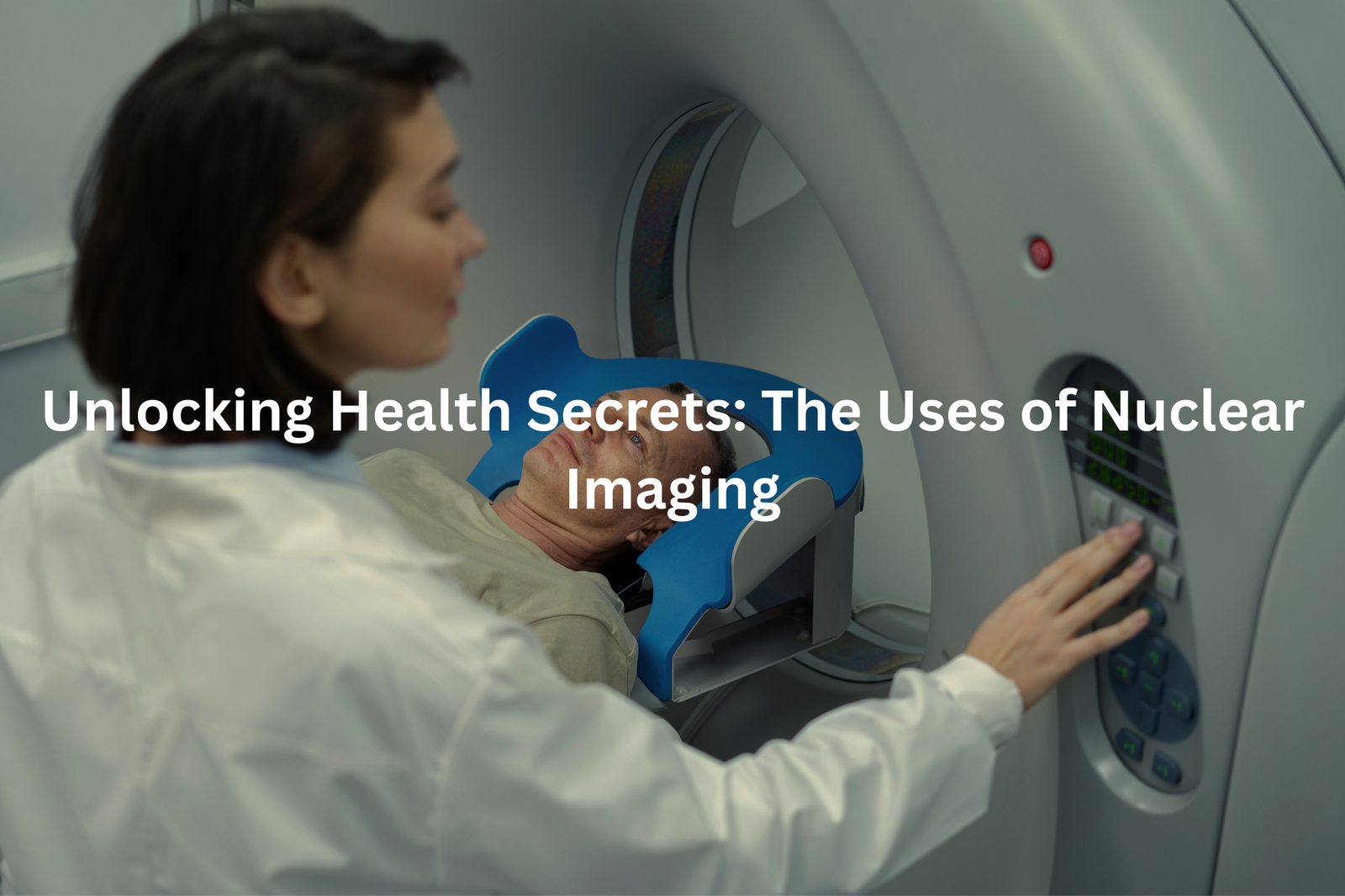
What is Nuclear Imaging?
Nuclear imaging scans let doctors see inside the body without making any cuts. These special medical pictures use tiny amounts of radioactive tracers, which work like glow-in-the-dark markers to show what’s happening inside.
Different types of scans serve different purposes:
- CT scans create detailed cross-section images using X-rays
- PET scans reveal how organs function in real-time
- Bone scans detect problems within the skeletal system
The process is straightforward (patients receive a small injection of the tracer), and most people don’t feel much during the procedure. The radiation exposure from these tracers equals roughly the same amount someone gets on a flight from Sydney to Perth.
Medical professionals use nuclear imaging to spot various health issues, from cancer to heart problems. The technology helps them make better decisions about treatment plans.
Patients should discuss any concerns about nuclear imaging with their healthcare provider, who can explain the process in detail.
Bone Scans: Finding Problems in Our Bones
Bone scans at Australian hospitals show the inner workings of a patient’s skeleton through a fascinating process(1). The procedure starts with a small injection of radioactive dye (technically called a radioisotope tracer) into the bloodstream.
The process follows these steps:
- A quick arm injection takes seconds
- The tracer moves through blood vessels for 2-3 hours
- Patient lies on scanning table for 30-60 minutes
- Special gamma camera captures detailed bone images
The scanner detects areas where bones might need extra attention, spotting issues that regular X-rays often miss. The dye collects more heavily in spots with increased bone activity, making problem areas stand out clearly on the scan images.
Most patients find the scan comfortable, as they can relax while the camera moves around them. The radioactive material leaves the body naturally within 24-48 hours. Doctors at major medical centres like Royal Melbourne Hospital use these scans to check for fractures, infections, and other bone concerns.y – it’s gone before you know it.
Thyroid Scans: Checking the Thyroid Gland
Sources: Radiology Video – Radiology Made Esay.
A thyroid scan reveals the inner workings of this butterfly-shaped gland that sits in the neck(2). The small but mighty organ controls how the body uses energy, making it crucial for daily function.
During a thyroid scan, patients receive a tiny dose of radioactive iodine (about 0.5 micrograms). The substance travels through the bloodstream straight to the thyroid gland. Special gamma cameras then track the iodine’s movement, creating detailed images of the gland’s structure and function.
The 30-minute procedure helps doctors spot various thyroid issues:
- Overactive thyroid areas
- Underactive regions
- Suspicious nodules
- Unusual growths
The amount of radiation used is minimal – similar to the exposure from a chest X-ray. Medical professionals monitor the entire process, ensuring patient safety throughout the scan. Regular thyroid scans serve as valuable tools for maintaining proper hormone balance and overall health.
Cardiac Imaging: Keeping Our Hearts Healthy

Heart scans give doctors a clear view of blood moving through the heart, much like watching traffic from above. These special pictures, called PET or SPECT scans, use a safe dye that lights up problem spots on the scanner’s screen.
During the scan, patients lie still while the machine takes detailed photos (about 15-20 minutes for most procedures). The heart keeps beating roughly 100,000 times each day, making these moving pictures quite remarkable.
The scans spot several heart issues:
- Blocked arteries
- Poor blood flow areas
- Heart disease signs
- Damaged heart muscle
Medical teams across Australia use these scans to find problems early, before symptoms start. The process doesn’t hurt – the dye goes in through a small needle, and the scanner moves quietly around the patient. Most Medicare plans cover cardiac scans when doctors think they’re needed, making them a practical choice for heart health checks.
Lung Scans: Breathing Easy
Lung scans at Royal North Shore Hospital in Sydney show doctors the inner workings of lungs without any cuts or surgery. VQ scans (ventilation-perfusion scans) track how air and blood move through the lungs, similar to monitoring traffic patterns(3).
The scanning process involves:
• A small amount of safe radioactive material injected into a vein
• Special gamma cameras that detect the material’s movement
• About 30 minutes of scanning time
During the scan, patients lie still on a table while cameras rotate around the chest. The procedure doesn’t cause pain, and patients can breathe normally throughout. These scans help doctors spot blood clots in the lungs, which can block airflow and need quick treatment.
The process might seem strange, but it’s as simple as taking a photograph. Patients don’t feel the radiation, and the amount used is quite small (about the same as 6 months of natural background radiation). Doctors use these scans to check lung function and find problems early, making treatment more effective.
PET Scans: Detecting Cancer Early
PET scans work like high-tech cameras that spot cancer cells in the body. These sneaky cells use more energy than normal ones, making them light up during the scan (technically called F-18 FDG uptake).
The process takes about 30-40 minutes. A small amount of radiotracer goes into the patient’s bloodstream through an IV, then spreads throughout the body. Areas where cells are burning through lots of energy – often cancer spots – show up brighter on the scanner’s screen.
Early detection with PET scans helps doctors find problems before they get worse. The technology picks up unusual cell activity that might not show up on regular X-rays or CT scans.
Patient preparation guidelines:
- No food for 6 hours before the scan
- Wear loose-fitting clothes
- Remove metal objects
- Stay hydrated with water
- Tell staff about medications
The scan itself doesn’t hurt, and the radiation exposure is quite low, similar to what someone gets from natural background radiation over 8-12 months.
Keeping It Safe: Safety Measures in Nuclear Imaging
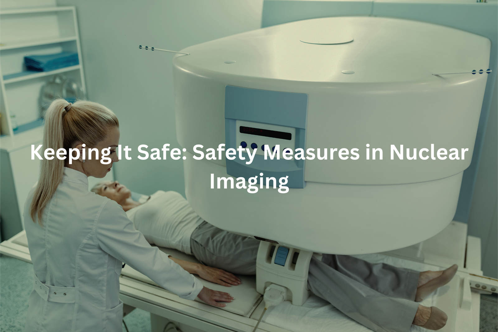
Nuclear scans at Royal North Shore Hospital in Sydney show how everyday medical procedures work without much fuss. The process takes about 45 minutes, with patients lying still while a gamma camera moves around them.
The radiation exposure during these scans ranges from 4 to 8 millisieverts, similar to a return flight from Sydney to London. Nuclear medicine specialists follow strict safety protocols set by ARPANSA (Australian Radiation Protection and Nuclear Safety Agency) for all Australian hospitals.
Before any scan, the medical team checks:
• Pregnancy and breastfeeding status
• Current medications
• Recent imaging procedures
Staff members wear radiation monitoring badges that change color if levels rise too high, though this rarely happens. These badges work as an extra safety measure in Australian hospitals. Patients can ask questions about the procedure anytime – medical staff regularly explain the process to help people feel more comfortable about their upcoming scans.
FAQ
What is a CT scan and how does it differ from nuclear imaging?
A CT (computed tomography) scan uses low-dose x-ray technology to take detailed images of the body’s internal structures. Unlike nuclear imaging tests that use gamma rays, CT scans create images by detecting the different ways x-rays pass through the body’s tissues. This allows CT scans to provide clear pictures of bones, organs, and other body systems.
How do nuclear imaging tests like PET and gamma scans work?
Nuclear imaging tests use small amounts of radioactive materials, called radiotracers, to create images of the body’s organs and tissues. These radioactive “gamma rays” are detected by a gamma camera, which takes pictures as the material travels through the body. This allows doctors to see how the body’s organs and tissues are functioning.
What are some common uses of nuclear imaging tests?
Nuclear imaging tests have a wide range of uses, from checking for heart disease to monitoring thyroid health to detecting cancer. They can provide information about blood flow, organ function, and metabolic activity, which can help doctors diagnose and treat a variety of conditions. Common nuclear scans include bone, cardiac, thyroid, and lymph node imaging.
How long does a nuclear imaging test take and what should I expect?
A typical nuclear imaging test takes 20-60 minutes, depending on the type of scan. During the procedure, the patient lies still on an exam table while the gamma camera takes images. Patients may receive an injection of a radioactive tracer, which allows the camera to detect and track the material as it moves through the body. While the test does involve small doses of radiation, the health benefits typically outweigh the minimal radiation risk.
Are there any side effects or risks with nuclear imaging tests?
Nuclear imaging tests are generally very safe, with only minimal side effects possible. The radioactive tracers used contain small amounts of radiation, but the doses are carefully controlled to be as low as possible. There is a small risk of allergic reaction to the tracer, but this is rare. Pregnant or breastfeeding women may need to take extra precautions. Overall, the benefits of these tests in diagnosing and monitoring health conditions outweigh the low-level radiation risks.
Conclusion
Nuclear imaging is a big part of medicine today. It helps doctors check how our organs work, like our bones and hearts. Using tools like bone scans, thyroid scans, and PET scans, they can find problems early and decide the best treatments. If you need a nuclear imaging test, talk to a healthcare provider. They can help answer your questions and make the process smoother. It’s always better to be prepared and informed!
References
- https://www.insideradiology.com.au/nuclear-medicine-bone-scan-hp/
- https://www.racgp.org.au/getattachment/980fb2e2-ccdd-4851-a2dd-393bf0f945c4/Thyroid-scans.aspx
- https://westernnuclearmedicine.com.au/lung-scan


