Doppler Ultrasound helps doctors see how your blood moves, making it easier to check your heart health. Learn why it’s an essential test!
Doppler Ultrasound is a cool test that helps doctors see how blood flows in your body. It uses sound waves to create pictures of moving blood. This is important because it can show doctors if your heart and blood vessels are working well.
They might use it to check for clots or other problems. The test is painless and quick, usually taking less than an hour. By understanding blood flow, doctors can help keep you healthy. So, if you want to learn more about how this fascinating test works and its benefits, keep reading!
Key Takeaway
- Doppler Ultrasound uses sound waves to see how blood moves in your body.
- It can help find problems like blood clots and heart disease.
- The test is safe and doesn’t hurt, making it easy for patients.
What is Doppler Imaging?
Doppler imaging helps doctors track blood flow through the body. The process uses sound waves (operating at frequencies between 2-18 MHz) that bounce off red blood cells in the bloodstream. When blood moves, these waves change pitch, similar to how a passing ambulance’s siren sounds different as it drives by.
The process works in three main steps:
- Sound waves travel into blood vessels
- Waves reflect off moving blood cells
- Equipment measures the pitch changes
Medical professionals use these measurements to detect problems like blocked arteries, blood clots, or aneurysms. The technology can spot blood flow changes as small as 1-2 millimetres per second, making it quite precise for diagnosis.
Patients don’t need to worry about pain during the procedure. The non-invasive scan doesn’t use needles or require incisions. While the machine might produce loud whooshing sounds, the examination typically takes just 15-30 minutes to complete. This diagnostic tool proves essential for monitoring circulation issues and cardiovascular health in modern medicine(1).
Vascular Doppler
Vascular Doppler testing shows blood flow patterns in arms and legs through sound waves(2). The device (operating at frequencies between 4-8 MHz) sends waves into blood vessels and measures how they bounce back.
Medical professionals use this non-invasive tool to:
- Check circulation problems
- Find blood clots
- Spot blocked arteries
- Measure blood flow speed
During the exam, patients lie on a table while a small probe glides across their skin. Clear gel helps the sound waves travel better. The machine turns blood flow sounds into pictures on a screen, letting doctors see any problems.
The test takes about 30 minutes, doesn’t hurt, and needs no special preparation. Patients might need to change positions – standing up or lying down – so doctors can check blood flow changes.
This test often helps explain symptoms like:
- Leg pain
- Swelling
- Cold hands or feet
- Skin colour changes
Medicare covers vascular Doppler when doctors think there might be circulation problems.
Doppler for Pregnancy
Doppler devices stand as essential tools in prenatal care, capturing the rhythmic sounds of a baby’s heartbeat within the womb. These handheld instruments (operating at frequencies between 2-3 MHz) transform sound waves into audible heartbeats, giving healthcare providers crucial information about fetal health.
The process works through simple physics. The device sends ultrasound waves through the mother’s abdomen, which bounce off the moving heart of the developing baby. These reflected waves create the distinctive whooshing sound that fills examination rooms across Australia.
Medical professionals typically detect heartbeats from 10-12 weeks of pregnancy, though timing varies. The normal fetal heart rate ranges from 120-160 beats per minute, about twice as fast as an adult’s.
The non-invasive nature of Doppler monitoring makes it a safe choice for regular check-ups. Healthcare providers use these readings to track fetal development and spot potential concerns early, making it a cornerstone of modern prenatal care.
How Doppler Works
Sources: Motivation Australia.
Doppler technology works through sound waves, much like a bat’s echolocation. The device sends out waves that bounce off moving blood cells, creating detailed maps of blood flow in the body (operating at frequencies between 2-18 MHz).
The process happens in simple steps:
- A small probe sends sound waves into the body
- Blood cells reflect these waves back
- The machine measures changes in wave frequency
- Software creates visual graphs of blood movement
The returning echoes change pitch based on blood cell movement – faster cells make higher pitches, slower ones make lower pitches. This helps doctors spot blood flow issues without any cuts or needles.
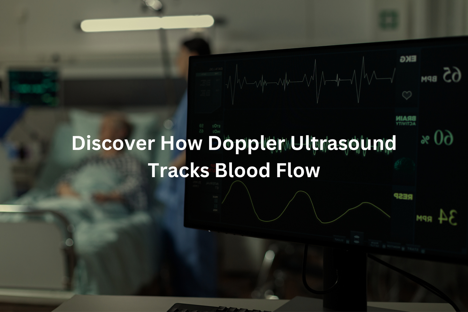
The graphs show blood speed and potential blockages in real-time. Medical professionals use these readings to check for conditions like deep vein thrombosis, arterial blockages, and heart valve problems. For patients worried about blood flow issues, Doppler scans offer quick answers without any pain or recovery time needed.
Arterial Doppler

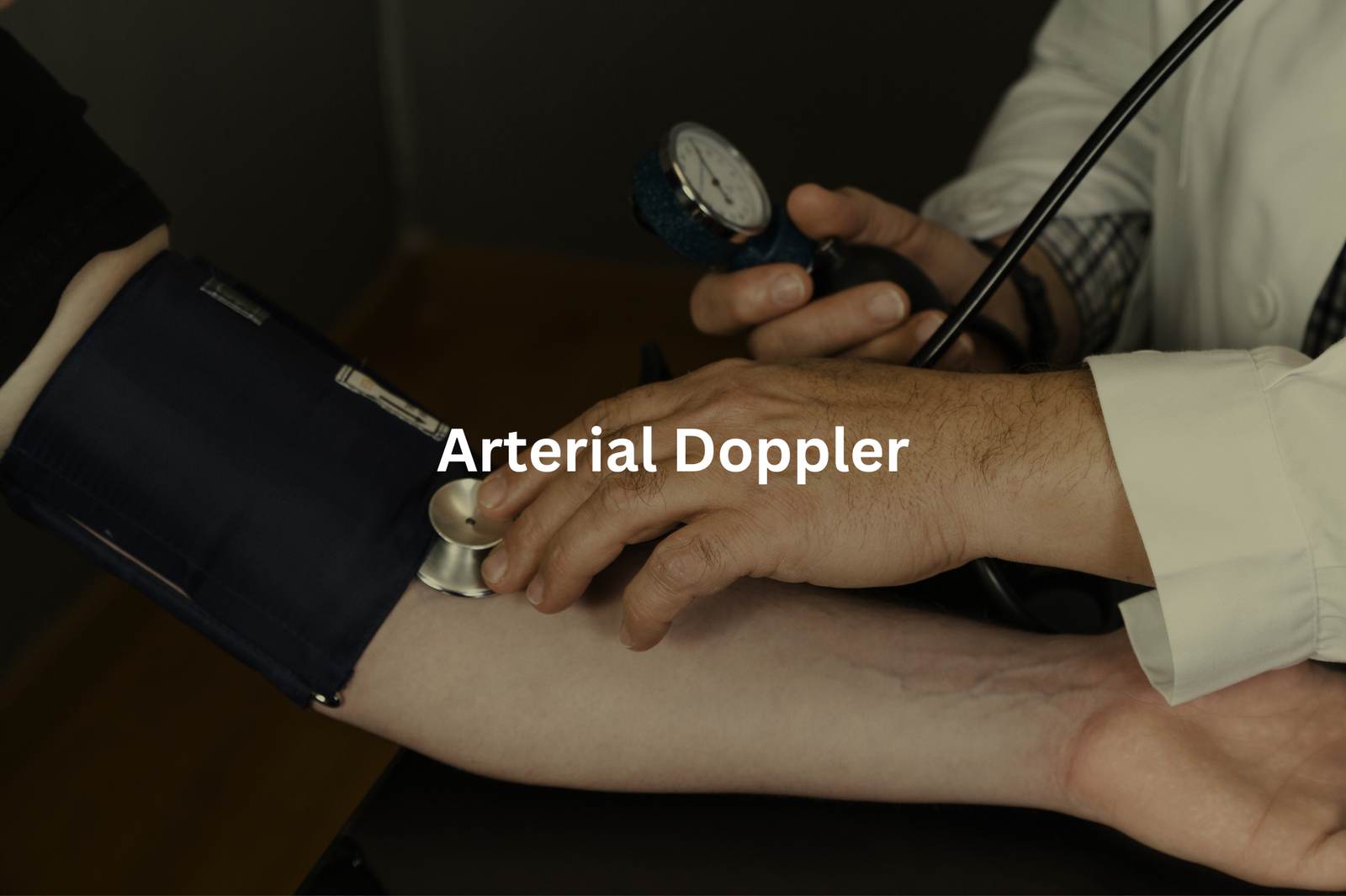
Arterial Doppler tests help medical professionals check blood flow in the arteries. The test uses sound waves (similar to ultrasound) to measure how blood moves through vessels that carry oxygen-rich blood from the heart to other body parts(3).
During the examination, a small handheld device sends sound waves into the arteries. These waves bounce back when they hit moving blood cells, creating detailed images of blood flow patterns. The machine then converts these patterns into graphs and measurements that doctors can analyse.
The test helps detect:
- Blocked arteries
- Narrowed blood vessels
- Poor circulation
- Blood clots
This non-invasive procedure takes about 30 minutes and doesn’t cause pain. Patients can wear regular clothes, though they might need to remove items covering the test area. The results show whether blood flow is normal or if there are circulation problems that need treatment.
Medical professionals often recommend this test for people with symptoms like leg pain, numbness, or signs of poor circulation.
Venous Doppler
Venous Doppler tests look at veins, which carry blood back to the heart. Unlike arterial Doppler that checks arteries, this test spots blood clots and blockages in veins (using sound waves at frequencies between 2-15 MHz).
The process takes about 30 minutes and works like this:
- A small device sends sound waves into the veins
- These waves bounce off moving blood cells
- A computer turns these echoes into pictures of blood flow
Common reasons for getting this test include:
- Swelling in legs or arms
- Unexplained leg pain
- Redness or warmth in limbs
- Checking for deep vein thrombosis (DVT)
The test helps doctors find problems before they become dangerous. When veins get blocked, blood pools up and forms clots that can break loose and move to the lungs. Quick treatment makes a big difference – most blood clots dissolve with proper medication (like blood thinners) and care.
Patients don’t need special preparation, and the test doesn’t hurt. They just lie still while the technician moves the device over their skin.
Doppler Imaging Safety
Doppler Ultrasound stands as one of the safest medical tests available in Australian healthcare. The technology works by sending sound waves through the body, much like sonar tracking fish in the ocean, and these waves bounce back to create detailed pictures of blood flow.
Unlike X-rays or CT scans (which use ionising radiation), Doppler Ultrasound relies on simple sound waves that pose no risk to patients. This makes it particularly valuable for:
- Pregnant women needing regular check-ups
- People with suspected blood clots
- Patients with circulation problems
- Heart condition monitoring
The procedure takes about 30 minutes (depending on the area being examined) and doesn’t need any special preparation. Medical professionals across Australia use this technology daily to check blood flow in arteries and veins, monitor unborn babies, and examine heart function.
Anyone worried about medical imaging should ask their GP about Doppler Ultrasound – it’s non-invasive, radiation-free, and provides quick results.
Doppler Ultrasound Preparation
A Doppler Ultrasound scan needs minimal preparation. The process takes about 30 minutes and doesn’t cause any pain (just a bit of cool gel on the skin).
Basic preparation steps include:
• Wearing loose, comfortable clothing
• Taking off jewellery near the scan area
• Following any fasting instructions from the doctor
During the scan, a sonographer applies a special conducting gel to the skin. This gel helps sound waves travel through the body’s tissues. The handheld device, called a transducer, moves across the gel-covered area to capture images of blood flow.
The procedure uses sound waves, not radiation, making it safe for all patients. Most people can drive themselves home afterward and return to normal activities straight away.
Some scans might need specific preparation, like fasting for 4-6 hours beforehand (especially for abdominal studies). The medical team will provide exact instructions based on the type of scan needed. Patients should arrive 10-15 minutes early to complete any required paperwork.
Doppler Results
After a Doppler ultrasound, medical professionals analyse blood flow patterns through vessels. The process takes several hours, sometimes up to a day, as specialists examine the detailed images for any irregularities.
The results typically show two main outcomes:
Normal Results:
- Blood flows at expected speeds
- No blockages present
- Regular follow-ups might be recommended
Abnormal Results:
- Blood clots (if present)
- Blocked or narrowed vessels
- Reduced blood flow in specific areas
The specialist explains these findings in clear terms, avoiding complex medical language. They’ll point out specific areas on the ultrasound images and discuss what the results mean for the patient’s health. If treatment is needed, they outline the next steps, which might include blood-thinning medications (like warfarin) or further tests.
Patients should write down questions before the follow-up appointment. This helps them remember important points to discuss with their healthcare provider about the results.
Doppler Applications

Doppler Ultrasound functions as a diagnostic tool that captures blood movement through vessels. The technology sends sound waves into the body and measures their reflection off moving blood cells (operating at frequencies between 2-18 MHz).
Medical professionals use this non-invasive method to detect several conditions:
Blood Flow Assessment
- Identifies blockages in arteries and veins
- Measures blood flow speed and direction
- Checks for blood clots in deep veins
Cardiovascular Monitoring
- Evaluates heart valve function
- Assesses blood flow in major arteries
- Detects arterial narrowing
The procedure takes about 30 minutes and doesn’t need special preparation. A sonographer applies gel to the skin and moves a small device called a transducer across the area. The machine creates images showing blood flow in real-time, using colours to indicate direction – red for flow toward the transducer, blue for flow away from it.
This technology helps doctors diagnose conditions like deep vein thrombosis, peripheral artery disease, and heart valve problems without using invasive methods.
FAQ
What is Doppler Ultrasound and how does it work?
Doppler ultrasound is a medical imaging technique that uses sound waves to measure the speed and direction of blood flow in your body. The ultrasound probe sends out sound waves that bounce off the moving blood cells, creating a change in pitch that can be detected and analysed. This allows healthcare providers to see how blood is flowing through your blood vessels, organs, and other soft tissues.
What are the different types of Doppler Ultrasound?
There are several types of Doppler ultrasound, including pulsed wave Doppler, colour Doppler, and power Doppler. Each mode provides different information about blood flow, allowing healthcare providers to get a wide range of details about blood movement in your body. Pulsed wave Doppler measures the speed of blood flow, colour Doppler shows the direction of flow, and power Doppler indicates the amount of blood flow.
How can Doppler Ultrasound help diagnose health issues?
Doppler ultrasound can help diagnose a variety of conditions, including blood clots in the arms or legs, heart disease, carotid artery disease, and arterial diseases in the major arteries. By detecting changes in blood flow, healthcare providers can identify blockages, narrowing, or abnormal patterns that may indicate a problem. Doppler can also measure blood pressure and assess the health of blood vessels, organs, and soft tissues.
What happens during a Doppler Ultrasound exam?
During a Doppler ultrasound exam, a healthcare provider will use a handheld ultrasound probe to send sound waves into your body. The probe is moved around and pressed against your skin, often with the use of ultrasound gel to help the sound waves travel. You may hear a “whooshing” sound as the Doppler measures the speed and direction of your blood flow. The exam is painless and typically takes 30-60 minutes to complete.
How can I prepare for a Doppler Ultrasound test?
To prepare for a Doppler ultrasound, you may need to fast for a certain period of time before the exam, and you’ll likely be asked to wear comfortable, loose-fitting clothing that provides easy access to the area being tested. You should also inform your healthcare provider about any medications you’re taking, as well as any known health conditions. Following the instructions provided by your healthcare team will help ensure the test goes smoothly.
Conclusion
Doppler ultrasound machines work like radar for the human body, tracking blood as it moves through vessels and organs (using high-frequency sound waves between 2-18 MHz). The test shows doctors exactly how blood flows, making it easier to spot problems in arteries and veins.
Patients simply lie still while a small device glides over their skin with some cool gel. The whole process takes about 30 minutes, and there’s no radiation involved – just sound waves bouncing back to create detailed pictures of blood movement.
References
- https://www.healthywa.wa.gov.au/Articles/A_E/Doppler-ultrasound
- https://www.nepeandiagnostics.com.au/doppler-vascular-ultrasound
- https://xray.com.au/service/arterial-and-venous-doppler-ultrasound/


