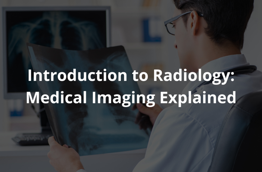
Radiology Overview: Discover its history, types, and how it helps doctors diagnose and treat diseases.
Radiology, a cornerstone of modern medicine, offers a window into the human body that was once unimaginable. Through advanced imaging techniques, it reveals the unseen—broken bones, hidden tumors, or even the rhythm of a beating heart. But behind every scan is a story: a patient waiting for answers, a radiologist interpreting shadows and light, and a history of innovation that shaped this field.
From X-rays to MRIs, radiology bridges technology and care, helping doctors diagnose and treat with precision. Curious about how these images come to life and the people who make it possible? Keep reading to uncover more.
Key Takeaway
- Radiology uses imaging tests like CT scans and MRIs to see inside the body.
- It helps doctors diagnose diseases and guide treatments.
- Understanding radiology can improve health outcomes for patients.
Definition of Radiology
Radiology is a part of medicine that helps doctors see inside the body without needing to cut it open. It uses special tools like X-rays, CT scans, and MRIs. Each of these tools is good for looking at different things. For example, X-rays are great for checking bones, while MRIs are better for soft stuff like muscles or the brain.
These imaging tests are super helpful for doctors. They let them look closely at organs, tissues, and bones. This helps doctors figure out what’s wrong and decide the best way to help. Plus, the tests are safe and don’t take much time. The pictures they make are really clear, which makes it easier for doctors to make good decisions.
Radiology is also great for catching problems early. Things like tumors or broken bones can be found faster, which means treatment can start sooner. It’s also useful for checking if treatments are working. With these tools, doctors can take better care of their patients.
History of Radiology
Radiology started back in 1895. A German scientist named Wilhelm Conrad Roentgen found something amazing while working with cathode rays. He noticed these rays could go through solid things. He even took the first X-ray picture of his wife’s hand, and you could see her bones! This discovery was so big that he won the first-ever Nobel Prize in Physics in 1901. [1]
After X-rays were discovered, doctors started using them pretty quickly. Then, in the 1970s, CT scans came along. These scans use X-rays too, but they make super detailed pictures of the body. It was a big deal because doctors could see inside the body much better without needing surgery. A little later, in the 1980s, MRI machines were invented. MRIs don’t use X-rays; instead, they use strong magnets and radio waves to make clear images of soft tissues.
These tools have made it easier for doctors to find and treat problems. For example, they can spot things like brain tumors or bleeding inside the body much more clearly now. Radiology keeps getting better, with new machines and methods being created all the time. This history shows how much radiology has changed medicine for the better.
Medical Imaging Basics
Medical imaging is like taking a peek inside the body without cutting it open. It gives doctors pictures of what’s going on under the skin, muscles, and bones. There are a few main ways to do this:
- X-rays: These are great for looking at bones. They can quickly show breaks or infections.
- CT Scans: A CT scan uses X-rays from different angles to make detailed pictures of the inside of the body. It’s helpful for seeing organs and blood vessels.
- MRI: This uses strong magnets and radio waves to get clear images of soft tissues, like the brain, spinal cord, or muscles.
- Ultrasound: Instead of radiation, this uses sound waves to create images. It’s often used during pregnancy but can also check other organs. [2]
These tests help doctors figure out what’s wrong and how to treat it. For example, if someone has a broken bone, an X-ray can show exactly where the break is. If a doctor suspects a tumor, they might use a CT scan or MRI to see how big it is and where it’s located. That information helps them decide the best way to treat it, like surgery or something else.
Medical imaging is a big deal in healthcare. It helps doctors make better decisions and gives patients a better chance at getting the right treatment.
Types of Radiology
Radiology is the area of medicine that uses these imaging tools. There are two main kinds:
- Diagnostic Radiology: This is about finding out what’s wrong. It uses imaging tests to spot diseases or see if treatments are working.
- Interventional Radiology: This is when doctors use imaging to guide them during procedures. For example, they might use it to place a catheter or take a biopsy. These procedures are usually less invasive than regular surgery.
Both types are super important. Diagnostic radiology helps catch problems early, which can make treatments work better. Interventional radiology gives doctors new ways to treat problems without big surgeries. That means less pain and quicker recovery for patients.
Together, these two areas of radiology are changing how doctors care for people. They make medicine safer, faster, and more effective.
Diagnostic Imaging
Diagnostic imaging is like taking pictures of the inside of your body to figure out what’s wrong. Doctors use different tools for this, like X-rays, CT scans, MRIs, and ultrasounds. Each one works a little differently and is good for specific things.
- X-rays are the simplest. They’re fast and great for checking bones. If someone breaks an arm or leg, an X-ray can show exactly where the fracture is. They’re also helpful for spotting infections in bones.
- CT scans (which stands for computed tomography) take things a step further. They give a much clearer picture of what’s going on inside. These are especially useful for looking at organs like the lungs or stomach.
- MRIs (magnetic resonance imaging) are even more detailed, but they’re best for soft tissues. For example, if a doctor needs to see the brain or muscles, they’ll probably go with an MRI.
- Then there’s the ultrasound. It’s super common during pregnancy because it’s safe and doesn’t use radiation. It lets parents and doctors get a peek at the baby before it’s born.
Doctors don’t just pick these tools randomly. They think about what the patient’s symptoms are and what they’re trying to find. Choosing the right method can make a big difference—it helps doctors figure out what’s wrong faster and plan the best treatment.
Interventional Radiology
Interventional radiology is like a mix of imaging and surgery, but it’s way less invasive. Instead of big cuts, doctors use tiny tools and guide them with imaging. This means patients heal faster and face fewer risks.
Here’s an example: If someone has a blocked artery, a doctor can use imaging to see exactly where the problem is. Then, they guide a thin tube (called a catheter) to the blockage and open it up. Most of the time, this is done with just local anesthesia, so the patient doesn’t feel much pain and recovers quicker.
This type of radiology is becoming more popular because it’s safer and easier for patients. It’s not just about fixing arteries, though. Doctors can also use it to take small tissue samples or drain fluid from the body.
Patients like these procedures because they’re less scary than traditional surgery, and they can get back to their normal lives sooner. It’s a win-win for everyone involved.
Role of a Radiologist
Radiologists are doctors who specialize in looking at medical images and doing certain procedures. They study pictures like X-rays, MRIs, and CT scans to figure out what might be wrong with a patient. They also help other doctors decide which tests are needed and sometimes treat patients using small, precise tools. Their job is really important for figuring out what’s going on and how to fix it.
Radiologists don’t work alone—they team up with other healthcare workers. They make sure the right tests are done and explain the results clearly. This teamwork helps patients get the best care possible.
Besides reading images, radiologists might also do hands-on procedures. For example, they can guide a needle during a biopsy or place a small tube (called a catheter) where it’s needed. These skills let them help in many ways, making them a key part of the medical team.
Importance of Imaging
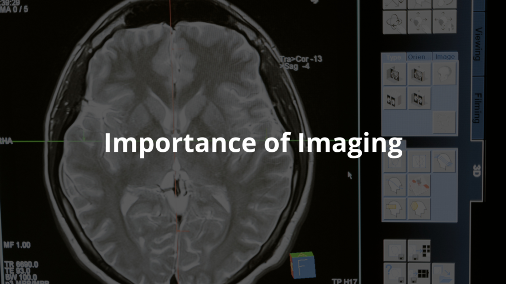
Imaging is a big deal in medicine today. It helps doctors find problems faster and treat them better. For instance, catching breast cancer early with an imaging test can save lives. Imaging also shows how well treatments are working, so doctors can make changes if needed to help patients get better.
Being able to see inside the body without surgery is a huge advantage. It lets doctors check things quickly and make smart decisions. Imaging can even help plan surgeries by giving doctors a clear picture of what they’re dealing with.
In short, medical imaging makes healthcare better. It helps doctors diagnose problems early and come up with strong treatment plans. With imaging, patients get safer and more effective care.
Radiology in Medicine
Radiology is a big part of healthcare now. It touches almost every area of medicine. Doctors don’t just use it to figure out what’s wrong—they also use it to plan treatments and even guide surgeries. As technology gets better, radiology keeps changing. New things, like artificial intelligence, are starting to help doctors make even better decisions. [3]
Different types of doctors depend on radiology for their work. Cancer doctors (oncologists) use scans to check if tumors are growing or shrinking. Heart doctors (cardiologists) look at images to see how well the heart is working. Bone and joint doctors (orthopedics) use X-rays to figure out what’s causing pain or injuries.
All these advances are changing how patients get treated. New tools and ideas are making care faster and safer. Being able to look inside the body without surgery is something that will always be super important in medicine.
FAQ
What exactly happens during magnetic resonance imaging and how does it use magnetic fields?
Magnetic resonance imaging (MRI) uses powerful magnetic fields and radio waves to create detailed pictures of your body. Unlike X-rays, it doesn’t use ionizing radiation. Instead, it aligns water molecules in your body using magnetic fields, then uses radio waves to create clear pictures of soft tissues and normal tissues. The whole process is painless and produces real time images your doctor can use right away.
How long does it take to become a diagnostic radiologist in the United States?
After finishing medical school, radiologists complete a five year residency program followed by an optional one year fellowship. This training covers everything from computed tomography to nuclear medicine. In the United States, they must then pass exams from the American Board of Radiology to become certified diagnostic radiologists. The whole journey takes about 13 years after high school.
What’s the difference between radiation therapy and diagnostic imaging tests?
While diagnostic radiologists use imaging tests like CT scans and PET scanning to find health problems, radiation oncology focuses on treating disease, especially cancer, using ionizing radiation. Radiation oncologists carefully plan treatments to target specific areas while protecting surrounding healthy tissue. It’s a key branch of medicine that works alongside diagnostic radiology to help with diagnosing and treating patients.
How do different imaging techniques like CT scan and positron emission tomography work?
CT scans (computerized axial tomography) use X-rays and computers to make detailed cross-section images at high speed. Positron emission tomography (PET) uses small amounts of radioactive isotopes to show how tissues are working. Sometimes healthcare providers use contrast agents to make certain areas show up better. These image guided procedures help doctors see inside your body in amazing detail.
What role does medical physics play in radiology and are there harmful effects to consider?
Medical physics ensures safe use of ionizing radiations in clinical radiology. When having imaging tests, you’re exposed to carefully controlled amounts of radiation based on established unit of measurement standards. While there can be harmful effects from radiation, doctors use the lowest doses needed. Sound waves (like in ultrasound) and magnetic resonance don’t use radiation at all, making them especially safe for imaging the alimentary tract and other body parts.
What kind of training do interventional radiologists need in health care?
Interventional radiologists are medical doctors who first complete their doctor of medicine or osteopathic medicine degree. After medical education at an accredited medical school, they go through specialized training focused on image guided procedures. The college of radiologists requires them to master both diagnostic skills and minimally invasive treatments. Their technical difficulties training covers everything from reading imaging tests to performing complex procedures.
Conclusion
Radiology’s come a long way—from the accidental discovery of X-rays back in 1895 to today’s high-tech MRIs and CT scans. It’s all about using imaging to figure out what’s going on inside the body and, honestly, it’s changed how doctors treat patients.
Better pictures mean better diagnoses, which means better care. And while it might sound complicated, at its core, it’s just about helping people heal. That’s what medicine should always be about, right?
References
- https://www.nde-ed.org/NDETechniques/Radiography/Introduction/history.xhtml
- https://en.wikipedia.org/wiki/Medical_imaging
- https://udshealth.com/blog/radiology-101-imaging-basics/