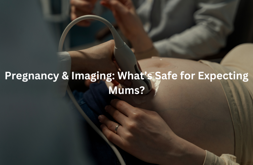
Learn about ultrasounds in pregnancy, their importance, and the types of scans available for expecting mothers.
Pregnancy ultrasounds: a quiet space, soft light. An expectant mother, hopeful, awaits her first glimpse of her baby. The machine emits a low hum; gel is applied, and an image appears. The doctor or sonographer uses a transducer, and the screen displays tiny fingers and toes. Obstetric ultrasounds (diagnostic imaging using sound waves) are standard.
These examinations are important, because they help monitor fetal growth and health. One scan typically occurs around 12 weeks gestation (to confirm dating) with another around 20 weeks (a detailed anatomy assessment). The latter evaluates major organ systems: brain, heart, kidneys, and more.
Average crown-rump length at 12 weeks is approximately 6cm. The aim is early detection of potential complications. Such information offers reassurance. Interested in learning more about ultrasound modalities and result interpretation? Continue reading.
Key Takeaway
- Ultrasounds help monitor the baby’s growth and health during pregnancy.
- There are different types of scans for various stages of pregnancy.
- Ultrasounds are very safe for both mothers and babies.
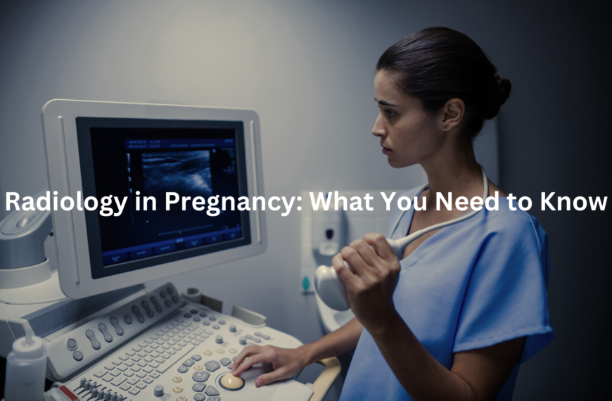
What are Ultrasounds in Pregnancy?
Ultrasounds in pregnancy, they’re like peeking into a secret world, using sound to paint a picture. Obstetric ultrasound, that’s the proper term, shows how the little tacker is growing. Trained sonographers, they’re the ones who usually do these scans. Important job, making sure both baby and mum are doing alright.
Different ultrasounds for different things, see?
- Dating Scan: Early days, confirms how far along the pregnancy is, and figures out the due date. Pretty crucial, that one.
- Morphology Scan: Later on, checks the baby’s development. Everything from head to toe, practically.
These scans use high-frequency sound waves (we’re talking beyond human hearing) to create those images. The sonographer moves a transducer (a wand-like device) over the belly. The sound waves bounce back, and the machine turns them into a picture. Simple as that, almost. With dating and morphology scans, parents are given crucial information throughout the pregnancy.
Why Are Ultrasounds Important?
The nuchal translucency scan, it’s done around 12 weeks, measures fluid at the back of the baby’s neck. This can help to assess the risk of certain chromosomal conditions. It offers parents a bit more information, a bit more certainty. One’s heard stories, of course.
Ultrasounds are vital for a few key reasons. It is known that baby growth is always crucial, so here’s the important reasons:
- Checking Baby Growth: Makes sure the little one is growing as expected. Averages are used, but every bub is different, eh?
- Detecting Problems Early: If there are potential issues, scans can help pick them up sooner rather than later. Time is of the essence.
- Monitoring Multiple Pregnancies: Twins, triplets… ultrasounds give info on each baby. Keeps track of everyone.
The nuchal scan is an important element during pregnancy. By getting this information, parents can determine the next steps for them and baby.
Types of Ultrasounds
Credits: Clarius Mobile Health
Expectant mothers might have a few different types of ultrasounds throughout their pregnancy. Each has a specific purpose, gives different information. It’s all about keeping an eye on things.
- Early Pregnancy Scan: Confirms the pregnancy. Checks for that first, important heartbeat.
- Dating Scan: Usually between 8-12 weeks, figures out the due date. Organises the calendar, so to speak.
- Nuchal Translucency Scan: Between 12-13 weeks, screens for risk of chromosomal variations. Gives some families extra knowledge.
- Morphology Scan: Around 18-20 weeks, a good look at the baby’s anatomy. Checks development from head to toe.
- Growth Scans: Third trimester, for high-risk pregnancies. Keeps tabs on growth.
These scans all use the same basic tech—sound waves—but look for different things. Sonographers are highly trained to get the best images. Ultimately, such tests allows medical teams to see inside and give the best care to the bub.
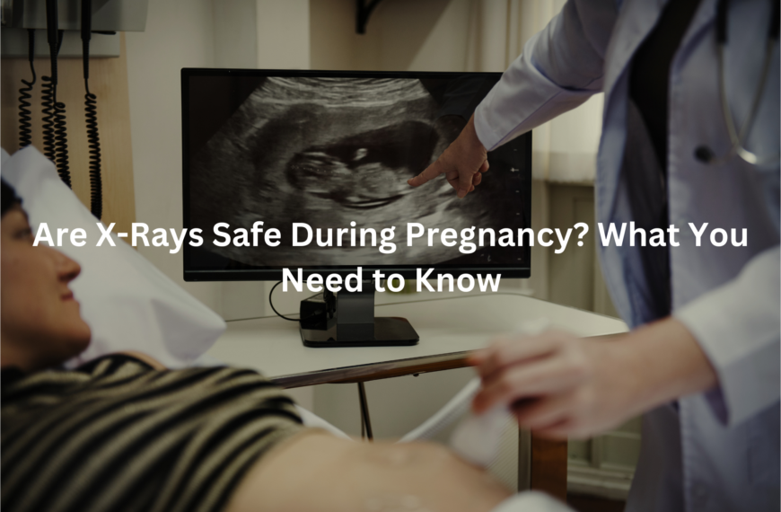
How Safe are Ultrasounds?
Safety of ultrasounds, it’s a common question. Understandable, too. Ultrasounds are generally considered safe for both mothers and babies. Unlike X-rays, they use sound waves. No harmful radiation involved. [1] That’s a relief.
The Royal Australian and New Zealand College of Radiologists (RANZCR) highlights the importance of using ultrasounds only when necessary. A good idea to limit exposure, even if it is considered safe. But, it is known that most women will have ultrasounds done as needed.
The scans are generally painless and non-invasive. Nothing to stress about there. A bit of gel on the belly and a transducer doing its thing. And you get to see your bub! Non-invasive procedures can save lives and ease minds.
What Happens During an Ultrasound?
Arriving for an ultrasound, a pregnant woman might feel a bit anxious. New experience, after all. The sonographer usually explains everything, puts minds at ease. Best to know what’s going on.
- Preparation: Might need to drink water beforehand. Full bladder helps get clearer images. Gotta plan ahead.
- The Scan: Gel goes on the belly. The sonographer moves the transducer around. Sends out sound waves, picks up the echoes. Makes a picture on the screen. Technology at work.
- Results: After the scan, the sonographer gives a quick rundown. Might send the images to a doctor for a proper report. Experts take a look.
It’s a window into the womb, showing the baby’s position, movements. The heartbeat, too. It may ease the minds of new parents as they look towards the future. Seeing is believing, as they say.
What Can Ultrasounds Show Us?
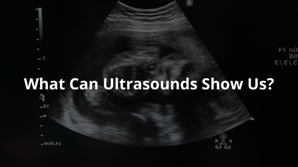
Ultrasounds reveal lots about the little bub. Doctors look for key things, information that helps guide care. It’s all about knowing what’s happening inside.
- Fetal Heartbeat Detection: Hearing that tiny heartbeat, it’s reassuring. A strong sign of life.
- Fetal Position: Knowing how the baby is positioned, helps with birth planning. Head down is ideal, of course.
- Fetal Measurements: Estimate the baby’s weight, check for growth problems. Keeping an eye on progress.
- Placental Location: Important for the mother’s health. Makes sure everything’s where it should be.
- Amniotic Fluid Measurement: Checks the fluid around the baby. Essential for development.
This information helps doctors and parents stay informed during the pregnancy. It is the little things and details that are worth the look. [2]
FAQ
When will I need scans during my pregnancy?
You’ll have several pregnancy sonograms throughout your journey. Most women get a dating scan at 8-12 weeks to check pregnancy dating and viability, a nuchal translucency scan at 11-13 weeks for first trimester screening, a morphology scan (also called an anomaly scan) at 18-22 weeks for second trimester screening, and growth scans in the third trimester screening if needed.
Your obstetrician might suggest more obstetric ultrasounds to watch foetal development tracking or check for pregnancy complications detection. How often you get antenatal scans depends on your needs.
What’s the difference between internal and belly ultrasounds?
These are two ways to get foetal imaging. A transvaginal ultrasound uses a wand that goes into the vagina, showing clearer pictures during an early pregnancy scan when the gestational sac visualisation is tiny.
A transabdominal ultrasound uses a probe moved over your belly and is used after the first few weeks. The sonographer picks the best method based on what they need to see. Both types are safe for pregnancy ultrasound safety and good for foetal wellbeing assessment.
What will I learn from my 20-week scan?
The morphology scan, done around 20 weeks, gives detailed foetal anatomy scan information. It checks for congenital anomalies detection by looking at the foetal spine examination, foetal brain development, foetal limb assessment, foetal kidney assessment, foetal bladder visualisation, and foetal stomach assessment.
The sonographer will take foetal measurements including head size (biparietal diameter measurement), leg length (femur length measurement), head circumference measurement, and belly size (abdominal circumference measurement) for foetal biometry. This scan also checks placental location, amniotic fluid measurement, and can often tell foetal sex if you want to know.
How do doctors check if my baby is growing well?
Doctors track foetal growth monitoring through regular ultrasounds. In early pregnancy, crown-rump length measurement helps with gestational age assessment. Later scans include foetal weight estimation by measuring your baby’s head, belly and legs.
If there are worries, special growth scans might use doppler ultrasound to check foetal blood flow assessment and umbilical cord assessment. These help find possible foetal growth restriction screening. Your doctor compares these results with what’s normal for your baby’s age.
What makes 3D and 4D ultrasounds special?
While regular obstetric ultrasounds show flat black and white pictures, 3D ultrasound creates a three-dimensional image of your baby, letting you see foetal facial features better. The 4D ultrasound adds movement, showing foetal movement and foetal breathing movements like a video.
These show foetal presentation and development in amazing detail, though they’re not more medically useful than normal scans. They’re usually extra and might not be covered by Medicare.
How do doctors check for twins or more?
If multiple pregnancy screening is needed, your sonographer will use foetal imaging to confirm how many babies you have. They’ll check if babies share a placenta (identical twins) or have separate placentas.
The scan looks at amniotic fluid index for each baby, checks foetal tone assessment, and watches each baby’s growth. For twins or more, you’ll have more ultrasounds to track foetal position of each baby and make sure all are growing well.
What happens during the neck fold scan?
This important first trimester screening happens between weeks 11-13 and measures fluid at the back of your baby’s neck (nuchal translucency). Along with blood tests and your age, it helps check the risk of chromosome problems.
The sonographer will also check foetal heartbeat detection, confirm pregnancy dating through gestational age assessment, and look at basic foetal anatomy. This non-invasive prenatal testing helps find possible problems early, letting you decide about more tests if needed. The scan takes about 30 minutes with results usually ready in a few days.
Conclusion
Ultrasounds, vital in pregnancy. They offer a safe way to check on the baby’s health. Each scan provides insights, helping to ensure everything is progressing well. Expectant parents can feel confident in the process. These scans are there to support them and their growing family. Embrace those ultrasound moments, they are precious. Filled with hope, it’s a peek into a new chapter. The experience allows parents to prepare for the future.
References
- https://www.pregnancybirthbaby.org.au/ultrasound-scan
- https://www.betterhealth.vic.gov.au/health/healthyliving/pregnancy-tests-ultrasound