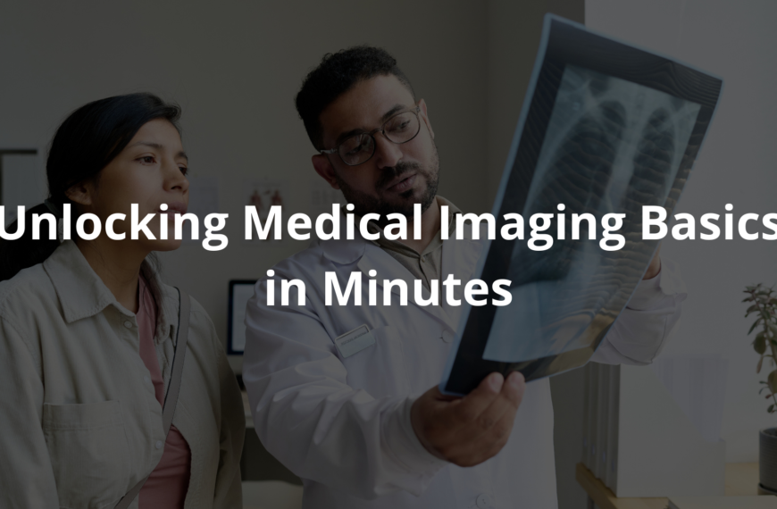
From diagnosis to treatment, the types of radiology play vital roles in healthcare. Discover how these imaging tests work and their benefits!
Radiology plays a crucial role in medicine, allowing doctors to peek inside the human body. There are several types, like X-rays, which help spot fractures, and MRIs, which see soft tissues. CT scans offer detailed pictures and can even help in emergency situations. Ultrasounds are great for viewing organs or monitoring pregnancies.
Each method has unique benefits, supporting diagnosis and treatment. I think understanding these different types can really help people appreciate how doctors use them. Curious to find out more about how these imaging tests work? Keep reading to uncover the details!
Key Takeaway
- Radiology has different types that help doctors see inside the body.
- Each type of radiology uses special machines to create images.
- Understanding these types can help patients feel more comfortable with their health care.
Diagnostic Radiology
Diagnostic radiology plays a crucial role in medicine, often working behind the scenes to help diagnose and treat patients.
Around 80% of medical decisions rely on imaging technology, making it an essential tool. Radiologists use non-invasive imaging techniques to see inside the body, providing valuable insights without the need for surgery.
X-rays are the most commonly used imaging tool. (1) They are quick and effective for identifying bone fractures. CT scans are more advanced, providing 3D images by taking multiple X-ray images from different angles.
They are useful for detecting internal bleeding or tumors. MRIs use magnets and radio waves, creating detailed images of soft tissues like muscles, brains, and joints. Unlike X-rays and CT scans, MRIs do not use radiation, making them a safer option for certain scans.
Each imaging method serves a specific purpose, helping radiologists select the right tool for the situation. It’s important to ask questions and understand what to expect during the procedure.
Interventional Radiology
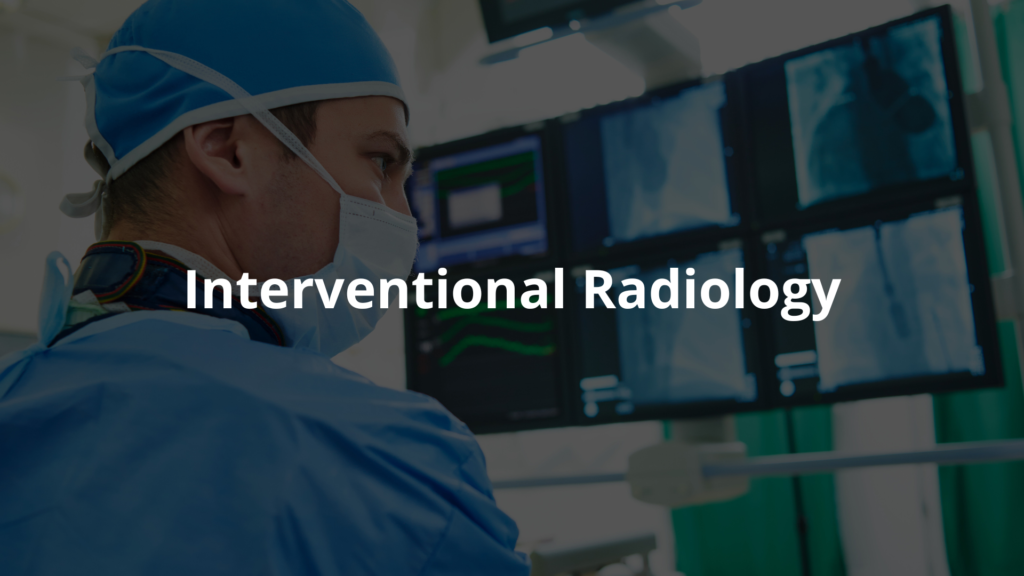
Interventional radiology is transforming medical procedures by using imaging techniques like X-rays, CT scans, and ultrasounds to guide tiny instruments through the body.
These procedures are minimally invasive, meaning patients experience less pain and shorter recovery times compared to traditional surgery. The tools used are often as small as a few millimetres wide, and the procedures are performed through small incisions or even pinholes.
Some common interventional radiology procedures include (2):
- Biopsies: Radiologists use imaging to guide a needle to a suspicious area, removing a tissue sample without major surgery. This allows for quicker recovery and often no stitches.
- Angioplasty and Stenting: For blocked blood vessels, radiologists use small balloons to open up the blockage and place a stent to keep it open. This is crucial for patients with heart or artery diseases.
- Tumor Embolization: Radiologists can inject medication or particles into the blood vessels of a tumour to cut off its blood supply, often used for liver cancer or fibroids.
- Thrombectomy: Special tools are used to remove blood clots from arteries or veins, a critical procedure for stroke or deep vein thrombosis patients.
These procedures are precise and effective, offering a less invasive alternative to traditional surgery with faster recovery times.
Nuclear Medicine
Source: Dr Christoph Agten
Nuclear medicine is a unique and underappreciated branch of radiology that uses small amounts of radioactive materials, called radiotracers, to observe how organs and tissues function.
Unlike traditional imaging, which shows structure, nuclear medicine reveals how well the body is working. This method is essential for diagnosing conditions like cancer, heart disease, and thyroid problems.
Some common types of nuclear medicine include:
- PET Scans (Positron Emission Tomography): These scans use radiotracers to highlight areas where cancer cells are active, helping doctors plan treatments.
- SPECT Scans (Single Photon Emission Computed Tomography): These scans focus on blood flow, especially in the heart. They can detect blockages and help prevent heart attacks.
Nuclear medicine is also used for treatment, such as radioactive iodine therapy for thyroid issues. The radiation doses used are small, and for most patients, the benefits outweigh the risks. It’s a highly targeted, effective, and non-invasive treatment option.
Pediatric Radiology
Pediatric radiology differs significantly from adult imaging due to the unique needs of children. Kids’ bodies are still growing, and their bones, organs, and tissues are more sensitive.
A radiologist must adjust settings, angles, and sometimes even the type of machine used to accommodate a child’s size and development. This requires precision and adaptability.
Radiation is a significant concern, as children are more vulnerable to damage due to their faster cell division.
Pediatric radiologists follow the ALARA principle—”As Low As Reasonably Achievable”—to minimise radiation exposure. They use shields, adjust settings, or opt for non-radiation methods like ultrasound or MRI when possible to protect the child.
Beyond technical expertise, pediatric radiologists also play a role in making kids feel comfortable.
They explain procedures in simple terms, use distractions like toys, and create a welcoming environment to ease anxiety. Their work is crucial in detecting everything from fractures to rare genetic conditions.
Breast Imaging
Breast imaging plays a crucial role in early detection of breast conditions. Digital mammography is a key tool, using X-rays to capture detailed images that can detect small changes like calcifications, which may indicate early-stage cancer. The ability to identify issues early greatly improves treatment outcomes.
Ultrasound is another valuable tool, often used when mammograms find unclear results. It uses sound waves, not radiation, to create images, offering a closer look at any lumps or abnormalities. This helps provide a more detailed view, especially in dense breast tissue.
MRI, reserved for high-risk cases or when other imaging methods are inconclusive, is highly sensitive. It offers an even more detailed view of the breast tissue, helping to identify potential issues that other scans might miss.
Breast imaging isn’t just about identifying problems; it provides women with options for treatment. Early detection leads to more treatment choices, improving health outcomes. Regular check-ups are essential for maintaining breast health.
FAQ
What is a CT scan and how does it work?
A CT (computed tomography) scan is a type of medical imaging that uses X-rays to create detailed images of the inside of your body. It can help doctors see your organs, bones, and other structures in a wide range of detail.
During a CT scan, you’ll lie on a table that moves through a large, circular machine. Small amounts of radiation are used to produce the images, which are then put together on a computer screen for the radiologist to analyze.
What is low-dose CT scanning and how can it help detect lung cancer early?
Low-dose CT scanning uses a small amount of radiation to create images of the lungs. This type of imaging test can help detect lung cancer in its early stages, before symptoms appear. It’s especially helpful for people at high risk of lung cancer, like long-time smokers.
The low-dose approach means less radiation exposure for the patient while still providing clear images that doctors can use to check for signs of cancer.
How can imaging tests help guide patient care and treatment?
Imaging tests like X-rays, CT scans, and MRIs allow doctors to see inside the body and diagnose conditions without having to perform surgery. These images can help guide treatment decisions, from choosing the right medications to planning surgical procedures.
Imaging also plays a crucial role in monitoring how a patient is responding to treatment over time. The ability to produce clear pictures of the body’s internal structures is invaluable for providing high-quality patient care.
What are the different types of medical imaging and how do they work?
There are many different imaging techniques used in healthcare, each with its own strengths. X-rays use small amounts of radiation to create two-dimensional images of bones and organs. CT scans use X-rays to produce more detailed, three-dimensional pictures.
MRIs use strong magnetic fields and radio waves to generate images without radiation. Ultrasound uses sound waves to look at soft tissues like the heart and developing babies. Nuclear imaging tests use small amounts of radioactive tracers to create pictures of organ function.
How can imaging help in the early detection and treatment of diseases?
Diagnostic imaging plays a crucial role in detecting health issues at their earliest stages. Tests like mammograms can find breast cancer before it causes symptoms. Bone density scans can identify osteoporosis before bones become dangerously weak. Heart disease can be caught early through cardiac imaging that reveals plaque buildup in arteries.
And CT scans of the lungs can detect lung cancer when it’s most treatable. Early detection through imaging allows for quicker, more effective treatment that can greatly improve patient outcomes.
Conclusion
Radiology plays a big part in medicine. There’s a lot of different types, each doing something special for patient care. You’ve got your basic X-rays, which most people know about, and then there’s the more advanced stuff like nuclear medicine.
These imaging methods can really help doctors figure out what’s going on and how to treat illnesses. If patients learn about the types of radiology, they might feel more at ease with their health care choices.
References
- https://asianheartinstitute.org/blog/what-is-radiology-and-types-of-radiology/
- https://blog.radiology.virginia.edu/interventional-radiologist-definition/
- https://www.ohsu.edu/school-of-medicine/diagnostic-radiology/pediatric-radiology-normal-measurements