
Uncover the history of radiology: Milestones that revolutionized medical imaging and advanced healthcare innovation.
Radiology’s story begins in a dimly lit lab in 1895, with Wilhelm Conrad Röntgen’s accidental discovery of X-rays. Imagine his astonishment when he saw the bones of his own hand glowing on a screen—an eerie, almost ghostly image.
That moment wasn’t just science; it was curiosity meeting luck, and it changed medicine forever. Suddenly, doctors could peer inside the human body without a single incision. Over the years, this field has grown into something extraordinary, from CT scans to MRIs, each step saving countless lives. There’s so much more to uncover about radiology’s evolution—stick around to explore it all.
Key Takeaway
- Wilhelm Conrad Röntgen discovered X-rays in 1895, starting a new era in medical imaging.
- CT scans and MRI scans are important tools that help doctors understand patients’ health better.
- Radiology continues to evolve with new technologies, improving patient care every day.
Early Discoveries and Developments
1895: Discovery of X-rays
In 1895, Wilhelm Conrad Röntgen stumbled upon something that would change medicine forever. He was working in his lab at the University of Würzburg, experimenting with cathode rays (basically, streams of electrons). That’s when he noticed something strange. These rays could pass through solid objects, like a hand, and leave an image on a photographic plate.
The first X-ray he ever made was of his wife’s hand. You could see the bones in her fingers and even the ring she was wearing. Imagine how wild that must’ve been back then—seeing the inside of a hand without cutting it open. [1]
This discovery was a big deal, and people knew it right away. Röntgen won the Nobel Prize in Physics in 1901 for his work. But it wasn’t just about taking cool pictures of bones. X-rays gave doctors a way to look inside the body without surgery. They could find broken bones, tumors, and other problems much faster. It made diagnosing illnesses so much easier and safer for patients.
1896: Rapid Adoption
Once Röntgen shared his discovery, hospitals all over the world jumped on it. By 1896, just a year later, many hospitals had already set up X-ray machines. Doctors were using them to figure out what was wrong with patients.
One of the first big uses of X-rays was in emergencies. Surgeons could find things like bullets or pieces of glass inside someone’s body without having to guess where they were. This made surgeries quicker and less risky.
By 1897, the British Röntgen Society was formed. It was one of the first groups to focus on radiology (the study of X-rays and medical imaging). As X-rays became more common, hospitals needed people who really knew how to use them. That’s how radiologists became a thing. These specialists learned how to read X-ray images and help other doctors figure out what was going on inside a patient.
Radiologists became an important part of medical teams. They weren’t just taking pictures; they were helping save lives by giving doctors the information they needed to make better decisions.
Early 20th Century Innovations
In the early 1900s, doctors started using new tools that made a big difference in how they cared for patients. One of these was the fluoroscope. This device let doctors see live images of what was happening inside a person’s body. Imagine being able to watch how someone’s organs moved while they swallowed or breathed. It was like a window into the body, and it helped doctors figure out what was wrong more quickly.
Then, in 1918, George Eastman (the guy behind Kodak) came up with film-based radiography. Before this, doctors used glass plates to take X-rays, which were heavy and easy to break. Switching to film made everything easier. The images were clearer, lighter to carry, and simpler to share with other doctors. This change didn’t just help doctors—it made things better for patients, too. Faster, clearer images meant faster, clearer answers about what was going on inside their bodies. [2]
Key Milestones in Radiology
World War I (1914–1918)
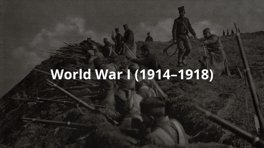
During World War I, X-rays became lifesavers on the battlefield. Doctors used them to find bullets or broken bones in injured soldiers. This was huge because it meant surgeons could see exactly where the problem was before operating.
Field hospitals had portable X-ray machines, which were a big deal back then. Soldiers who might’ve been too hurt to survive without quick help got the care they needed faster. It wasn’t perfect, but it saved lives.
After the war, people realized how important X-rays were—not just for soldiers but for everyone. Hospitals started investing more in X-ray machines, and doctors got better training in how to use them. It was the start of radiology becoming a big part of medicine, both for emergencies and everyday care.
1920s: Better X-Ray Pictures and New Groups for Radiologists
In the 1920s, a man named André Bocage came up with a way to make X-rays better. His moving X-ray techniques gave doctors clearer pictures of bones and organs. This was a big deal because it helped doctors see what was going on inside the body more easily.
At the same time, groups like the Radiological Society of North America (RSNA) and the American Registry of Radiologic Technologists (ARRT) were formed. These groups focused on teaching people about radiology and setting rules so everyone could do their job better.
The moving X-rays were special because they could take pictures while a person was moving. This helped doctors see how things like the heart or lungs worked while they were in action. And with the new organizations, radiologists started sharing ideas and learning from each other. This teamwork made care for patients a lot better.
1940s–1950s: New Ways to See Inside the Body
In the 1940s and 1950s, scientists made even more progress. In 1946, they discovered something called nuclear magnetic resonance (NMR). This discovery led to new ways to take pictures of the body.
Then, in the late 1950s, a doctor named Ian Donald introduced ultrasound. Ultrasound used sound waves to make pictures of the inside of the body. It became especially popular for checking on babies during pregnancy.
Another big thing during this time was nuclear medicine. This method used tiny amounts of radioactive material to help doctors find and treat diseases. It let them see how organs were working and catch problems early.
Both nuclear medicine and ultrasound were great because they didn’t require surgery. That meant patients didn’t have to go through as much pain or risk. These tools made it easier and safer for doctors to help people.
1970s: CT and MRI Innovations
The 1970s brought big changes to how doctors looked inside the human body. In 1972, a man named Godfrey Hounsfield invented the CT scanner. This machine could take pictures of the body in thin slices, almost like looking at the layers of a cake. Just a few years later, in 1977, Raymond Damadian made the first MRI scan happen. Both of these inventions changed the way doctors figured out what was wrong with their patients.
CT scans were a game-changer because they showed details that regular X-rays couldn’t. They helped doctors find things like tumors or hidden injuries. MRIs, on the other hand, used magnets and radio waves to take super-clear pictures of soft tissues, like the brain or muscles. This made it easier to spot problems in places that were hard to see before. Together, these two tools made diagnosing illnesses faster and more accurate, and they became must-haves in hospitals everywhere.
Modern Era and Future Directions
Fast forward to today, and radiology has only gotten better. Digital imaging tools, like something called PACS (Picture Archiving and Communication Systems), let doctors save and share pictures easily. They don’t have to deal with stacks of film anymore. And now, artificial intelligence (AI) is stepping in to help. AI can look at images and point out things that might take a human longer to notice.
With digital imaging, doctors can pull up a patient’s old scans in seconds and compare them to new ones. This helps them see if a condition is getting better or worse. AI tools are still being worked on, but they’re already helping radiologists find things like tiny tumors or other problems faster. As technology keeps improving, radiology is expected to do even more for doctors and patients. It’s making healthcare smarter and giving doctors better ways to understand diseases without needing to cut anyone open.
If there’s one takeaway, it’s this: radiology is always moving forward. And that’s good news for everyone.
FAQ
How did Marie Curie and Henri Becquerel’s discoveries shape early radiation science?
Marie Curie and Henri Becquerel laid the groundwork for radiation science. Becquerel discovered radioactive elements in 1896 when he found uranium salts could expose photographic plates without light. Marie Curie took this further by isolating radium and polonium, leading to better understanding of radiation types. Their work helped create the foundation for using ionizing radiation in medicine.
What role did Thomas Edison and early innovators play in developing ray tubes for medical use?
Thomas Edison and his team experimented with cathode ray tubes and fluorescent screens in the 1890s. They created images using ray beams, leading to better ray tubes for medical use. This work helped establish early imaging techniques that doctors could use in real time to see inside patients.
How did computed tomography (CT scanning) revolutionize diagnostic imaging?
CT imaging transformed healthcare by creating detailed cross-sectional images of the body. Unlike regular ray images, CT scans use electron beams and high energy radiation to produce 3D pictures. This imaging modality helps doctors spot problems that might be missed on regular x-rays, improving diagnostic accuracy and patient care.
What advances led to modern diagnostic radiology becoming a distinct medical specialty?
Diagnostic radiology evolved through improved imaging techniques and imaging informatics. Medical schools in the United States developed specific training programs, while the American College helped establish standards. The field grew to include specialized areas like pediatric radiology and emergency medicine.
How has digital technology changed clinical radiology and image quality?
Digital radiography replaced photographic film, making radiology departments more efficient. Modern imaging modalities provide better image quality and use less radiation. This shift helped make minimally invasive procedures possible and improved how doctors diagnose health problems.
What safety measures protect patients during diagnostic imaging?
Radiation safety and radiation protection are crucial in healthcare. Doctors carefully control radiation exposure using modern imaging techniques. They might use contrast agents when needed for better pictures while following strict safety rules. The field keeps improving ways to get good diagnostic images while keeping radiation doses low.
How do different imaging modalities work together in modern medicine?
Today’s diagnostic imaging uses many tools like CT scans, MRI scans, and ultrasound technology. Each type of radiation and imaging technique has specific uses in medicine. Industrial radiography has also influenced medical imaging development. This mix of technologies helps doctors provide better patient care.
Conclusion
Radiology’s history stretches from Röntgen’s first X-ray in 1895 to today’s advanced imaging like MRIs and CT scans. Each step has changed how doctors see inside the human body, making diagnosis faster and treatment more precise. It’s wild to think how far we’ve come—from blurry shadows on glass plates to detailed 3D images. As technology keeps evolving, radiology will keep shaping medicine, helping doctors figure out what’s wrong and how to fix it.
References
- https://4rai.com/2021/04/30/history-of-radiology/
- https://www.arrt.org/pages/arrt-timeline