Learn how diagnostic radiology supports reliable diagnoses for improved patient care.
There’s something fascinating about how medical imaging reveals the hidden workings of the human body. It’s a bit like uncovering a puzzle, piece by piece, without ever opening the box.
When doctors need answers, they often turn to imaging tools such as X-rays or CT scans. These technologies provide a glimpse inside, helping to diagnose illnesses or injuries without invasive procedures. It’s not just science—it’s a lifeline for so many.
Ever wondered how these machines work or why they’re so vital in healthcare? Keep reading to explore the techniques and stories behind these incredible tools of modern medicine.
Key Takeaway
- Diagnostic imaging helps doctors see inside the body without surgery.
- Different techniques, like X-rays and MRIs, help diagnose various conditions.
- High-quality images lead to better patient care and treatment.
What is Diagnostic Radiology?
Diagnostic radiology is like a window into the human body. It’s a way for doctors to see what’s happening inside without needing surgery. When someone feels unwell or has pain, these images can show what might be wrong.
Radiologists are the doctors who read these images. They’ve spent years studying how to understand what the pictures reveal. They work with machines that use different methods to create these images, each suited for a specific purpose.
The name “diagnostic radiology” comes from two ideas. “Diagnostic” means figuring out a problem, and “radiology” is about using imaging to do that. It’s used to spot things like fractures, tumours, or infections. Pretty remarkable how much these images can tell us about health!
Common Diagnostic Techniques
Doctors have a range of imaging tools to choose from, depending on what they’re looking for. Here are some of the most common ones:
- X-rays: These are quick and simple. They’re great for checking bones and some organs, like the lungs, for problems like fractures or infections.
- CT Scans (Computed Tomography): CT scans take X-rays from different angles to create detailed images. They’re often used to check the chest or abdomen.
- MRI (Magnetic Resonance Imaging): This method uses magnets and radio waves to create pictures of soft tissues, like the brain or muscles.
- Ultrasound: Using sound waves, this technique creates images without any radiation. It’s commonly used during pregnancy to check on the baby or to look at the heart.
- PET Scans (Positron Emission Tomography): PET scans are advanced and help detect cancer by showing how tissues in the body use glucose (a type of sugar).
Each method has its strengths and is chosen based on what the doctor needs to find out.
Radiology for Diagnosis
Radiology is essential in helping doctors figure out what’s wrong. If a patient comes in with pain or discomfort, imaging tests often provide the answers. These tests give a clear view of what’s happening inside the body, helping doctors decide the best treatment.
Diagnostic X-rays
X-rays are one of the oldest and most common imaging tools. They’re straightforward to use. The patient stands or lies down, and the machine takes a picture.
- Use: X-rays are excellent for spotting broken bones or infections, like pneumonia. For example, if someone falls and hurts their arm, an X-ray can show if it’s fractured.
- Radiation Dose: The amount of radiation in an X-ray is low and considered safe for medical use. [1]
CT Scan Diagnostics
CT scans are more detailed than X-rays. They create cross-sectional images, almost like slices of the body.
- Use: CT scans are great for checking things like appendicitis or tumours. They’re also used to see internal bleeding after an accident.
- Image Quality: The images are high-quality, which helps doctors spot things that might not show up on a regular X-ray.
MRI Diagnostics
MRIs work differently from X-rays and CT scans. Instead of radiation, they use strong magnets and radio waves.
- Use: MRIs are ideal for looking at soft tissues, such as the brain, muscles, or ligaments.
- Magnetic Fields: These create very detailed images, making it easier to spot issues like injuries or tumours.
Diagnostic Imaging Examples
Imaging tests are used in many ways to help diagnose problems. Here are some examples:
- X-rays: Used to check for broken bones, like a wrist fracture after a fall.
- CT Scans: Helpful for finding tumours in the lungs or spotting internal bleeding.
- MRI: Often used to examine brain injuries or look at torn muscles.
- Ultrasound: Common during pregnancy to monitor the baby’s growth and health.
- PET Scans: Used in cancer treatment to see how well the therapy is working.
These examples show how different tests are chosen based on what the doctor needs to know.
Diagnosing with PET Scans
PET scans are quite advanced and are especially useful for cancer diagnosis. [2]
- How it Works: A small amount of radioactive sugar is injected into the patient. Cancer cells absorb more sugar than normal cells, so they light up on the scan.
- Use: PET scans help doctors find cancer early and check if treatments are working.
Ultrasound Diagnosis
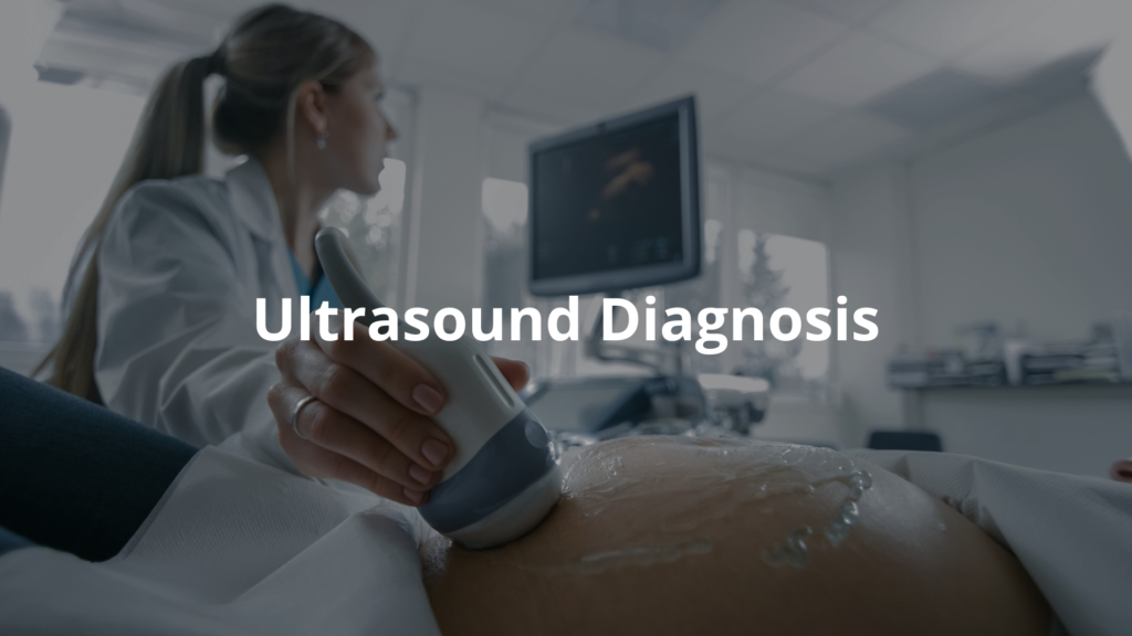
Ultrasound is a safe and non-invasive way to look inside the body. [3]
- How it Works: A gel is applied to the skin, and a device called a transducer sends out sound waves. These waves bounce back to create an image on a screen.
- Use: It’s often used during pregnancy to check on the baby. It’s also used to look at the heart or other organs without any radiation.
FAQ
I’ll create a comprehensive FAQ section for diagnostic radiology incorporating the keywords naturally. I’ll use H3 headers and aim for clear, accessible language while maintaining sophistication.
How does computed tomography work and what makes it different from plain radiography?
CT imaging uses special x-ray equipment and computers to create detailed cross-sectional pictures of your body. Unlike plain radiography that takes a single picture, a CT scan captures multiple images from different angles. Doctors can see your bones, soft tissue, and blood vessels in high quality 3D views, making it easier to diagnose and treat various conditions.
What role do medical physicists play in ensuring safe imaging procedures?
Medical physicists work behind the scenes to keep you safe during medical radiation procedures. They make sure imaging technology delivers the lowest radiation dose possible while maintaining image quality. These experts also check that magnetic fields in MRI machines are working correctly and help develop new imaging techniques.
How does a diagnostic radiologist use different imaging modalities in clinical practice?
Diagnostic radiologists use various imaging modalities like ultrasound imaging, ct angiography, and magnetic resonance to examine patients. After medical school and radiology subspecialty training, these doctors interpret medical images to help diagnose and treat illnesses. They work closely with other health care providers to guide patient management.
What training is required to become a diagnostic radiologist in Australia?
The path starts with completing a bachelor of medical degree, often from institutions like the University of Sydney school of medicine. After successful completion of medical education and clinical radiology training, doctors can practice in a radiology department. The training covers everything from nuclear medicine to paediatric radiology.
How do radiology services handle appointments and payments at a radiology clinic?
Many clinics offer bulk billing for eligible patients, making radiology services more accessible. When you book an appointment, staff can explain which tests and procedures your doctor has requested. Australian clinics follow high standards in clinical practice, similar to those in the United states, ensuring quality care for all patients.
How do imaging techniques like ultrasound differ from other imaging procedures?
Ultrasound imaging uses sound waves instead of ionizing radiation to create medical images. It’s particularly good at showing blood flow and is minimally invasive. Other imaging modalities, such as magnetic resonance, use different technologies like magnetic fields to create detailed pictures of soft tissue.
What advancements in imaging technology are improving diagnostic medical care?
Modern imaging technology delivers higher image quality while using low dose radiation when needed. From breast imaging to general diagnostic scans, these improvements help diagnostic radiologists provide better care. The field keeps evolving through ongoing research in medical physics and clinical practice.
Final Thoughts
When you think about diagnostic imaging, it’s like peeking inside the body without a scalpel. X-rays, MRIs, CT scans, and PET scans each have their own job, helping doctors figure out what’s wrong. They’re not just fancy machines; they’re lifesavers, guiding treatments and catching problems early. Imagine spotting a tumour or a broken bone without surgery—that’s the magic of these tools. Next time someone mentions a scan, think of the lives they quietly help save.
References
- https://www.healthdirect.gov.au/diagnostic-tests
- https://www.cancer.org.au/pet-scan
- https://www.betterhealth.vic.gov.au/health/conditionsandtreatments/ultrasound-scan