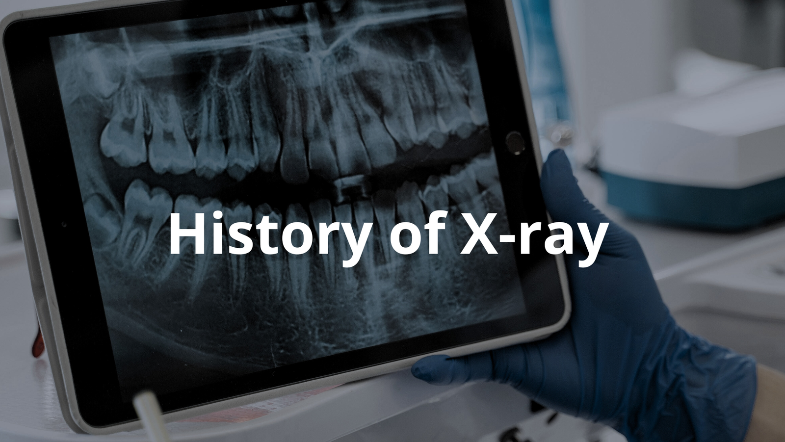
Learn how X-rays came to be and their impact on medicine. Discover their journey from discovery to modern use.
X-rays have a fascinating history. They were discovered in 1895 by a curious German physicist named Wilhelm Conrad Röntgen. While he was working with something called cathode rays, he stumbled upon a new kind of ray. This ray could pass through things, and it was invisible! It helped doctors see inside the human body without needing to cut it open.
That’s amazing, right? Keep reading to learn more about the history of X-rays, their development, and how they help us today(1)!
Key Takeaways:
- X-rays were discovered in 1895 by Wilhelm Conrad Röntgen while he was experimenting with cathode rays.
- The first X-ray image was of Röntgen’s wife’s hand, showing her bones and a ring she wore.
- Today, X-rays are vital in medicine for diagnosing many conditions, including broken bones and soft tissue injuries.
The Discovery of X-Rays
The discovery of X-rays is quite a remarkable story. Back in 1895, a German physicist named Wilhelm Conrad Röntgen was doing experiments at the University of Würzburg. He was using a cathode ray tube, which is a device that creates images using rays. Suddenly, he noticed something out of the ordinary.
A screen nearby was glowing, even though it was covered. It wasn’t getting any light from the outside! This made him curious. He thought, “What could be causing this glow?”
Röntgen didn’t just stop there. He kept digging, thinking like a detective. He soon realised that this was a new type of radiation that he couldn’t see. He decided to call it “X-rays” because the “X” meant unknown. Picture him in a dimly lit room, eyes wide with wonder, as he thought about this mysterious ray that could penetrate objects. It was a huge breakthrough.
To prove his discovery, Röntgen took the first-ever X-ray photograph. He aimed the rays at his wife Anna Bertha’s hand, and what did he see? Her bones, clear as day, along with her wedding ring! This was astonishing, not just for him but for everyone. People were amazed that they could see inside a living person without needing to make a single cut.
Can you imagine how thrilling that must’ve felt? Röntgen’s work opened up a whole new world of possibilities in medicine.
Early Applications of X-Rays
Just a year later, in 1896, doctors began to see the value of X-rays. They quickly figured out that X-rays could show broken bones. Imagine being a doctor back then, filled with excitement over this new tool! They could now diagnose fractures without needing to guess or operate. It must’ve felt like magic! But there was a catch.
At that point, people still didn’t know about the possible risks of X-ray exposure. They were so eager to use this technology that they overlooked the dangers.
At the time, radiation wasn’t well understood. Doctors probably thought it was just a helpful light that could help them see better. Many believed it was harmless. Sadly, some doctors didn’t take proper precautions, which could lead to overexposure. They didn’t realise that too much radiation could be harmful. Despite this, X-rays were quickly used to help wounded soldiers during wars and to diagnose various illnesses(2).
The excitement of this new medical tool spread like wildfire. Hospitals started investing in X-ray machines. They wanted to be on the cutting edge of medical technology. The ability to see inside the human body was a game changer. It was a time of great hope and innovation.
X-rays allowed doctors to identify issues that were previously invisible, like fractures or even signs of diseases like tuberculosis. This was a huge step forward in medical care and changed how doctors treated patients forever.
The Invention of the X-Ray Machine
In the same year that Röntgen made his discovery, a British physicist named Sir William Crookes invented the first X-ray machine. This machine made it much easier for doctors to create X-ray images. Before that, it was all a bit of a guessing game. Now, with Crookes’ invention, they could see much more clearly. Imagine a room filled with excitement as doctors gathered around this new machine, eager to see what it could do!
With the new X-ray machine, doctors could see fractures in bones and other medical conditions much more clearly than before. This new tool became a common sight in hospitals. It helped doctors to diagnose diseases like tuberculosis, which was a serious illness at the time. The ability to look inside the body without surgery was revolutionary.
The X-ray machine used high voltage to produce X-rays, which would then pass through the body and hit a photographic plate. This created an image of the internal structures. It was a bit like developing a photograph, but instead of capturing a moment in time, it captured the hidden parts of a person’s body.
This innovation changed the face of medicine. Doctors could now see what was going on inside their patients without having to perform risky surgeries. It made diagnosing conditions faster and more accurate. As X-ray technology became widespread, it helped save countless lives. It was a time of hope, discovery, and the beginning of a new era in medical practices. And who knows? Perhaps if you asked Röntgen, he might say he was just a curious man looking for answers, but he ended up changing medicine forever.
Advancements in X-Ray Technology
The journey of X-ray technology didn’t stop there. Throughout the 20th century, many improvements were made.
- In 1918, George Eastman introduced a special film for taking X-rays. This film replaced the older glass plates, making X-ray images better and clearer.
- By 1946, scientists discovered nuclear magnetic resonance (NMR), which helped develop better imaging technologies.
- Then in the 1970s, something called Computed Tomography (CT) scans came along. These scans changed everything by allowing doctors to see detailed images of the body in slices, making it easier to diagnose complex problems.
- Finally, in the 1980s, Magnetic Resonance Imaging (MRI) was introduced. This technology used magnets and radio waves to create images without any radiation exposure, continuing to improve how we see inside the body.
The Digital Revolution in Radiography

As time marched on, X-ray technology transformed dramatically. Traditional film-based X-rays gave way to digital radiography. This shift changed everything for doctors and patients alike. Now, instead of waiting for film to develop, images were created electronically in mere moments. It’s like stepping from an old black and white photo into a vibrant colour picture.
The benefits of digital radiography are numerous:
- Less Radiation Exposure: Patients receive much lower doses of radiation. This is especially important for those needing multiple scans.
- Faster Imaging: Doctors can take images quickly, which means less time waiting for results. Imagine a doctor getting an answer in minutes instead of hours!
- Improved Image Quality: The quality of images is significantly better. They can be adjusted easily, allowing doctors to zoom in on specific areas for a clearer view.
Digital X-rays have truly revolutionised the way medical imaging is done. It’s exciting to think about how this technology continues to make patient care faster and safer.
Recent Innovations in X-Ray Technology
Today, X-ray technology keeps pushing boundaries with new innovations. Each advancement brings fresh possibilities to improve diagnostics. For instance, 3D imaging allows doctors to see inside the body in three dimensions. This helps them understand complex conditions more clearly. With 3D imaging, it’s like viewing a sculpture instead of a flat painting; every detail can be examined from different angles.
Another recent advancement is Dual-Energy X-ray Absorptiometry (DEXA). This technology is mainly used to measure bone density. Doctors can quickly determine if someone has weak bones, which is crucial for preventing fractures.
Additionally, Hybrid Imaging Systems combine different techniques, like positron emission tomography (PET) and CT scans. This offers a complete picture, helping doctors make more informed decisions about treatment.
- 3D Imaging: Offers a detailed view of the body’s internal structures.
- DEXA: Measures bone density to assess fracture risk.
- Hybrid Systems: Combine imaging techniques for better diagnoses.
With these advancements, it’s clear that the future of X-ray technology is bright, and it’s exciting to think about the continued improvements in medical care!
Conclusion
In wrapping up, the history of X-rays is really fascinating! From their accidental discovery by Wilhelm Conrad Röntgen to their essential role in medicine today, X-rays have changed how we diagnose and treat many conditions. They’ve made it possible for doctors to see inside the human body safely and effectively. And as technology continues to improve, we can only imagine the exciting possibilities ahead in medical imaging!
So, next time you see an X-ray image, remember the incredible journey it took to get there. The next time you visit a doctor, you might just encounter this amazing technology that has been helping people for over a century!
FAQ
How did Wilhelm Conrad Roentgen discover X-rays using ray tubes and fluorescent screens?
In 1895, German physicist Wilhelm Conrad Roentgen noticed that a barium platinocyanide screen glowed while experimenting with a crookes cathode ray tube. He found that unknown type of rays could pass through black cardboard, creating visible light on the screen. He later took the first ray image of his wife’s hand, showing her wedding ring and bones.
What were the early years of X-ray imaging like in the medical field?
During the early years, ray machines became popular in medical applications. Doctors used ray imaging to examine wounded soldiers and diagnose conditions. The technology spread quickly through the United States and Europe, though the harmful effects of radiation exposure weren’t fully understood yet.
How did scientists like Marie Curie and Thomas Edison contribute to ray technology?
Marie Curie furthered our understanding of radiation while Thomas Edison experimented with ray fluorescence. Both made significant contributions to basic science around electromagnetic radiation. Edison later stepped back from ray experiments after his assistant developed health problems from radiation exposure.
How do modern medical imaging technologies like CT scans work with human tissue?
CT scans use ray sources to create detailed images of soft tissues and blood vessels in the human body. This form of electromagnetic radiation passes through living tissue at different rates, allowing doctors to see inside the body. The technology combines multiple ray images to create computed tomography scans.
What safety measures evolved to protect against ray exposure in medical and industrial settings?
The medical society developed radiation protection standards after learning about the biological effects of ionizing radiation. Today, health physics guidelines control radiation dose levels. Medical imaging uses the lowest possible radiation while maintaining quality control in diagnostic procedures.
How did X-rays transform beyond medical applications to other scientific fields?
X-rays found uses in industrial radiography and ray diffraction studies. Scientists use high energy and low energy rays across the electromagnetic spectrum. Space telescopes and ray observatories study gamma rays from space, while airport security uses ray technology for screening.
What earned Wilhelm Conrad Roentgen the first Nobel Prize in Physics?
Roentgen received the nobel prize in physics in 1901 for his discovery of rays that could pass through solid objects. His work at the University led to revolutionary applications in physiology or medicine, fundamentally changing diagnostic capabilities in the life sciences.
References
- http://starlingdiagnostics.com/the-history-of-x-rays-from-discovery-to-modern-medical-imaging/
- https://en.wikipedia.org/wiki/X-ray