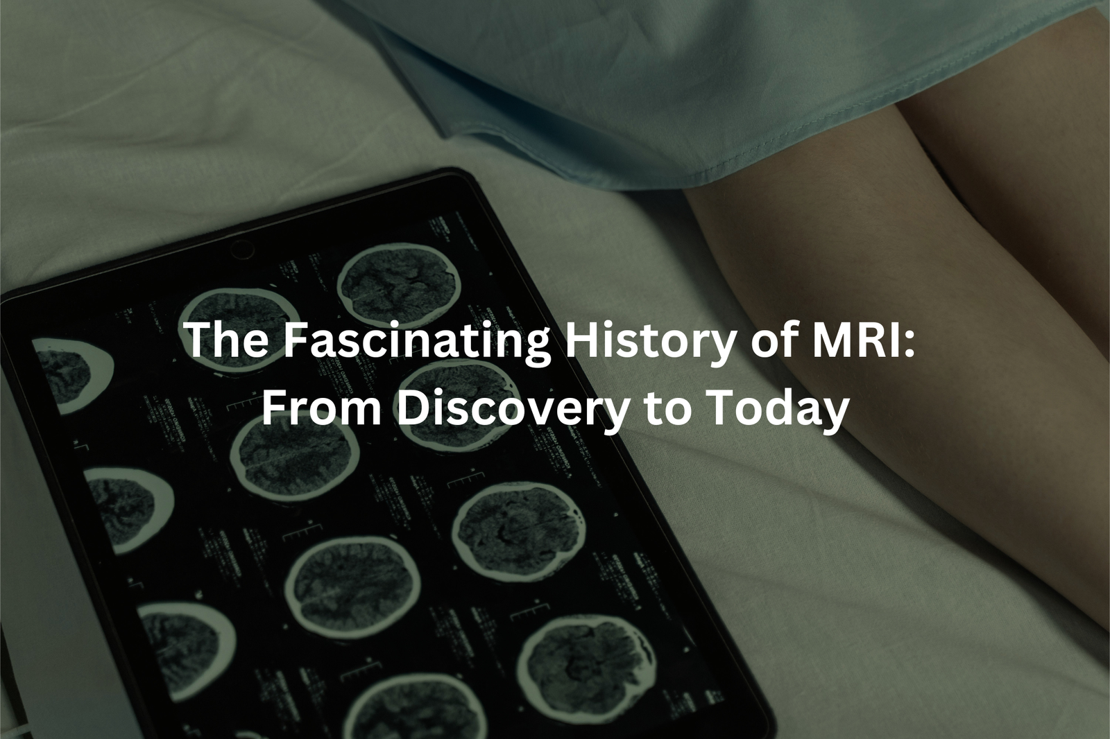
MRI history explained: How this amazing machine became a key tool for diagnosing and saving lives.
The first time I saw an MRI machine, it was impressive. It really looked like a giant doughnut with a tunnel. This machine, known as Magnetic Resonance Imaging, or MRI, can take pictures of the inside of a body without any cuts. MRIs were invented in the late 1970s and changed medicine forever.
Doctors use MRIs to find out what’s wrong inside people’s bodies. They can see everything from a brain to bones in great detail. If you’re curious to learn more about how MRIs work and why they’re so helpful, keep reading for all the details!
Key Takeaways
- MRI was invented in the late 1970s and early 1980s.
- It helps doctors see soft tissues like the brain and muscles without using radiation.
- New technology, like AI, is making MRI even better every day.
The Early Days of MRI in Australia
Back in the early days, MRI machines in Australia were huge and slow. Patients had to lie still for over an hour just to get a clear image. Imagine trying to keep a kid still for that long! It must’ve been a real challenge. But over time, Aussie scientists and doctors worked hard to improve things.
They came up with new scanning methods that made the process quicker. These days, many MRI scans take only 15 to 30 minutes, sometimes even less. My friend had one recently, and he was done in about 12 minutes. He said it wasn’t nearly as bad as he expected. That’s a big deal, especially for kids or people who get nervous in tight spaces.
The images are also much sharper now. Doctors can see tiny details in organs, muscles, or even the brain. This helps them figure out what’s wrong and decide on the best treatment faster.
MRI technology has come a long way since it first arrived in Australia in the 1980s. Aussie researchers have played a big role in making scans faster and clearer(1). It’s made a huge difference in healthcare, helping doctors catch problems early and treat patients better. If you ever need an MRI, you’ll probably appreciate how much easier it is now!
How MRI Works: The Science Behind the Machine
Source: Translational Medicine, Monash University.
People often ask, how does this big doughnut-shaped machine called an MRI work? It’s actually pretty fascinating. When someone lies inside, the machine uses a powerful magnet and radio waves to create detailed pictures of the inside of their body. Here’s how: the magnet aligns the water molecules in your body (and yes, most of your body is water—about 60%!), then the radio waves bounce off those molecules. These bounces send back signals, which the machine turns into images.
What’s really cool is that these images are super clear and can show things like muscles, organs, and even tiny problems that might be hiding. Unlike X-rays or CT scans, MRIs don’t use radiation, which makes them safer, especially for kids or pregnant women. Doctors often choose MRIs when they need a closer look without the risks of radiation.
It’s kind of amazing, isn’t it? A machine like this can help doctors find problems early, which means people can get the right treatment sooner. If you ever need an MRI, just remember—it’s all about magnets, water, and some clever science.
The Importance of Early Detection
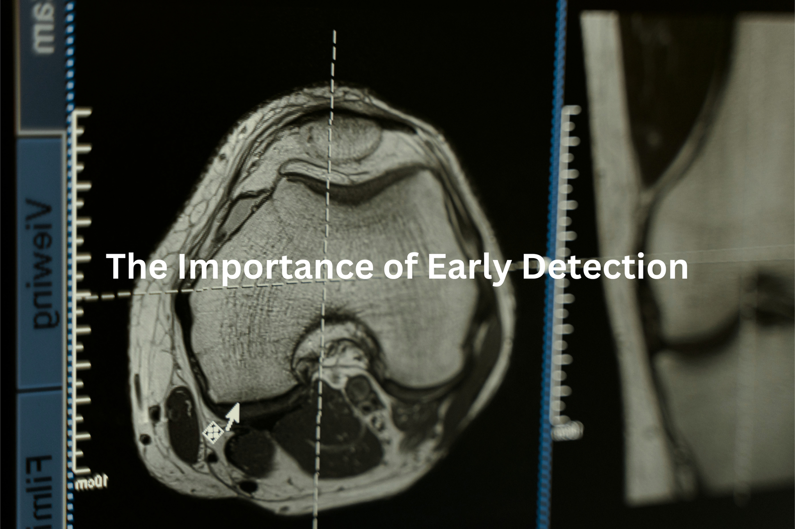
One of the most useful things about MRI is how it can spot health problems early on. For example, if a doctor thinks someone might have cancer, an MRI can show if there are any cancer cells hiding in the body. Catching these issues early often means better chances for treatment to work. The quicker someone starts getting help, the better their chances of recovering.
Imagine this: A patient goes in for an MRI, and the scan shows a tumour that no one knew was there. Because it’s found early, the doctors can start treatment right away. This could mean the difference between life and death. It’s why hospitals here in Australia and all over the world use MRI machines so often. They’re like a secret weapon for doctors, helping them see inside the body without surgery.
So, if you ever need an MRI, remember—it’s not just a machine. It’s a lifesaver.
AI and MRI: A New Partnership
AI and MRI technology working together is pretty fascinating, isn’t it? Think about it—AI (artificial intelligence) is now helping doctors read MRI scans faster and more precisely. It’s like giving radiologists (the specialists who interpret these scans) a super-smart helper that never gets tired or distracted.
Here’s what happens: the AI scans through MRI images and flags anything unusual, like areas that might need extra attention(2). This doesn’t replace the doctor’s expertise, of course, but it speeds things up and makes sure nothing important gets missed. For example, if there’s a tiny tumour hiding in the scan, AI might spot it quicker than a human eye alone. That could mean earlier treatment, which is a big deal.
I reckon this combo of AI and MRI is just the start. As AI keeps improving, we might see even smarter MRI machines that can do more than we ever thought possible. It’s exciting to think about where this could lead.
Training and Safety
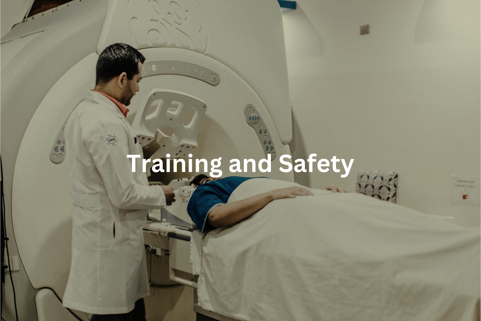
MRI machines are fascinating, aren’t they? They use powerful magnets to take detailed pictures of the inside of your body, but with that power comes the need for strict safety rules. In Australia, the Royal Australian and New Zealand College of Radiologists (RANZCR) works hard to make sure doctors and technicians are properly trained to use these machines safely.
Before a scan, the technician will ask the patient a lot of questions. They’re checking for things like metal implants, pacemakers, or even metal fragments from old injuries. Why? Because the MRI’s magnet is so strong it can interfere with these devices or even move metal objects inside the body. That’s why it’s so important to double-check.
This careful process helps patients feel safe. Knowing there’s a team of professionals following strict guidelines gives peace of mind. If you ever need an MRI, remember to tell the technician everything about your medical history. It’s all part of keeping you safe.
The Future of MRI
MRI machines are like magic windows into the human body. They don’t use X-rays or anything harmful, just strong magnets and radio waves to take pictures of what’s inside us. But what’s really fascinating is how they’re getting even better. There’s something called functional MRI (fMRI), which doesn’t just take pictures—it watches the brain in action(3). Imagine seeing which part of your brain lights up when you’re solving a puzzle or feeling happy. That’s what fMRI can do.
Doctors and scientists use this to study things like strokes, Alzheimer’s, or even depression. It’s helping them understand how the brain works and what goes wrong when someone’s sick.
And the machines themselves? They’re improving too. Researchers are working on making them faster and sharper, so doctors can get clearer images in less time. That means quicker diagnoses and better treatment plans.
If you ever get the chance, ask your doctor about how MRI works. It’s a bit like science fiction, but it’s real—and it’s saving lives.
Conclusion
MRI, or Magnetic Resonance Imaging, has a fascinating history that started in the late 1970s. It’s about more than just taking clear pictures of inside our bodies; MRIs help doctors find problems early and treat illnesses better, saving lives. With ongoing improvements, this technology continues to be vital in healthcare. So next time you hear about an MRI, think of its remarkable journey and how it plays a big part in keeping us healthy.
FAQ
What is a CT Scan and how does it differ from MRI?
CT scans use X-rays to create detailed images of the body, while MRI (Magnetic Resonance Imaging) uses powerful magnets and radio waves to generate images. MRI can provide better contrast for soft tissues like the brain, heart and other organs compared to CT scans.
What is Body MRI and how is it used?
Body MRI scans the entire body to examine organs, muscles, tendons and other structures. It is commonly used to diagnose and monitor diseases, injuries or other conditions affecting the abdomen, pelvis, chest or other body regions. Body MRI provides high-quality images without using ionizing radiation like X-rays.
What are the benefits of Open MRI compared to traditional MRI?
Open MRI scanners have a more open design that can accommodate larger patients and those with claustrophobia. They also allow for easier access during the scan. However, open MRI systems typically have lower magnetic field strengths, which can result in lower image quality compared to closed, high-field MRI machines.
How has Fast MRI technology improved imaging?
Fast MRI techniques like echo planar imaging have dramatically reduced scan times, from over an hour to just a few minutes. This allows imaging of moving organs like the heart and blood vessels in real-time. Faster scans also improve patient comfort and reduce the need for sedation.
How has MRI data analysis and visualization advanced over time?
Sophisticated MRI data processing and visualization software have revolutionized how MRI images are analyzed. Radiologists can now extract detailed quantitative information about brain structure, blood flow, and other physiological parameters. Advances in 3D rendering also allow for more intuitive interpretations of complex MRI data.
Who were some of the key pioneers in the development of MRI technology?
Nobel Prize winners Paul Lauterbur and Felix Bloch made crucial early contributions to the fundamental principles of MRI. Le Bihan’s work on diffusion MRI further expanded the capabilities of the technology in the 1980s and 1990s. These and other scientists laid the groundwork for modern clinical and research MRI systems.
How has MRI use evolved in medical diagnostics over time?
MRI has become an essential diagnostic tool across many medical fields, from neurology and cardiology to orthopaedics. Advances in high-field, fast and hybrid MRI systems allow clinicians to image an ever-wider range of body tissues and physiological processes with exceptional detail and clarity. MRI is now a routine part of clinical care in the 21st century.
References
- https://onlinelibrary.wiley.com/doi/pdf/10.5694/j.1326-5377.1986.tb112392.x
- https://www.canhealth.com/2024/01/31/australia-deploys-ai-in-radiology-across-the-country/
- https://pmc.ncbi.nlm.nih.gov/articles/PMC9529952/