
Learn how common imaging techniques help doctors spot health issues like broken bones or cancer. Discover how they work to keep you healthy.
When we visit the doctor, they often want to see inside our bodies. Common imaging techniques help with this. They include X-rays, which show broken bones, CT scans that give detailed pictures of organs, and MRIs that look at soft tissues like the brain.
Each test works differently but they all help doctors find problems like injuries or even diseases. These tools are very important for keeping us healthy. If you want to learn more about how these techniques work and why they matter, keep reading!
Key Takeaway
- Imaging techniques help doctors see inside our bodies.
- Different tests are used for different parts of the body.
- These tests can help find problems early and make better treatment choices.
X-rays
It’s kind of amazing how x-rays work, isn’t it? They let doctors see inside you without needing to cut you open(1). X-rays are used everywhere—hospitals, clinics, even at the dentist. They’re quick, painless, and really helpful.
Here’s how they work: the x-ray machine sends out invisible rays (they’re like light but you can’t see them). These rays pass through soft stuff like skin and muscles easily, but bones are denser and block more rays. That’s why bones look bright white on the images.
Doctors use x-rays for all sorts of things:
- Finding broken bones, like a fractured arm.
- Checking lungs for problems like pneumonia.
- Spotting joint issues or even swallowed objects.
A mate of mine had one done after a bad cough. It showed fluid in his lungs, and he got treated right away. If you ever need one, don’t stress. It’s quick, safe, and gives doctors the info they need.
CT Scans
CT scans are like looking inside a book without opening it. Instead of one picture like an x-ray, they take heaps—hundreds, actually. These images, taken from different angles, are pieced together by a computer to create detailed slices of your body.
Doctors use CT scans for lots of reasons:
- Finding internal bleeding after accidents.
- Spotting tumours in organs like kidneys or lungs.
- Checking for hidden fractures in bones.
- Monitoring conditions like lung or heart disease.
Sometimes, they use a dye called contrast material (injected or swallowed) to make organs and blood vessels pop in the images. A mate of mine had one after a footy injury—thought it was a sprain, but the scan showed a hidden fracture.
It’s quick, about 20–30 minutes, and painless. Yes, there’s some radiation, but it’s managed carefully. If you’re having one, ask the radiographer a question or two—it helps to know what’s coming.
MRI Scans
The MRI machine hums softly, like it’s gearing up for something important. It’s not just a big scanner—it’s a tool that shows doctors detailed images of soft tissues, nerves, and the brain without using x-rays. Instead, it relies on strong magnets and radio waves to create pictures.
An MRI scan can take 30 to 60 minutes, and you’ll lie inside a snug tube. It’s noisy, with clanging sounds (some people find it calming, others not so much). Doctors often use MRIs to find problems like:
- Persistent headaches or back pain.
- Ligament injuries or tumours.
- Conditions like multiple sclerosis.
I remember my sister’s MRI after a car accident. It showed a slipped disc that x-rays missed. Before the scan, they’ll check if you’ve got metal on you—magnets and metal don’t mix. Wear comfy clothes, and if you’re nervous, ask for music or tips to relax. It helps.
PET Scans
The room feels still, almost like it’s holding its breath. A PET scan (positron emission tomography) isn’t like a regular X-ray or MRI. It doesn’t just show what things look like—it shows how they’re working inside.
It starts with a small injection of a radioactive tracer (don’t worry, it’s safe in tiny amounts)(2). This tracer moves through your blood and collects in areas with high activity, like fast-growing cancer cells. On the scan, these areas light up.
Doctors use PET scans for many reasons:
- To find cancer or see if treatment like chemo is working.
- To check for heart problems or brain disorders.
- To plan surgeries with more accuracy.
My aunt had one when her doctor found a lung issue. It showed exactly where the problem was, helping her team plan treatment. If you ever need one, wear comfy clothes, skip big meals, and know it’s more than a picture—it’s a guide.
Ultrasound Imaging
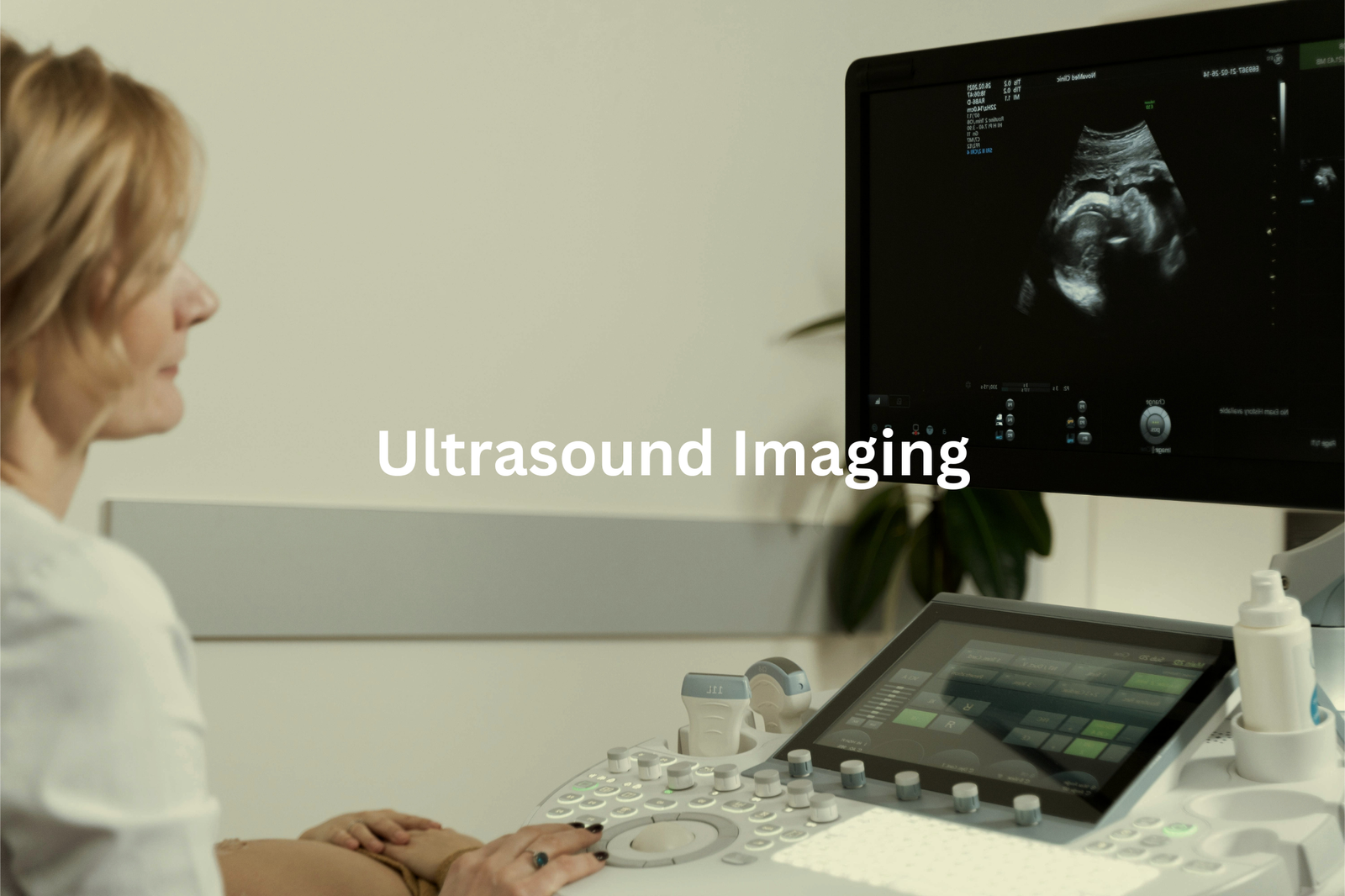
The steady hum of the machine fills the room, broken by the soft clicking of keys. Ultrasounds feel a bit like magic, don’t they? No needles, no radiation—just sound waves bouncing around to create pictures. A safe, simple way to look inside the body.
Here’s how it works:
- A transducer sends out high-frequency sound waves.
- These waves bounce off tissues like echoes.
- A computer turns those echoes into images.
Most people think of pregnancy scans—tiny feet, flickering heartbeats—but ultrasounds do more. They check hearts, kidneys, blood flow, and even spot gallstones or thyroid issues. All without harmful radiation.
I remember my cousin showing me her baby’s ultrasound. She kept pointing at the little nose, though it looked like a blob to me. Still, incredible. Appointments are quick, about 30 minutes. Drink water beforehand for clear images. If you’re nervous, ask if ultrasound’s an option—it’s painless and fascinating.
Nuclear Medicine
There’s something a bit strange about injecting something radioactive into your body. Sounds like a sci-fi movie, right? But in nuclear medicine, it’s just another day at the office—and it’s actually really useful(3).
Here’s the gist:
- A tiny amount of radioactive material (called a tracer) is injected.
- It travels through your bloodstream to specific areas.
- Gamma cameras pick up the radiation and turn it into detailed images.
What’s amazing is these scans don’t just show what an organ looks like—they show how it’s working. A bone scan, for instance, can spot healing fractures or bone diseases. A heart scan can track blood flow. It’s like a sneak peek inside your body.
I remember my aunt’s bone scan after a hip fracture. It even revealed early osteoporosis, which helped prevent future breaks. Don’t stress about the radiation—it’s minimal and clears out quickly. It’s clever science, plain and simple.
Mammography
Mammograms are a bit odd, aren’t they? Not painful, just a little uncomfortable. But they’re so important. That short time in a chilly room with a machine pressing down could save a life.
Here’s how it works: mammograms use low-dose x-rays to take detailed pictures of breast tissue(4). Most women start at 40 (younger if there’s a family history of breast cancer). The goal? To find tiny lumps or changes you can’t feel yet.
Breast cancer often doesn’t show symptoms early, which makes these tests crucial. A mammogram can detect something as small as 1 millimetre—about the size of a grain of rice. Early detection means better treatment options.
Worried about radiation? Don’t be. The dose is very low—like taking a long-haul flight. If it’s time for your mammogram, book it. It’s quick, and it could make all the difference. Peace of mind is worth it.
Bone Density Scans
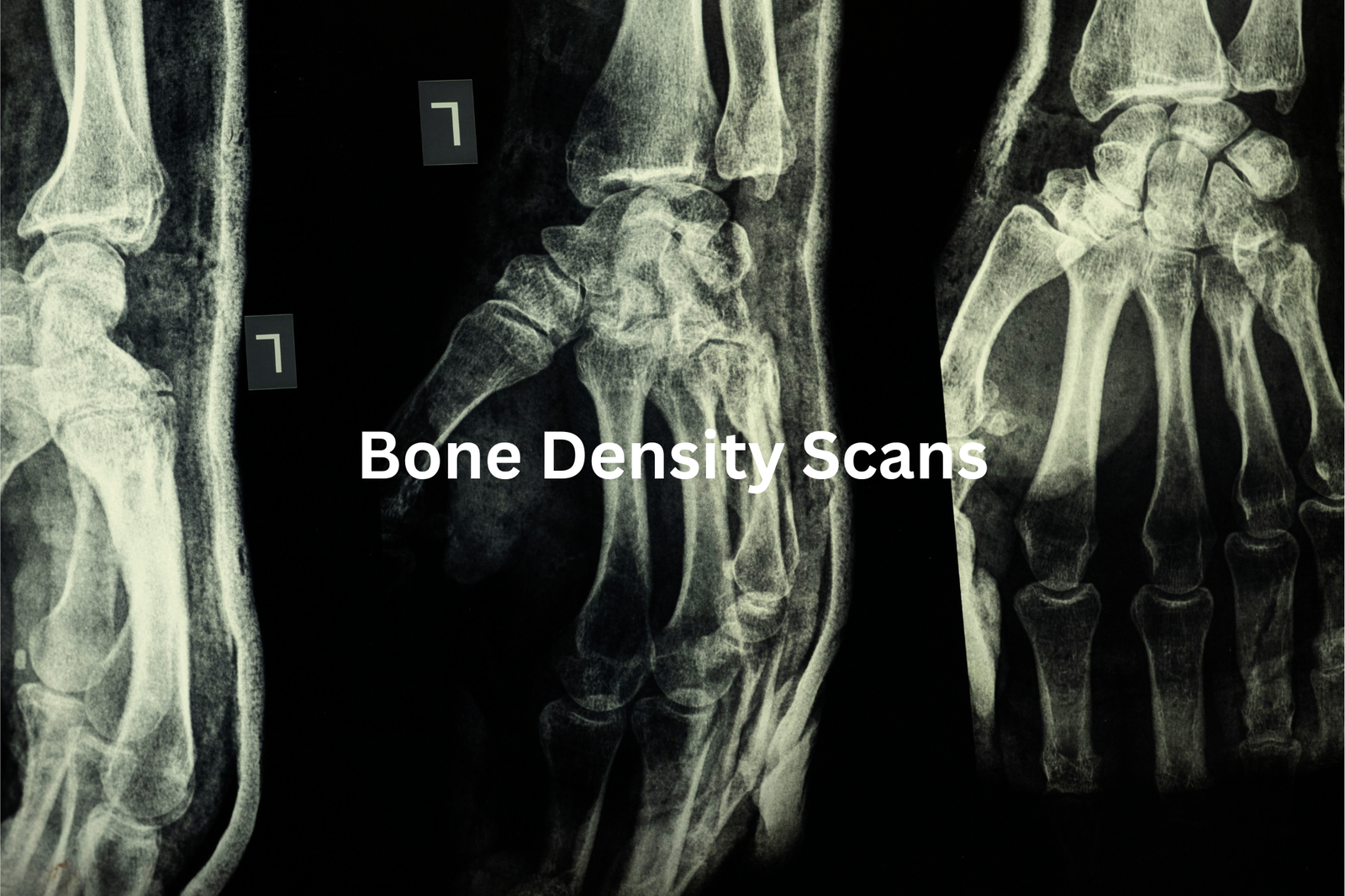
Bones seem so strong, don’t they? But they can weaken quietly over time. My nan learned this when a simple fall left her with a fractured hip. Her bones weren’t as strong as she thought.
A DEXA scan (dual-energy x-ray absorptiometry) is a quick, painless test that checks bone density. It uses a low-dose x-ray to measure bone strength, usually in the hips or spine. The results, called T-scores, compare your bone density to healthy young adults. A score of -2.5 or lower means osteoporosis, while -1 to -2.5 signals osteopenia, an early stage of bone loss.
Osteoporosis often gives no warning until a bone breaks. But it’s preventable. Eating calcium-rich foods like yoghurt or leafy greens, getting vitamin D from sunlight, and doing weight-bearing exercises can help. If you’re worried about your bones, ask your doctor about a scan. It’s quick and could save you a lot of trouble later.
Fluoroscopy
Sources: The Royal Children’s Hospital Melbourne.
It’s a bit odd, seeing someone’s insides move on a screen, but that’s what fluoroscopy does. It’s like an x-ray video, showing how things work in real-time.
Here’s the gist:
- X-rays pass through the body, creating live images.
- A contrast agent (like barium) highlights areas for better visibility.
- It’s used to check swallowing, blood flow, joints, or guide procedures like catheter placement.
My uncle once had trouble swallowing, so they gave him barium to drink. On the screen, you could see it slide down his oesophagus. The doctor quickly spotted a narrowing that needed treatment.
Radiation’s involved, but it’s minimal—similar to a few chest x-rays. Precautions are taken, especially for kids or pregnant women. If you’re having one, wear loose clothes, avoid jewellery, and follow any prep instructions. It’s quick and painless. Watching your body work? Pretty fascinating, really.
Doppler Ultrasound
It’s a bit odd, hearing your own blood moving, but that’s what a Doppler ultrasound lets you do. Unlike regular ultrasounds that show pictures, Doppler shows blood flow in real time.
Here’s how it works:
- The machine sends out sound waves (no radiation involved).
- These waves bounce off moving blood cells, then return to the machine.
- The data creates an image of how your blood flows.
Doctors use it to spot things like blood clots, blocked arteries, or heart problems. When my aunt had one, she was always feeling drained, and her doctor wanted to check her circulation. The test was simple—just some gel, a handheld probe, and soft beeping sounds. It didn’t hurt, and it only took about 30 minutes.
You might need to wear loose clothes or stay still for a bit. It’s safe, painless, and gives doctors a clear view of your blood flow.
FAQ
What is Magnetic resonance imaging (MRI) and how does it work?
Magnetic resonance imaging (MRI) is a medical imaging technique that uses strong magnetic fields and radio waves to create detailed 3D images of the body’s internal organs and tissues. MRI scans provide cross-sectional images that allow health care providers to examine the body in great detail without using radiation. MRI is commonly used to diagnose a variety of conditions, including cancer, heart and blood vessel diseases, and problems with the brain, spine, and other internal organs.
What are the different types of medical imaging tests?
There are several common types of medical imaging tests, each with its own advantages and uses. These include X-rays, computed tomography (CT) scans, magnetic resonance imaging (MRI), nuclear scans, ultrasound, and others. Each test provides different information about the body’s internal structures and can be used to diagnose a wide range of medical conditions, from broken bones to organ diseases to cancer. Your health care provider will recommend the most appropriate imaging test based on your medical history and symptoms.
How do medical imaging tests work?
Medical imaging tests use various technologies to create detailed pictures of the inside of the body. X-rays and CT scans use small doses of radiation to produce images. MRI scans use strong magnetic fields and radio waves. Ultrasound uses high-energy sound waves. Nuclear scans involve injecting a small amount of radioactive material. Each method has unique advantages and is suited for diagnosing different medical conditions. Your health care provider will choose the most appropriate imaging test based on your specific situation and health needs.
What are the risks of medical imaging tests?
Most medical imaging tests carry a small amount of risk, usually related to the use of radiation or contrast dyes. X-rays and CT scans use ionising radiation, which can slightly increase the risk of cancer over time with repeated exposures. Nuclear scans also involve radiation. Contrast dyes used in some tests can occasionally cause allergic reactions. However, the benefits of these tests in diagnosing and monitoring medical conditions usually outweigh the small risks. Your health care provider will discuss the risks and benefits with you before ordering any imaging tests.
How can I prepare for a medical imaging test?
Preparing for a medical imaging test usually involves following some simple instructions from your health care provider or the imaging facility. This may include fasting before the test, drinking extra fluids, or avoiding certain medications. You may need to remove any metal objects, such as jewellery or piercings, before an MRI. It’s important to let your provider know about any medical conditions, allergies, or pregnancy, as this can affect the type of test performed. Following the preparation instructions carefully will help ensure the test goes smoothly.
What can medical imaging tests diagnose?
Medical imaging tests can be used to diagnose a wide variety of conditions, including cancer, heart disease, neurological disorders, musculoskeletal injuries, and problems with internal organs. X-rays can identify broken bones, tumours, and fluid buildup. CT scans provide detailed 3D images that allow providers to see organs, blood vessels, and other structures. MRI scans are especially useful for examining the brain, spine, and soft tissues. Ultrasound uses high-energy sound waves to create real-time images of organs and blood flow. Nuclear scans can identify abnormalities in organ function. Imaging tests give health care providers crucial information to make accurate diagnoses and develop effective treatment plans.
How are medical imaging tests performed?
The specific process for undergoing a medical imaging test depends on the type of test. For X-rays and CT scans, you’ll be positioned on a table and the machine will move around you to capture images. MRI scans have you lie inside a large, tube-shaped machine. Ultrasound involves a technician moving a small handheld device over your skin to produce real-time images. Nuclear scans may require an injection of a small amount of radioactive material before the imaging is done. In all cases, you’ll be given instructions on how to prepare and what to expect during the test. The imaging staff will guide you through the process.
What safety precautions are taken during medical imaging tests?
Medical imaging facilities take several safety precautions to protect patients and staff. For tests involving radiation, such as X-rays and CT scans, the amount of radiation exposure is carefully monitored and kept as low as possible. Technicians use shielding devices and position patients to minimise radiation exposure. Facilities also have strict protocols for safely handling and disposing of radioactive materials used in some tests. For MRI scans, strong magnetic fields are contained within the machine. Patients are screened for any metal objects that could be pulled into the machine’s magnetic field. Ultrasound and other non-invasive tests do not use radiation at all. Overall, modern imaging facilities prioritise patient safety through rigorous protocols and careful oversight.
How will I receive the results of my medical imaging test?
After your medical imaging test, the images will be reviewed by a specialist physician called a radiologist. They will analyse the images and provide a detailed report to your primary care provider or the physician who ordered the test. Your provider will then discuss the results with you, explain what they mean, and outline any next steps or treatment recommendations. This may happen during a follow-up appointment or your provider may contact you by phone or mail. If there are any urgent findings, the radiologist and your provider will make sure you receive that information promptly. You should receive the final report and have the opportunity to discuss the results thoroughly.
How much do medical imaging tests cost?
The cost of medical imaging tests can vary quite a bit depending on factors like the type of test, your location, your health insurance coverage, and the facility where the test is performed. In general, X-rays and ultrasounds tend to be less expensive, while advanced tests like MRI and CT scans can be more costly. Prices also depend on whether the test is performed in a hospital, outpatient imaging centre, or doctor’s office. Your health insurance plan will cover at least part of the cost of medically necessary imaging tests, but you may have to pay a deductible or coinsurance amount. Check with your insurance provider to understand your specific coverage and out-of-pocket costs.
Conclusion
When doctors need to see inside our bodies, they use imaging techniques like x-rays and MRI scans. Each test serves a unique purpose. X-rays are great for seeing bones, while MRIs show soft tissues and organs in detail. These imaging tests give doctors important information to help them make better choices about our health. Whether it’s a quick x-ray or a detailed MRI, remember these tests play an important part in helping us stay well!
References
- https://www.healthdirect.gov.au/x-rays
- https://www.canberrahealthservices.act.gov.au/services-and-clinics/services/positron-emission-tomography
- https://aanms.org.au/what-is-nuclear-medicine/
- https://www.cancer.org.au/mammogram


