This article explores dynamic imaging, a fascinating way to see the body’s processes in real time, helping doctors diagnose and treat better.
Dynamic imaging is a pretty cool idea. It’s like watching a movie of what happens inside our bodies. Instead of just looking at still pictures, doctors can see how everything works while it’s moving. This can help them figure out what might be wrong. Imagine being able to see how blood flows through your heart or how your stomach digests food! Keep reading to discover how dynamic imaging helps with different medical needs.
Key Takeaway
- Dynamic imaging shows moving parts inside the body, helping doctors see what’s going on.
- It plays an important role in diagnosing and treating many health issues.
- This kind of imaging can help with everything from heart problems to joint injuries.
What is Dynamic Imaging?
Dynamic imaging is a technique that captures images of things inside our bodies while they move. This is super helpful because our bodies don’t stay still. They’re always working and changing. For example, when you eat, your stomach and intestines are busy breaking down the food. Doctors need to see these changes to understand how our bodies work.
Why Use Dynamic Imaging?
Dynamic imaging is especially useful during procedures. During cardiac catheterisations, doctors can watch blood flow in and out of the heart. They also use it to see how joints move during orthopaedic treatments.
Seeing these movements helps doctors decide what’s working and what’s not. If a treatment needs adjusting, they’ll know without waiting for more static images.
Benefits of Dynamic Imaging
- Real-time Observation: Doctors can see changes as they happen.
- Better Diagnoses: Watching motion helps uncover issues that static images might miss, like how blood flow slows under stress.
- Condition Monitoring: It tracks progress over time, catching slow-healing injuries.
One patient with shoulder pain had endless static X-rays—all of them normal. Dynamic imaging finally showed the problem: her shoulder joint was slipping out of place with every movement. Knowing this meant proper treatment, and her pain finally eased. (1)
Different Types of Dynamic Imaging
There are several ways dynamic imaging can be done. Each method has its own special benefits. (2)
Dynamic Digital Radiography (DDR)
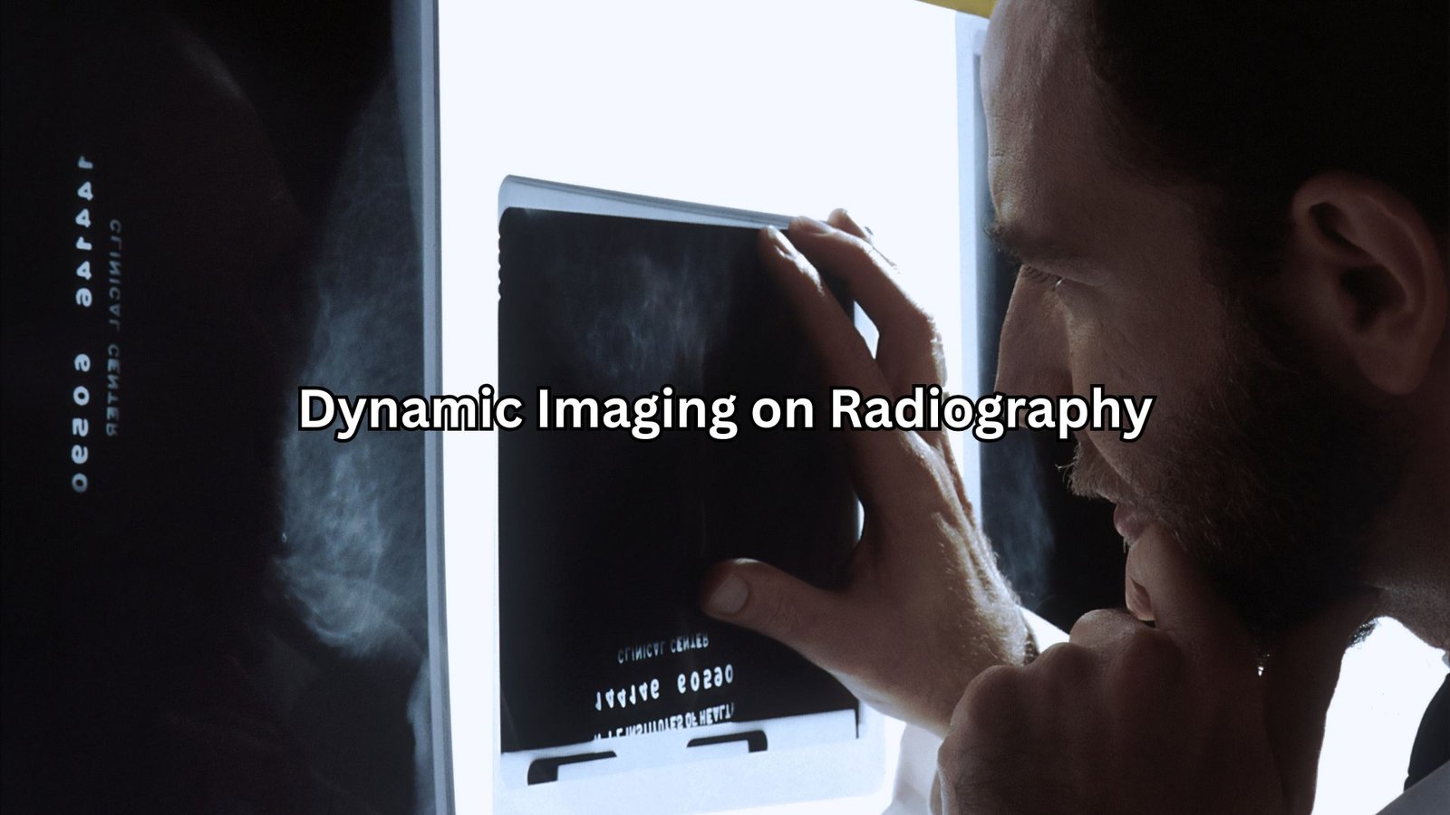
This is a newer way to take X-ray images. It captures up to 15 pictures every second! Imagine watching a slow-motion video of your bones moving. This helps doctors see how stable your spine is or how well your joints are working.
Nuclear Medicine
In this method, doctors give a patient a tiny bit of radioactive medicine. This medicine helps show how fluids move in the body. It’s like a special light that helps reveal how organs and tissues are working.
MRI and CT Scans
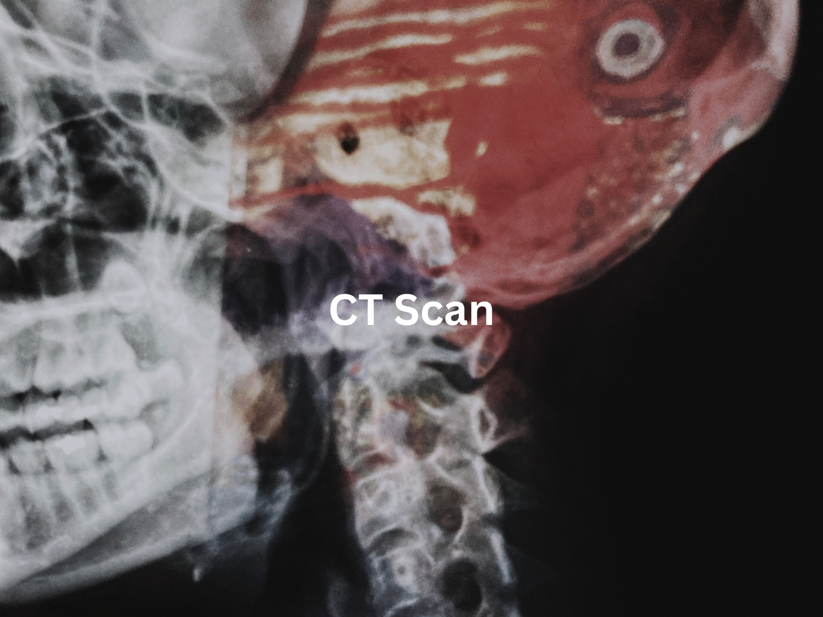
Both of these tools can also be used in dynamic imaging. They help doctors look at soft tissues and organs in detail, revealing how they behave over time. This can be super useful for cardiac imaging or looking at muscles and joints.
How Does Dynamic Imaging Help?
Credit: Summit Medical Institute
Dynamic imaging is here to help doctors understand our bodies better. Here are some ways it does this:
Watching the Body in Motion
Ever wondered how doctors peek inside without opening us up? Dynamic imaging is like watching a video instead of a photo. It captures how things move and flow. Blood flow, for instance, can be seen traveling through veins and arteries in real time, often measured in centimeters per second. If there’s a blockage, it’s spotted quickly.
It’s the same with digestion. When you eat, your stomach and intestines push food along through peristalsis. Dynamic imaging shows that movement, helps doctors find sluggish spots or blockages.
Spotting What Static Images Miss
Static images capture one moment, but dynamic imaging works like a movie. In the heart, coronary artery disease might not show up at rest. Under stress, though, blood flow changes. Dynamic imaging can reveal these changes and help doctors decide if surgery, medication, or lifestyle changes are needed.
Movement Matters for Muscles and Joints
Joint or muscle issues? Dynamic imaging captures motion. When a knee acts up, doctors can observe how it bends and bears weight. One runner had knee pain, but static images showed nothing. Dynamic imaging revealed her kneecap sliding off-track with each bend. That discovery led to the right fix, offering relief and a clear path to recovery.
Examples of Dynamic Imaging Procedures

Watching the human body in motion can be revealing. Dynamic imaging is often the go-to tool when doctors need to see how something works rather than just how it looks.
Barium Studies
Ever had trouble swallowing or stomach issues? Doctors often turn to barium swallow studies for answers. A patient drinks a thick liquid (barium sulphate) that lights up under X-rays. It’s not the tastiest, but it’s effective. Once the barium coats the digestive tract, doctors can watch how food moves from the oesophagus to the stomach. If there’s a blockage or sluggish movement, they’ll spot it right away.
Cardiac Imaging
Dynamic imaging is a lifesaver for heart patients. It shows the heart beating in real time, revealing how blood flows through the heart chambers. During a cardiac catheterisation, doctors thread a thin tube into the heart to get even clearer images. Watching the heart work this way helps catch issues like narrowed arteries. Early diagnosis can mean fewer complications down the road.
Orthopaedic Interventions
Surgery on bones and joints requires precision. Dynamic imaging helps doctors check that everything is aligned properly. Imagine a surgeon placing a screw to hold a fractured bone together. With dynamic imaging, they can see if the screw is straight and stable. Even the smallest misalignment could cause future pain or reduced mobility.
The Future of Dynamic Imaging
The future of dynamic imaging looks bright. Advances in technology might bring even better ways to capture these moving images. Artificial intelligence (AI) and robotics could step in to help doctors analyze images faster and with more accuracy.
AI has a few standout benefits:
- Faster Analysis: AI can process images in seconds, spotting potential issues immediately. That means quicker diagnoses and, sometimes, life-saving speed.
- Automated Feedback: With AI reviewing images, doctors can receive real-time suggestions—like alerts about irregular blood flow or joint misalignments.
- Improved Accuracy: By helping reduce human error, AI may make diagnoses even more reliable.
Practical Takeaway
If your body feels off, dynamic imaging can offer a clearer view. It helps doctors see your body working—or not—leading to more accurate treatment decisions. With AI in the mix, the process might only get better.
FAQ
What is real-time dynamic imaging, and how does it help with medical visualisation?
Real-time dynamic imaging lets doctors see how anatomical structures move, making it easier to understand physiological activities. It’s a key tool in medical visualisation, especially when examining dynamic processes like blood flow or gastrointestinal motility. This helps identify issues that static images might miss.
How is fluoroscopy used in gastrointestinal studies and barium swallow studies?
Fluoroscopy is often used in gastrointestinal studies like barium swallow studies. Patients drink a contrast agent, allowing doctors to watch the digestive tract in motion. This helps diagnose swallowing disorders and digestive tract blockages more accurately by visualising real-time movement.
How does dynamic imaging improve cardiac catheterisations and angiography?
Dynamic imaging in cardiac catheterisations and angiography helps doctors see blood flow through the chambers of the heart. It’s crucial for diagnosing coronary artery disease, stent placement, and other conditions requiring precise treatment. It’s also used for detailed blood flow visualisation.
What’s the role of dynamic imaging in orthopaedics and fracture reduction?
Dynamic imaging is vital in orthopaedics for procedures like joint arthroscopy, screw placement, and fracture reduction. By showing joint motion and spine stability in real time, doctors can ensure optimal alignment during minimally invasive procedures and improve surgical precision.
How are AI and robotics changing dynamic imaging for healthcare interventions?
Artificial intelligence (AI) and robotics are enhancing dynamic imaging by speeding up image analysis and providing automated feedback. This reduces human error, improves diagnostic detail, and ensures better outcomes for healthcare interventions like oncology, neurology, and musculoskeletal conditions.
How does ultrasound imaging help with musculoskeletal and obstetric conditions?
Ultrasound imaging is often used to examine soft tissues and joint motion. Musculoskeletal ultrasound helps guide tendon and joint injections, ensuring accurate placement. Obstetric ultrasound monitors fetal movement in real time, providing crucial information about pregnancy health and development.
Conclusion
Dynamic imaging is an amazing way for doctors to see what’s happening inside our bodies while everything is moving. This technique helps with diagnosing and treating various medical conditions, from heart problems to joint injuries. It’s all about watching the body in action to make better health decisions. Whether it’s through X-rays, MRIs, or other methods, dynamic imaging gives doctors a clearer picture of how to help their patients.
So, next time you hear about dynamic imaging, remember it’s not just about pictures; it’s about understanding the story of our bodies in motion.
References
- https://www.wehi.edu.au/collaborative-centre/centre-for-dynamic-imaging/
- https://www.lanl.gov/engage/organizations/physical-sciences/physics/dynamic-imaging-radiography


