Explore the fascinating journey of imaging technology from X-rays to modern scans and how it changed healthcare.
Imaging has come a long way, hasn’t it? I mean, just think about how we can now see inside our bodies with amazing clarity. It all started many years ago when Wilhelm Conrad Röntgen discovered X-rays in 1895.
This discovery opened the door to a world where doctors could see what was happening inside without even needing to cut anyone open! In this article, we’ll look at the evolution of imaging, from the first ray images to the incredible technology we have today. Let’s get started!
Key Takeaway
- Medical imaging started with X-rays in the late 19th century.
- New technologies like CT scans and MRIs have improved diagnosis.
- The future of imaging continues to evolve with innovations like PET scans.
The Beginning of Medical Imaging
It’s interesting to think about how we used to guess what was wrong inside our bodies. There were some brave doctors who had to rely on their instincts and what they could feel. But then came Wilhelm Conrad Röntgen in 1895. He discovered something amazing: X-rays. This discovery opened a door that changed medicine forever. Imagine, for the first time, doctors could see bones and other structures inside the human body without surgery!
X-rays quickly became a tool for finding broken bones and diagnosing diseases. During World War I, they saved countless lives because they helped doctors see injuries more clearly. Soldiers who had been hurt could be examined quickly, and doctors could treat them more effectively. This was a significant moment in the history of medical imaging(1).
Later on, as time marched forward, the mid-20th century introduced ultrasound. This technology was revolutionary. Doctors could now use sound waves to see inside pregnant women. They could watch babies move in real-time! This was not only exciting but also safe. Ultrasound didn’t use radiation, so it became widely accepted. Doctors began using it to look at organs like the heart and liver, making it a vital tool in medical diagnostics.
Ultrasound helped many families. Imagine a father seeing his baby’s heartbeat for the first time. It brought joy and relief. The technology was growing, and the possibilities seemed endless.
The Rise of CT Scans
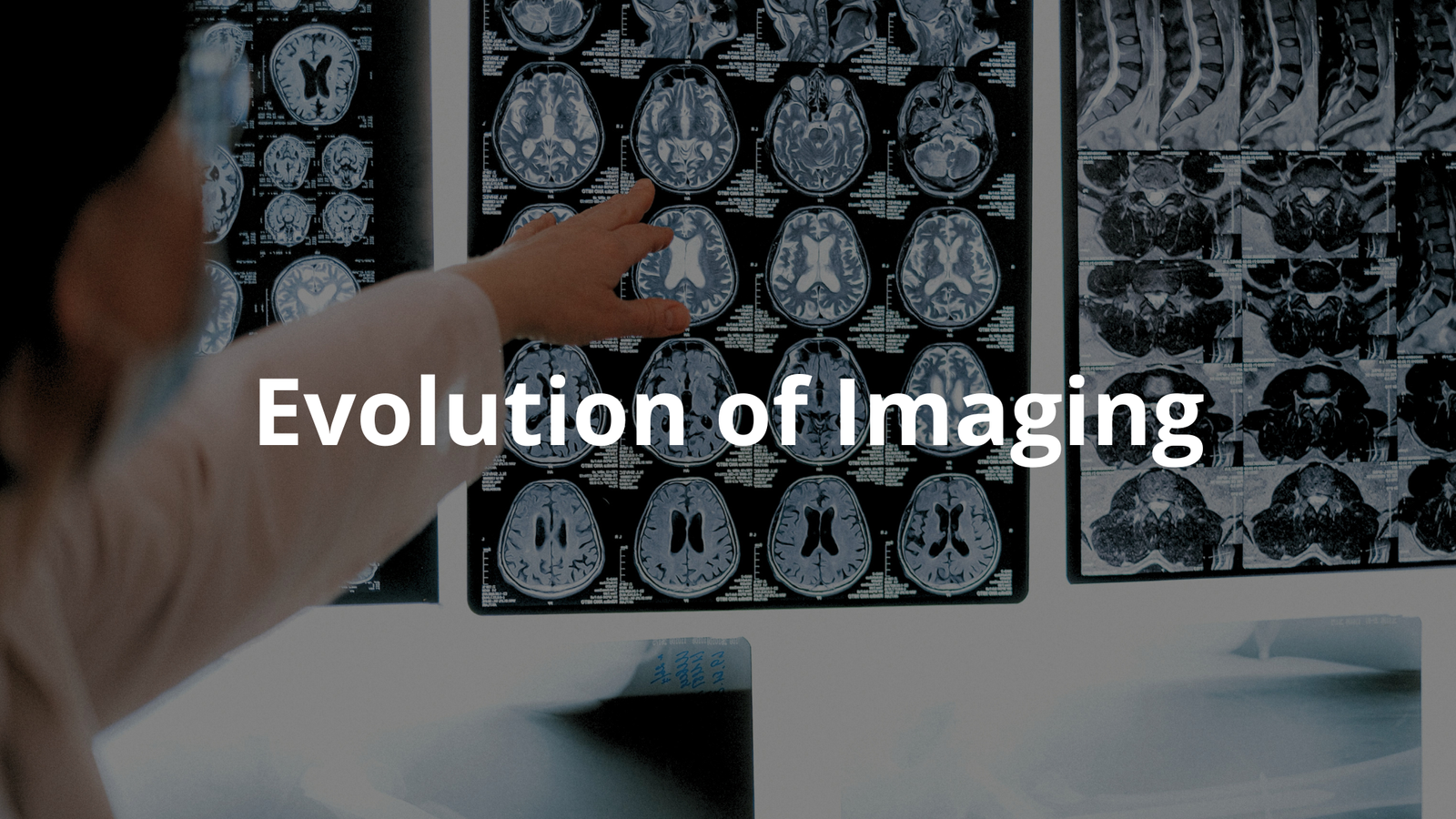
In the 1970s, a remarkable breakthrough occurred: the CT scan, also known as computed tomography. This advanced machine revolutionised imaging by creating detailed 3D images of the human body. It was astonishing! With CT scans, doctors could finally see tumours and blood clots that regular X-rays could miss.
- Enhanced Diagnosis: Allowed for early detection of serious conditions like cancer.
- Improved Patient Care: More accurate diagnoses brought comfort to patients and their families.
Then came MRI, or magnetic resonance imaging, in the 1980s. This technology was yet another leap forward. By using radio waves and strong magnets, MRI could produce stunning images of soft tissues without any ionising radiation.
- Safety First: No harmful radiation exposure.
- Detailed Imaging: Vital for diagnosing issues in the brain, muscles, and organs.
Each of these innovations brought new hope and possibilities, paving the way for an exciting future in medical imaging. The journey was just beginning.
Molecular Imaging and PET Scans
As the 21st century unfolded, the world of medical imaging kept growing more exciting. One of the most remarkable advancements was Positron Emission Tomography, commonly known as PET scans. These scans allow doctors to see how organs and tissues are functioning in real-time. A PET scan can reveal metabolic activity, which is especially helpful for diagnosing conditions like cancer. Imagine being able to see how your body is working on a cellular level!
- Complete Picture: Doctors often combine PET scans with CT or MRI to get a more comprehensive view of a patient’s health.
- Growing Demand: Between 2000 and 2021, imaging studies in Australia increased by over 864,000 each year!
New facilities, like the Total Body PET Facility that opened in Sydney, are pushing boundaries. This place focuses on research into diseases while keeping radiation exposure low. It’s clear that imaging services are becoming more important in understanding what happens inside the body.
The Role of Radiographers

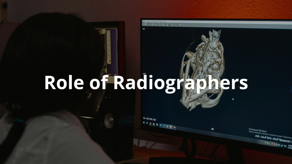
Now, let’s turn our attention to the unsung heroes of medical imaging: radiographers. These professionals are the ones behind the machines, ensuring everything runs smoothly. In Australia, their role has evolved significantly as technology advanced. Radiographers now check images before the doctors see them, which helps reduce mistakes.
- Error Reduction: They signal when something looks abnormal, improving the accuracy of diagnoses.
- Teamwork: The collaboration between radiographers and doctors is essential for effective patient care.
Imagine a radiographer swiftly reviewing scans, knowing that their keen eye could make a difference in someone’s life. This teamwork is vital for accurate diagnosis and treatment, making it easy to see how important radiographers have become in the modern healthcare landscape. With technology continuing to advance, their role will likely grow even more pivotal in the future(2).
The Future of Imaging
The future of imaging looks incredibly bright! There’s a sense of excitement in the air as technology keeps pushing boundaries. With the rise of AI algorithms and machine learning, imaging technology is becoming sharper and more precise. These tools help doctors analyse images better. They can detect things that might have gone unnoticed before.
- AI in Imaging: These systems can learn from past images to improve accuracy.
- Personalised Medicine: Doctors can tailor treatments based on individual patients’ unique imaging data.
Imagine a world where a doctor can look at a scan and know exactly what it means, thanks to advanced technology. This kind of innovation will probably help with early diagnosis and treatment planning.
The evolution of imaging has been remarkable. From the early X-ray images that transformed medical diagnosis to the high-tech scans of today, the journey remains fascinating. Imaging technology not just helps doctors identify diseases but also plays a crucial role in overall patient care.
As the field continues to grow, the possibilities are endless. Each new method brings fresh hope and opportunities for better health outcomes. The excitement surrounding the future of imaging is palpable, and it’s thrilling to think about what might come next.
Conclusion
To wrap things up, the evolution of imaging has transformed healthcare in many ways. From the early days of X-rays to the advanced technologies like CT scans and PET scans, we’ve seen incredible progress. The role of radiographers has also become more important, ensuring that patients receive the best care possible.
As technology continues to grow, we can expect even more exciting developments in the future of medical imaging!
FAQ
How did imaging technology revolutionise medical diagnoses in the 19th century?
Wilhelm Conrad Röntgen’s discovery of ray technology with photographic plates in 1895 transformed medical imaging. His work earned him the Nobel Prize in Physics and marked the beginning of diagnostic imaging. For the first time, healthcare professionals could see inside the human body without surgery, leading to more accurate diagnoses.
How has digital imaging changed patient care since the 20th century?
The evolution of medical imaging progressed from basic ray images to advanced imaging technologies like computed tomography, magnetic resonance imaging, and positron emission tomography. Digital imaging and picture archiving and communication systems revolutionised image quality and accessibility, while artificial intelligence and machine learning enhanced diagnostic accuracy.
What makes ultrasound imaging unique in medical diagnosis?
Ultrasound imaging, pioneered by Ian Donald, uses high-frequency sound waves instead of ionising radiation to create real-time images. This imaging technique safely visualises soft tissues, blood flow, and blood vessels. Healthcare professionals rely on ultrasound technology for early diagnosis and minimally invasive procedures.
How do modern imaging techniques assist in cancer diagnosis?
Medical imaging technology helps detect cancer cells at the cellular level. Positron emission tomography tracks metabolic activity, while magnetic resonance imaging provides detailed images of soft tissues. These imaging techniques enable early detection of breast cancer, prostate cancer, and disease progression, improving treatment planning and patient care., prostate cancer, and disease progression, improving treatment planning and patient outcomes.
References
- https://onlinelibrary.wiley.com/doi/full/10.1002/jmrs.610
- https://onlinelibrary.wiley.com/doi/abs/10.1111/1754-9485.13591


