
Learn about fluoroscopy, its uses, safety, and preparation for procedures.
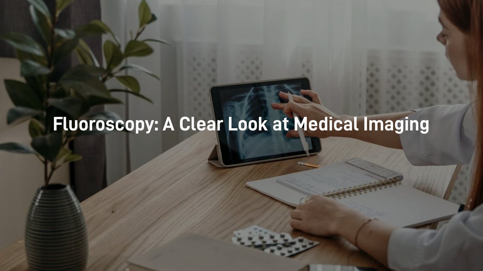
Fluoroscopy is a specialised type of X-ray imaging that provides real-time views of the body’s internal functions. By capturing continuous X-ray images, it allows doctors to see how organs and systems move and work together, making it an invaluable tool for diagnosing medical conditions.
This technology is used in a wide range of medical procedures, from examining the digestive system to guiding treatments like catheter placements. Understanding how fluoroscopy works can help ease concerns and prepare patients for what to expect.
Curious to learn more about how fluoroscopy plays a role in modern medicine? Keep reading to find out!
Key Takeaway
- Fluoroscopy provides real-time images for doctors to see inside the body.
- Safety is important, and proper preparation helps keep patients safe.
- There are many uses for fluoroscopy, from checking the gastrointestinal tract to guiding surgeries.
Fluoroscopy Imaging

Fluoroscopy imaging allows doctors to see inside the body in real time, providing a continuous stream of X-ray images. This advanced technique is especially useful for observing movement within the body, such as food passing through the gastrointestinal tract. By capturing live images, fluoroscopy helps diagnose conditions quickly and effectively. (1)
Key details about fluoroscopy:
- Real-time imaging – Uses a specialised X-ray beam to produce continuous images.
- Dynamic observation – Ideal for watching movement, such as swallowing or blood flow.
- Medical applications – Commonly used in gastrointestinal studies, joint injections, and cardiac procedures.
- Quick diagnosis – Helps identify issues efficiently without the need for exploratory surgery.
Fluoroscopic images provide invaluable insights into how the body functions, aiding in both diagnosis and treatment planning. With its ability to capture live motion, fluoroscopy remains a crucial tool in modern medicine.
Barium Swallow
A barium swallow is a common fluoroscopy test used to examine the throat and oesophagus. During the procedure, the patient drinks a thick, chalky liquid called barium.
This special contrast substance coats the lining of the oesophagus, making it easier for doctors to detect any abnormalities on X-ray images. While swallowing barium may feel a bit strange, the test is essential for diagnosing swallowing difficulties and other related conditions.
Key details about a barium swallow:
- Purpose – Helps detect swallowing problems, strictures, or reflux.
- Barium contrast – A thick, white liquid that highlights the oesophagus on X-rays.
- Fluoroscopy imaging – Captures real-time movement as the patient swallows.
- Patient experience – May feel slightly heavy or chalky but is generally well tolerated.
This quick and effective test provides valuable insights into a patient’s swallowing function, helping guide accurate diagnosis and treatment planning.
Fluoroscopy for Surgery
Credits: Rayus Radiology
Fluoroscopy is also widely used during surgeries, helping doctors guide their instruments with precision. This real-time imaging ensures that medical devices, such as catheters, are placed accurately within the body. By providing continuous X-ray images, fluoroscopy supports a range of interventional procedures, improving both safety and effectiveness.
Key uses of fluoroscopy in surgery:
- Instrument guidance – Helps doctors position tools like catheters and stents correctly.
- Real-time imaging – Provides continuous visual feedback during procedures.
- Interventional applications – Used in cardiac catheterisation to assess blood flow in the heart.
- Enhanced accuracy – Reduces the risk of complications by ensuring proper placement.
This advanced imaging technique plays a crucial role in modern surgery, allowing for minimally invasive procedures with greater precision. Whether guiding a catheter in the heart or assisting in orthopaedic surgeries, fluoroscopy helps improve patient outcomes. (2)
Dynamic Imaging
Dynamic imaging captures moving images, allowing doctors to see organs in action. Unlike standard X-rays, which provide still pictures, fluoroscopy offers a real-time view of how the body functions. This dynamic perspective helps doctors assess how well organs are working, making it an essential tool for diagnosing various conditions.
Key benefits of dynamic imaging with fluoroscopy:
- Real-time movement – Shows organs in motion, such as the heart beating or food passing through the digestive system.
- More detailed assessment – Helps doctors evaluate function, not just structure.
- Improved diagnosis – Useful for detecting issues like swallowing disorders or joint problems.
- Versatile application – Used in gastrointestinal studies, cardiac tests, and orthopaedic evaluations.
By watching organs move in real time, doctors can gain a clearer understanding of a patient’s health. This advanced imaging technique plays a crucial role in accurate diagnosis and effective treatment planning.
Fluoroscopy Safety
Safety is a key concern when using fluoroscopy. Since it involves radiation, doctors take careful steps to minimise exposure by using the lowest dose possible. They follow strict guidelines from the National Council on Radiation Protection and Measurements to ensure patient safety. Understanding these precautions can help patients feel more comfortable during their procedure.
Key fluoroscopy safety measures:
- Lowest possible dose – Doctors adjust settings to keep radiation exposure to a minimum.
- Strict guidelines – Procedures follow national safety standards to protect patients.
- Patient awareness – Patients are encouraged to ask about radiation levels and safety precautions.
- Risk management – The benefits of the procedure are carefully weighed against potential risks.
By following these safety protocols, fluoroscopy remains a safe and effective imaging tool. Patients can feel reassured knowing that every precaution is taken to keep radiation exposure as low as possible.
Contrast Fluoroscopy
Contrast fluoroscopy uses special dyes to improve the visibility of different parts of the body during imaging. These contrast agents, often called X-ray dyes, help highlight structures like the stomach and intestines, making it easier for doctors to identify potential issues. By enhancing image clarity, contrast fluoroscopy plays a vital role in diagnosing conditions that may not be visible on standard X-rays.
Key benefits of contrast fluoroscopy:
- Improved visibility – Contrast dyes make organs and tissues stand out more clearly.
- Better diagnosis – Helps detect blockages, abnormalities, or other medical concerns.
- Common uses – Often used for gastrointestinal studies, blood vessel imaging, and urinary tract exams.
- Safe and effective – Most contrast agents are well tolerated, with minimal side effects.
This advanced imaging technique allows doctors to get a clearer view of internal structures, ensuring more accurate diagnoses and effective treatment plans.
Fluoroscopy Preparation
Before a fluoroscopy exam, patients receive specific instructions to ensure clear and accurate images. Preparation steps vary depending on the procedure, but following these guidelines is essential for the best results. In some cases, patients may need to fast or adjust their medications before the appointment.
Key preparation steps for fluoroscopy:
- Fasting requirements – Patients may need to avoid eating or drinking for several hours beforehand.
- Medication adjustments – Certain medications might need to be paused or changed; always check with your doctor.
- Clothing and accessories – Loose clothing is recommended, and jewellery or metal objects may need to be removed.
- Special instructions – Some procedures require drinking a contrast dye or following additional steps.
By carefully following these instructions, patients can help ensure their fluoroscopy exam provides the most accurate results, leading to a more effective diagnosis and treatment plan.
Fluoroscopy Uses
Fluoroscopy is a highly versatile imaging technique used across various medical procedures. By providing real-time moving images of the body, it helps doctors diagnose conditions, guide treatments, and assess organ function. Whether checking for blockages or assisting with complex interventions, fluoroscopy is an essential tool in modern healthcare.
Key uses of fluoroscopy:
- Gastrointestinal diagnosis – Helps detect issues such as ulcers, reflux, and blockages.
- Interventional procedures – Guides doctors in placing catheters, stents, and other medical devices.
- Blood flow assessment – Used in cardiac procedures to monitor how blood moves through the heart and vessels.
- Organ function monitoring – Provides live imaging of organs like the lungs, stomach, and kidneys.
With its ability to capture dynamic images, fluoroscopy plays a vital role in both diagnosing and treating medical conditions, ensuring more accurate and effective patient care.
Risks of Fluoroscopy
Like any medical procedure, fluoroscopy comes with some risks, the main one being exposure to radiation. While the radiation levels are generally low, repeated exposure can accumulate over time.
Certain groups, such as pregnant women and young children, may require extra precautions to minimise potential risks. Discussing these concerns with a doctor can help patients make well-informed decisions about their care.
Key considerations for fluoroscopy safety:
- Radiation exposure – While doses are low, unnecessary exposure should be minimised where possible.
- Pregnancy precautions – Pregnant women should inform their doctor, as alternative imaging methods may be recommended.
- Children’s safety – Young children are more sensitive to radiation, so doctors may adjust techniques or explore other options.
- Informed decision-making – Patients should discuss potential risks and benefits with their doctor.
By taking the necessary precautions and following medical advice, fluoroscopy remains a safe and effective tool for diagnosis and treatment.
Interventional Fluoroscopy
Interventional fluoroscopy is an essential imaging technique that helps doctors perform procedures with greater accuracy. By providing real-time X-ray images, it allows precise guidance for placing medical devices or taking biopsies. This improves both the safety and effectiveness of many treatments, reducing risks and ensuring better outcomes for patients.
Key benefits of interventional fluoroscopy:
- Real-time guidance – Helps doctors navigate instruments accurately, reducing errors.
- Precise device placement – Used for inserting catheters, stents, and other medical devices.
- Biopsy assistance – Ensures accurate needle placement for tissue sampling.
- Improved safety – Reduces complications by allowing doctors to see exactly where they are working.
With its ability to provide clear, live imaging, interventional fluoroscopy plays a crucial role in modern medicine, making many procedures safer, more efficient, and less invasive.
Conclusion
In summary, fluoroscopy is a vital medical imaging tool that provides real-time views inside the body. It helps doctors diagnose conditions and perform procedures with greater accuracy.
Proper preparation and safety measures ensure the best results for patients. Understanding how fluoroscopy works can help people feel more at ease when undergoing these exams. With its ability to improve diagnoses and treatments, fluoroscopy continues to play an essential role in modern healthcare.
FAQ
What is fluoroscopy and how does it work?
Fluoroscopy is a type of medical imaging that uses an X-ray beam to produce fluoroscopic images in real time. Unlike standard X-ray imaging exams, which capture still images, fluoroscopy provides continuous X-ray images, allowing health care providers to see internal structures in motion. This is especially useful for guiding medical procedures, such as fluoroscopic guidance for catheter placements and other interventional procedures.
How does fluoroscopy compare to other imaging modalities?
Fluoroscopy differs from other diagnostic X-ray systems like magnetic resonance imaging (MRI). While MRI uses magnetic fields to create detailed images, fluoroscopy provides real-time X-ray imaging, making it ideal for guiding interventional procedures. Other imaging modalities, such as digital fluoroscopy, enhance image quality while reducing doses to patients.
How is image quality maintained during fluoroscopy examinations?
Fluoroscopy units must meet Medical X-Ray Imaging Devices Conformance standards to ensure high diagnostic quality. Facility accreditation, quality assurance programs, and regular checks of X-ray Imaging Devices help maintain clear fluoroscopic views. Additionally, using a single fluoroscopy frame technique can help reduce patient dose while still producing high-quality images.
References
- https://www.svpr.com.au/our-services/fluoroscopy
- https://lakeimaging.com.au/services/fluoroscopy/


