
This article discusses fluoroscopy safety measures to protect patients and staff from radiation during medical procedures.
Fluoroscopy is like having a TV that shows what’s happening inside your body. Doctors can see bones, organs, and even blood flow in real time. But there’s a catch. This amazing technology uses radiation, and while it can be very helpful, it also requires some serious safety measures.
Imagine being a superhero but needing to wear protective gear to keep yourself safe while saving the day! Let’s explore how we keep both patients and medical staff safe during fluoroscopy.
Key Takeaway
- Fluoroscopy uses radiation, which can be harmful if not handled carefully.
- Safety measures include protective gear and keeping a safe distance from the radiation source.
- Following guidelines helps ensure everyone’s safety during procedures.
What is Fluoroscopy?
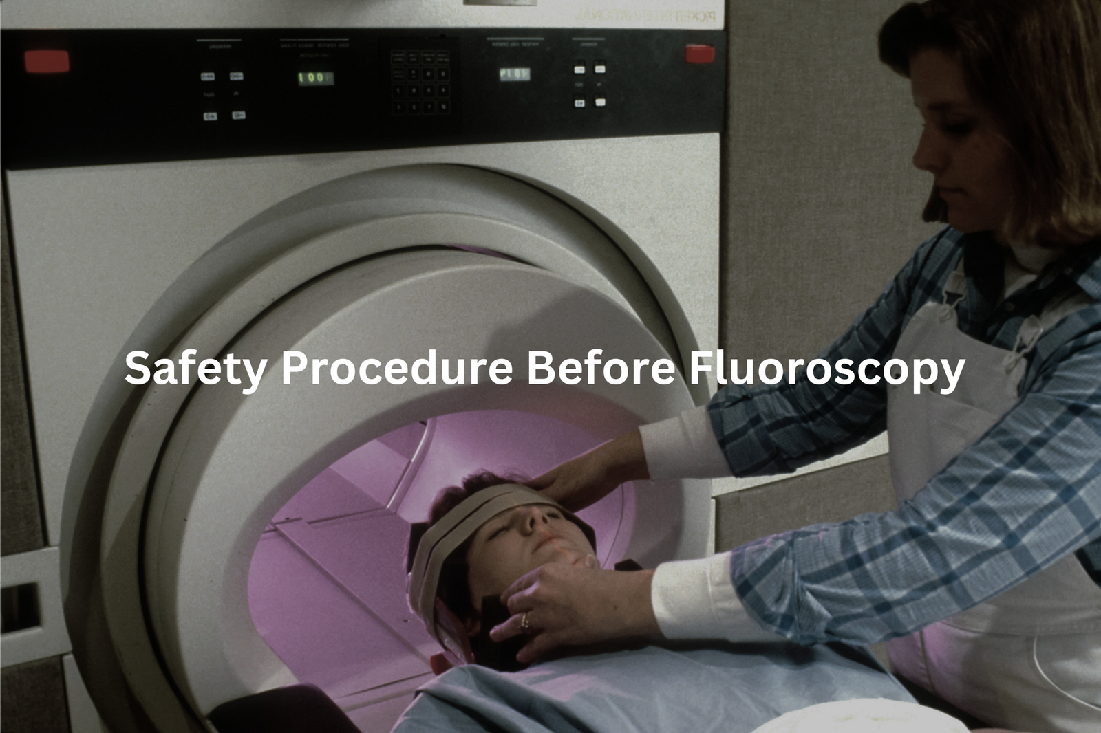
Fluoroscopy lets doctors see the body’s inner workings in real time—like a moving X-ray film. A machine beams X-rays through the body, and a monitor shows the results live. This can reveal how blood moves through vessels or how the digestive tract functions during a barium swallow. The ability to observe movement, not just still images, is what makes fluoroscopy so powerful. But there’s a trade-off: radiation exposure.
Even short procedures can use a decent amount of radiation (measured in milligrays). For longer or complex ones—like placing a kidney stent—exposure can increase significantly. Fluoroscopy’s benefits are huge, but safety is still key.
The Risks of Fluoroscopy
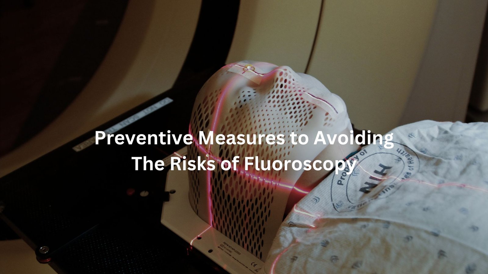
Radiation exposure is the biggest concern with fluoroscopy. While the doses used are generally safe, too much can damage tissue. Doctors monitor the time and intensity to limit unnecessary exposure.
Tissue damage happens when radiation disrupts cells’ DNA. It’s usually rare but can cause burns if exposure is high. Long-term effects? They include a slightly raised risk of cancer—especially after repeated exposures. Think of the thyroid and bone marrow (they’re more sensitive to radiation).
The takeaway: fluoroscopy is essential, but caution makes all the difference. Ask questions. Shorten procedure time if possible. Safety matters more than speed. (1)
Safety Measures and Protective Equipment
Credit: Corwin Health Physics Inc.
To keep everyone safe during fluoroscopy, doctors and medical teams use several important safety measures. Here’s a look at how they do this:
Protective Gear
Fluoroscopy rooms can be pretty intense places, but the staff are prepared. They wear layers of gear that may look clunky but could mean the difference between safe and harmful radiation levels. Lead aprons (weighing around 5 to 7 kg) cover most of the body and block stray X-rays.
Thyroid shields wrap around the neck, protecting the thyroid gland—a sensitive area when it comes to radiation exposure. Lead glasses, though not exactly a fashion statement, shield the eyes from unnecessary exposure. Wearing this gear every day isn’t glamorous, but it’s essential.
Distance Matters
If you ever watch a procedure from the corner of a fluoroscopy room, you’ll see staff keeping a safe distance. It’s not just for comfort—it’s the inverse square law in action. Simply put, standing twice as far from the X-ray source cuts the radiation exposure down by four times.
Even a metre’s difference makes a big impact. Doctors and techs often use mobile lead shields when they can’t step away, keeping themselves as far from the beam as possible.
Time is Key
Minimising time under the fluoroscope is a golden rule. For every extra second a patient is exposed, radiation dose increases. Complex procedures—stenting or catheter placements—can last for 20 to 60 minutes, but reducing any unnecessary time helps limit exposure.
Doctors are trained to plan out each step before hitting the pedal. That means less guesswork during the procedure. And when the beam isn’t needed, it’s turned off immediately. Every second saved counts.
Proper Positioning
The C-arm, named for its curved frame, can be a tricky machine to position. But placing it just right can drastically reduce unnecessary exposure. A common practice is to keep the X-ray source below the patient and the image detector above—this minimises scatter radiation.
Rotating or tilting the C-arm is common, but small changes in angle can change how much radiation hits the patient and staff. Careful adjustments protect everyone in the room without compromising image quality.
Equipment Maintenance
Imagine driving a car that hasn’t been serviced for years—you’d worry about it breaking down. The same goes for fluoroscopy machines. Regular check-ups (usually every six to twelve months) help spot any issues with radiation output.
Technicians inspect for weak shielding, calibration errors, or any small leaks. A machine that’s running well reduces the risk of unexpected high doses. Maintenance isn’t glamorous, but it’s one of the most important parts of keeping fluoroscopy safe. (2)
Monitoring and Training
Tracking radiation exposure is like watching the clock—essential, yet easy to overlook without reminders. Medical staff wear dosimeters (tiny radiation-measuring devices), usually clipped to their uniforms. These gadgets record how much radiation they’ve absorbed over time, helping ensure exposure stays within safe limits. A dosimeter reading of 20 millisieverts per year is typically the upper limit for healthcare professionals.
But gear isn’t enough. Training matters, too. Every staff member needs to know the safety guidelines—how to position patients, use shielding techniques, and limit fluoroscopy time. Without this knowledge, even the best equipment won’t protect anyone.
Cultivating a “safety-first” mindset at work helps, too. By making protection a shared priority, staff can create a culture where questions about safety aren’t seen as overkill—they’re just part of the job. A simple reminder in the break room, or even a quick huddle before a procedure, can make all the difference.
FAQ
What are the common safety measures for fluoroscopy?
Fluoroscopy safety includes wearing personal protective devices like lead aprons, thyroid shields, and lead glasses. Staff follow the ALARA (as low as reasonably achievable) principle to minimise radiation exposure. Proper equipment maintenance and positioning of the C-arm can also reduce radiation risks. Medical facilities may use lead barriers and gloves to limit scatter radiation.
How do fluoroscopy operators monitor radiation exposure?
Operators often use dosimeters to track absorbed doses for both patients and staff. Regular monitoring ensures compliance with safety guidelines. Facilities may also conduct safety checks to prevent equipment malfunctions or unexpected radiation leaks, reducing risks like skin injury or cataracts.
How is patient safety maintained during procedures like EVAR or kidney stent placement?
For complex procedures like endovascular aneurysm repair (EVAR) and kidney stent placement, fluoroscopy operators aim to minimise the dose through careful C-arm positioning, time management, and kVp/mAs settings. Following safety protocols can reduce deterministic and stochastic effects.
What are the risks of prolonged fluoroscopy exposure?
Prolonged exposure to fluoroscopy can increase the risk of skin injuries, cataracts, and radiation-related cancers. Both patients and staff face risks from absorbed doses. Safety culture training and using personal protective devices can help mitigate these risks.
Why is equipment maintenance important for fluoroscopy safety?
Proper maintenance ensures fluoroscopy units are functioning correctly, reducing the chance of unexpected radiation leaks or malfunctions. Regular inspections of shielding and calibration checks help optimise patient and staff safety. Quality improvement efforts focus on catching issues early.
How does time and distance affect fluoroscopy safety?
Minimising time under the X-ray beam and maximising distance from the source reduce radiation exposure. The inverse square law means that doubling the distance from the source significantly lowers radiation dose. Using timers helps operators manage exposure times better.
What is the role of medical physics in fluoroscopy safety?
Medical physics professionals play a key role in dose estimation, radiation biology assessments, and optimising safety standards. They help facilities develop guidelines for protective barriers, ALARA practices, and equipment monitoring to improve safety during fluoroscopy procedures.
How do safety checks and training enhance fluoroscopy safety?
Routine safety checks and ongoing training foster a strong safety culture in medical facilities. Operators learn optimisation techniques to reduce scatter radiation and absorbed doses. Following standards and checklists during procedures, like ERCP or bile duct stenting, helps minimise risks.
What is the purpose of lead aprons, thyroid shields, and lead glasses during fluoroscopy?
Lead aprons, thyroid shields, and lead glasses provide essential protection by blocking X-ray beams and reducing scatter radiation exposure. These personal protective devices protect sensitive organs like the thyroid gland, breast tissue, and eyes, helping lower the risk of radiation injuries.
How do fluoroscopy operators optimise dose during procedures like ERCP or renal angioplasty?
Operators use techniques like proper C-arm positioning, limiting fluoroscopy time, and adjusting kVp and mAs settings to optimise the dose. Following ALARA principles ensures absorbed doses remain as low as reasonably achievable, protecting both patients and staff.
How can fluoroscopy pose risks to skin and organs?
Excessive fluoroscopy exposure may lead to skin injuries or damage to sensitive organs due to high absorbed doses. Careful monitoring, time management, and safety standards help reduce these deterministic effects, especially in longer procedures like angiography or neuraxial injections.
What are the benefits of safety culture and training in fluoroscopy?
A strong safety culture, combined with regular training, ensures operators follow guidelines for positioning, shielding, and dose optimisation. This reduces radiation risks, promotes the use of lead barriers and dosimeters, and fosters consistent quality improvement in medical facilities.
Conclusion
Fluoroscopy is a powerful tool in medicine that helps doctors see inside the body while it’s moving. But with great power comes great responsibility! Safety measures are essential to protect both patients and medical staff from radiation risks.
By using protective gear, maintaining distance, and following safety guidelines, everyone involved can stay safe during these important procedures. So, the next time you hear about fluoroscopy, remember that safety is always the number one goal!
References
- https://www.ncbi.nlm.nih.gov/books/NBK570567/
- https://ajronline.org/doi/10.2214/AJR.16.16556


