
Interventional fluoroscopy helps doctors treat patients with less pain and faster recovery. Find out how this advanced imaging method works.
Interventional fluoroscopy is a tool doctors use to see inside our bodies while helping us get better. It uses ionising radiation, which is a kind of energy, to create real-time images. This technique helps doctors perform procedures that often cause less pain than traditional surgery, which is really important for patient comfort.
For example, it can help fix problems in organs without big cuts. If you want to learn more about how this works and what it means for you or someone you care about, keep reading!
Key Takeaway
- Interventional fluoroscopy helps doctors see inside the body in real time.
- It offers benefits like smaller cuts and faster healing.
- There are some risks, like radiation exposure, that doctors work to manage.
What is Interventional Fluoroscopy?
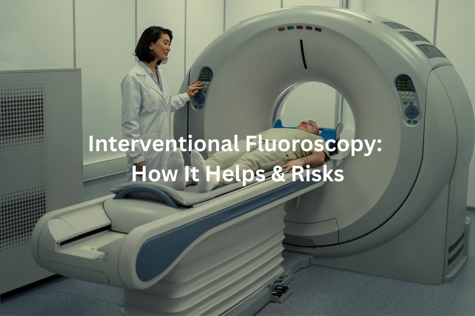
Interventional fluoroscopy creates moving X-ray pictures that doctors use to see inside patients during medical procedures. Like a video camera for the body’s interior, this technology shows blood vessels, organs, and medical tools in real-time on a screen.
The process helps doctors perform complex procedures without making big cuts. They insert small tubes called catheters through blood vessels while watching their progress on the screen (these tubes measure just 2-3 millimeters wide).
Key benefits of fluoroscopy:
- Shows blockages in blood vessels
- Guides stent placement
- Finds internal bleeding spots
- Helps fix heart problems
The radiation dose from each scan stays low (about 0.1-2 millisieverts), and doctors use protective shields to keep patients safe. Modern machines automatically adjust radiation levels based on body thickness.
While patients lie still under the scanning machine, doctors can spot and fix problems that once needed major surgery. This means shorter hospital stays and quicker recovery times for most people.
Why Do Doctors Use Interventional Fluoroscopy?
Fluoroscopy shows the body’s inner workings in real time on a screen, like a moving X-ray(1). The technology helps medical staff see organs, blood vessels, and other structures while they work.
Medical teams use fluoroscopy for several procedures:
• Heart treatments – placing stents in blocked arteries
• Blood vessel repairs – fixing circulation problems
• Digestive system studies – tracking food movement with special dyes
• Pain treatments – guiding injections to specific spots
The equipment gives doctors clear views inside the body (down to 0.1 millimetres), which leads to more precise treatments and smaller cuts. While fluoroscopy does use radiation, medical centres follow strict safety rules. Protective gear like lead aprons and careful timing keep exposure low.
Patients should ask their doctors about:
• Length of the procedure
• Expected radiation dose
• Other testing options
• Recovery time
Most fluoroscopy procedures take 20-30 minutes, though this varies based on what needs examining. The benefits typically outweigh any risks when used properly.
Benefits of Interventional Fluoroscopy
Sources: Physics for Radiology Residents.
Interventional fluoroscopy uses tiny incisions, often smaller than 5mm, unlike traditional surgeries that leave noticeable scars. Medical professionals prefer this method for several reasons:
Benefits of fluoroscopy:
- Creates minimal cuts
- Patients return home within hours
- Lower chance of infection
- Real-time X-ray guidance
The procedure works like a moving X-ray, helping doctors see inside the body while they work. They can place stents, fix aneurysms, and clear blocked arteries without making large cuts(2).
While effective, fluoroscopy does have limitations. Medical staff must monitor radiation exposure carefully, using protective shields and keeping scan times brief. Some medical conditions still require traditional surgery.
For suitable cases, fluoroscopy offers:
- Less post-procedure pain
- Shorter hospital stays
- Quick recovery time
Patients can discuss with their healthcare providers whether fluoroscopy suits their medical needs. Many procedures allow same-day discharge with only a small bandage to show for it.
Risks with Interventional Fluoroscopy

Radiation in medical procedures works like an invisible force, passing through body tissues during diagnostic imaging. Interventional fluoroscopy, a common medical tool, uses this radiation to guide doctors during treatments.
The risks of radiation exposure need careful management(3). Medical staff keep doses low, but multiple procedures can increase total exposure (typical single-procedure dose ranges from 15-70 mGy). Proper protection includes:
• Lead aprons (0.5mm lead equivalent)
• Thyroid shields
• Distance from X-ray source
• Time limits per procedure
Modern fluoroscopy machines use pulsed radiation instead of continuous beams, cutting exposure by 40-60%. These short bursts create clear images while reducing patient risk.
Patients should discuss radiation safety with their healthcare providers. Questions about scan numbers and safety measures help ensure proper care. Medical centres must track exposure levels, particularly for patients needing repeated procedures, to prevent radiation-related complications like skin burns.
How is Interventional Fluoroscopy Used in Australia?

Fluoroscopy safety in Australian healthcare needs more attention. The radiation-based imaging method requires proper training and understanding from medical staff.
Australian hospitals and clinics are making changes to protect patients and staff. Medical centers now push for additional education, with many requiring special radiation safety courses for doctors and radiographers.
Modern fluoroscopy machines (using pulsed radiation technology) cut down exposure while keeping image quality clear. ARPANSA sets strict rules about radiation levels in medical procedures – something more facilities follow these days.
The spread of fluoroscopy to smaller clinics means new safety steps:
- Time limits on procedures
- Regular equipment checks
- Protective gear requirements
- Staff training programs
Patients should ask about:
- Radiation dose levels
- Frequency of needed scans
- Safety equipment used
- Staff qualifications
Medical centers must track exposure times and review alternative options for procedures lasting over 10 minutes. This approach reduces unnecessary radiation without affecting diagnostic results.
FAQ
What is the purpose of the x-ray beam in interventional fluoroscopy?
The x-ray beam is used in interventional fluoroscopy to create real-time images of the patient’s internal structures. The x-ray tube generates the x-ray beam, which interacts with the patient’s body and is detected by the image receptor to produce the images. Understanding the x-ray beam and how it interacts with the patient is crucial for optimising image quality and minimising radiation exposure.
How can medical physicists help with interventional fluoroscopy?
Medical physicists play a vital role in interventional fluoroscopy. They can assist in measuring and evaluating radiation dose levels, optimising equipment settings to deliver the lowest dose possible, and developing protocols to ensure the safety of patients and healthcare workers. Their expertise in understanding radiation physics and dose management is invaluable for delivering high-quality, low-dose interventional procedures.
What is the importance of low-dose techniques in interventional fluoroscopy?
Reducing radiation dose is a top priority in interventional fluoroscopy. Low-dose techniques, such as using copper filters, adjusting the x-ray beam energy and pulse rate, and optimising the field of view, can significantly lower the radiation exposure to patients and medical staff. Implementing these techniques helps minimise the potential health risks associated with radiation while maintaining the necessary image quality for diagnosis and treatment.
How can interventional fluoroscopy procedures affect patient skin?
Interventional fluoroscopy procedures can sometimes deliver high radiation doses to small areas of the patient’s skin, potentially leading to skin injuries like burns or redness. Factors like the procedure duration, x-ray beam angle, and patient size can influence the skin dose. Healthcare providers must carefully monitor and record skin doses to identify and manage any potential skin effects, ensuring the safety and well-being of their patients.
What is the role of air kerma and dose rates in interventional fluoroscopy?
Air kerma and dose rates are crucial metrics in interventional fluoroscopy. Air kerma measures the amount of radiation energy absorbed in the air at a specific location, while dose rates indicate the radiation dose delivered over time. Understanding and monitoring these values helps healthcare providers assess radiation exposure, optimise equipment settings, and ensure the safety of patients and staff during interventional procedures.
How can 3D imaging be used in interventional fluoroscopy?
Advances in interventional fluoroscopy technology have enabled the integration of 3D imaging capabilities. 3D imaging can provide healthcare providers with more detailed, volumetric information about the patient’s anatomy, potentially improving the precision and effectiveness of interventional procedures. However, the use of 3D imaging may also increase radiation exposure, so it’s essential to carefully balance the benefits and risks for each patient.
What is the role of the NCRP report in interventional fluoroscopy?
The National Council on Radiation Protection and Measurements (NCRP) has published reports that provide guidance on radiation protection in interventional fluoroscopy. These reports, such as NCRP Report 168, offer recommendations on dose management, patient and staff safety, quality assurance, and other best practices. Healthcare providers can refer to these authoritative resources to ensure their interventional fluoroscopy procedures adhere to the highest standards of radiation safety and quality.
How can healthcare providers reduce radiation doses in interventional fluoroscopy?
Healthcare providers can employ various strategies to reduce radiation doses in interventional fluoroscopy. These include using low-dose imaging modes, optimising the x-ray beam parameters, minimising the field of view, and implementing dose-saving techniques like the use of copper filters. Additionally, careful patient positioning, monitoring radiation exposure, and following established protocols can help healthcare teams deliver high-quality interventional procedures while prioritising patient and staff safety.
Conclusion
Doctors use interventional fluoroscopy to look inside patients’ bodies during medical treatments. This x-ray based tool (operating at 70-100 kVp) creates real-time moving pictures on a screen. The method needs smaller cuts than regular surgery and patients heal faster. While radiation exposure exists as a risk, trained medical staff follow strict safety protocols. Patients should ask their doctors about the procedure’s details, expected outcomes, and safety measures before treatment starts.
References
- https://www.alfredhealth.org.au/services/radiology-at-alfred-health/fluoroscopy
- https://www.epa.nsw.gov.au/-/media/epa/corporate-site/resources/radiation/24p4505-standard-6-part-4-fluoroscopy.pdf
- https://www.insideradiology.com.au/radiation-risk-hp/


