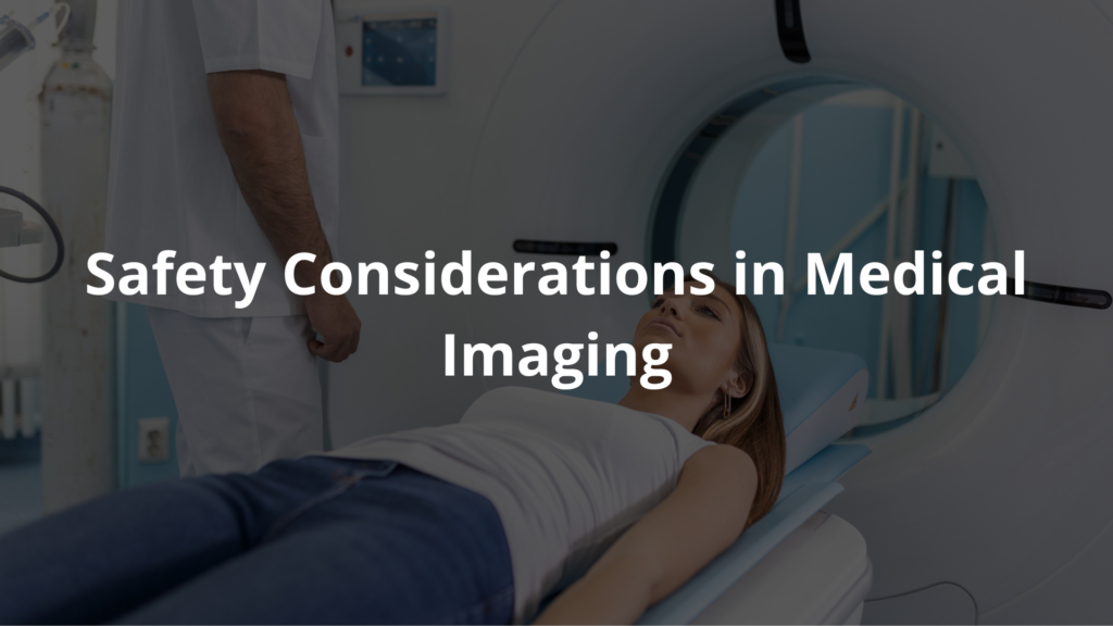
Medical imaging basics: Understand its types and how it revolutionizes safe health diagnostics.
Medical imaging is like a window into the human body, offering doctors a way to figure out what’s going on without making a single cut. It’s a field that blends science with care, where machines like X-rays, MRIs, and ultrasounds become tools for understanding—not just technology.
Each type of imaging has its own story, its own way of helping patients, and its own risks and benefits. But what makes them work? And how do they keep patients safe? These are questions worth exploring. So, stick around—there’s more to this than meets the eye. Literally.
Key Takeaway
- Medical imaging helps doctors see inside the body.
- There are different types of imaging like X-rays, CT, MRI, and ultrasound.
- Patient safety is a top priority when using imaging techniques.
Understanding Types of Medical Imaging
Medical imaging comes in all sorts of forms, and each one works a little differently to take pictures of the inside of the body.
1. X-rays
X-rays are probably the most basic kind of medical imaging. They use something called ionizing radiation (a type of energy) to create pictures. These pictures help doctors see bones and sometimes other parts of the body. The whole process is super quick—usually just a few minutes. Most of the time, patients lie down on a table while the machine takes the images. [1]
Doctors use X-rays a lot. They’re great for spotting broken bones, finding infections, and even checking for some cancers. After the X-ray is taken, the machine saves the image on a special film or a digital screen. This way, doctors can look inside the body without needing to do surgery.
2. Computed Tomography (CT)
CT scans are like a more advanced version of X-rays. Instead of just one picture, a CT scan takes a bunch of X-ray images from different angles. Then, it puts them together to make detailed pictures of the organs and tissues. It’s kind of like slicing a loaf of bread—you can see each layer one at a time.
CT scans are really helpful for finding things like tumors, checking blood vessels, or helping doctors during certain procedures. The whole test usually takes about 10 to 15 minutes. Patients lie on a table that slides into a big, round machine (it looks like a giant doughnut). The machine works fast, snapping pictures so doctors can figure out what’s going on inside the body.
3. Magnetic Resonance Imaging (MRI)
MRI is a way to take pictures of the inside of the body without using radiation. Instead, it relies on strong magnets and radio waves. The images it produces are super detailed, especially for soft tissues like the brain, muscles, and organs. Doctors often use MRIs to find tumors or check for injuries.
The whole process can feel a bit long—it usually takes about 45 minutes to an hour. Patients lie down on a table, and that table slides into a big, tube-shaped machine. While it’s working, the machine makes some pretty loud noises (kind of like banging or knocking). But most people are okay during the scan. They can even listen to music to pass the time. [2]
4. Ultrasound
Ultrasound works differently. It uses sound waves—really high-frequency ones—to create pictures of what’s inside the body. This method is especially common during pregnancy because it lets doctors check on the baby. But it’s also used for other things, like looking at the heart or other organs.
Here’s how it works: A gel is spread on the skin, and then a small device called a transducer is moved over the area being checked. The sound waves bounce off the tissues, and that creates live images on a screen. The whole thing usually takes about 30 minutes. And since there’s no radiation involved, it’s completely safe.
5. Nuclear Medicine
Nuclear medicine is like a detective for the body, using tiny amounts of radioactive material to figure out how organs are working. Sometimes, doctors inject this material into a vein, or patients might drink or swallow it. Once inside, the material (called a tracer) moves through the body and makes certain areas light up on special cameras.
This is especially helpful for spotting cancer or checking how well an organ is doing its job. It’s not a quick process, though. Patients might spend about 1.5 to 2 hours in the testing room because the doctors need to take pictures at different times to see how the tracer moves around.
The Physics Behind Imaging Techniques
Medical imaging isn’t magic—it’s science. Each method works because of specific physical principles that make it possible to see inside the body.
Ionizing Radiation
X-rays and CT scans both use something called ionizing radiation. This kind of radiation can be harmful if you’re exposed to too much of it, so doctors are careful about how often they order these tests. They try to balance getting the information they need while keeping the exposure as low as possible.
Magnetism and Radio Waves
MRI machines are a bit like giant magnets. They use strong magnets to line up tiny hydrogen atoms in your body (we’ve all got them because water has hydrogen). Then, radio waves are sent through the body, and the machine picks up signals from those atoms. These signals are turned into super-clear images. The best part? No radiation is involved.
Sound Waves
Ultrasound is another way to see inside the body, but it uses sound waves instead of radiation. A machine sends sound waves into the body, and when they bounce back, the machine turns them into pictures. It’s a safe option for everyone, even pregnant women, because there’s no radiation at all.
If you’re ever in one of these machines, it might feel a little strange, but knowing how they work can make it less intimidating. Whether it’s magnets, sound waves, or tiny tracers, each method has its own job to do. And they’re all here to help doctors figure out what’s going on inside without needing to cut you open.
Safety Considerations in Medical Imaging

Safety is a big deal when it comes to medical imaging. Different types of tests have different safety levels, and doctors think carefully about these before deciding which one to use.
MRI: This kind of scan is pretty safe for almost everyone. It doesn’t use radiation, which makes it a good option for people who might need extra care, like pregnant women. But there’s a catch—if someone has certain metal implants or devices in their body, an MRI might not be safe because of the strong magnets it uses.
X-rays and CT Scans: These tests do use radiation, so doctors have to be careful. They only order these scans when they really need to figure out something important about a patient’s health. It’s okay for patients to ask their doctor why they need the test and if there are any risks involved.
Knowing a little about how these tests work can make them less scary. Patients should feel free to talk to their doctor about any worries they have. When people understand more about these tests, it can help them feel calmer and more prepared.
FAQ
What makes magnetic resonance imaging different from other medical imaging modalities?
Unlike ray imaging techniques, magnetic resonance imaging uses magnetic fields and radio frequency pulses to create detailed medical images. The rf pulse excites water molecules in soft tissue, producing signals that create high resolution images without radiation exposure. This imaging modality is especially good at showing subtle differences in tissues through t1 weighted and other weighted imaging techniques.
How do computed tomography scans work and what safety measures are in place?
Computed tomography, or CT, uses a rotating ray tube to take multiple radiographic images from different angles. While radiation dose is a concern, modern imaging equipment includes safety features to protect patient safety. The image acquisition process creates detailed 3d images that help doctors see inside the body. Australian health care facilities follow strict guidelines for radiation exposure.
What role does artificial intelligence play in diagnostic imaging today?
AI is transforming image analysis and processing in diagnostic radiology. Machine learning helps with everything from image quality improvement to faster acquisition time. While clinical applications are still being tested through clinical trials, AI shows promise in helping doctors interpret medical images more accurately. Many international journal articles in computer science discuss this emerging technology.
What are the main differences between imaging modalities used in diagnostic radiology?
Each imaging technology serves different clinical applications. Ultrasound imaging uses high frequency sound waves, while nuclear medicine imaging and positron emission tomography use small amounts of radioactive materials. Optical coherence tomography provides detailed images of small structures, while electrical impedance tomography maps tissue conductivity. The choice depends on what kind of images need to be acquired.
How is image data stored and analyzed in modern medical facilities?
Hospitals use specialized image processing systems to handle medical images from various imaging studies. These systems allow for high resolution image analysis and help maintain image quality. While some facilities use autorenew packs for storage, most modern systems integrate with broader health care record systems. The technology continues advancing, as shown in research from google scholar and publications by cambridge university press and crc press.
What advances in imaging techniques are improving diagnostic capabilities?
New developments in spatial resolution and imaging modalities help doctors see more detail in diagnostic imaging. Advanced contrast agents help highlight specific tissues, while faster acquisition times make imaging studies more comfortable. The medicine and biology field continues to advance imaging technology through medical physics research and rigorous clinical trials published in the international journal of medical devices.
Conclusion
Medical imaging is like a window into the body, showing doctors what’s hidden beneath the surface. From X-rays to MRIs, each type has its own purpose (and quirks). Safety’s always a big deal—no one wants unnecessary radiation. As tech keeps advancing, these tools get sharper, faster, and smarter. It’s not just about fancy machines; it’s about helping people heal. If you ever need one, ask questions. Knowing what’s happening can make it less intimidating.
References
- https://blog.radiology.virginia.edu/different-imaging-tests-explained/
- https://udshealth.com/blog/radiology-101-imaging-basics/


