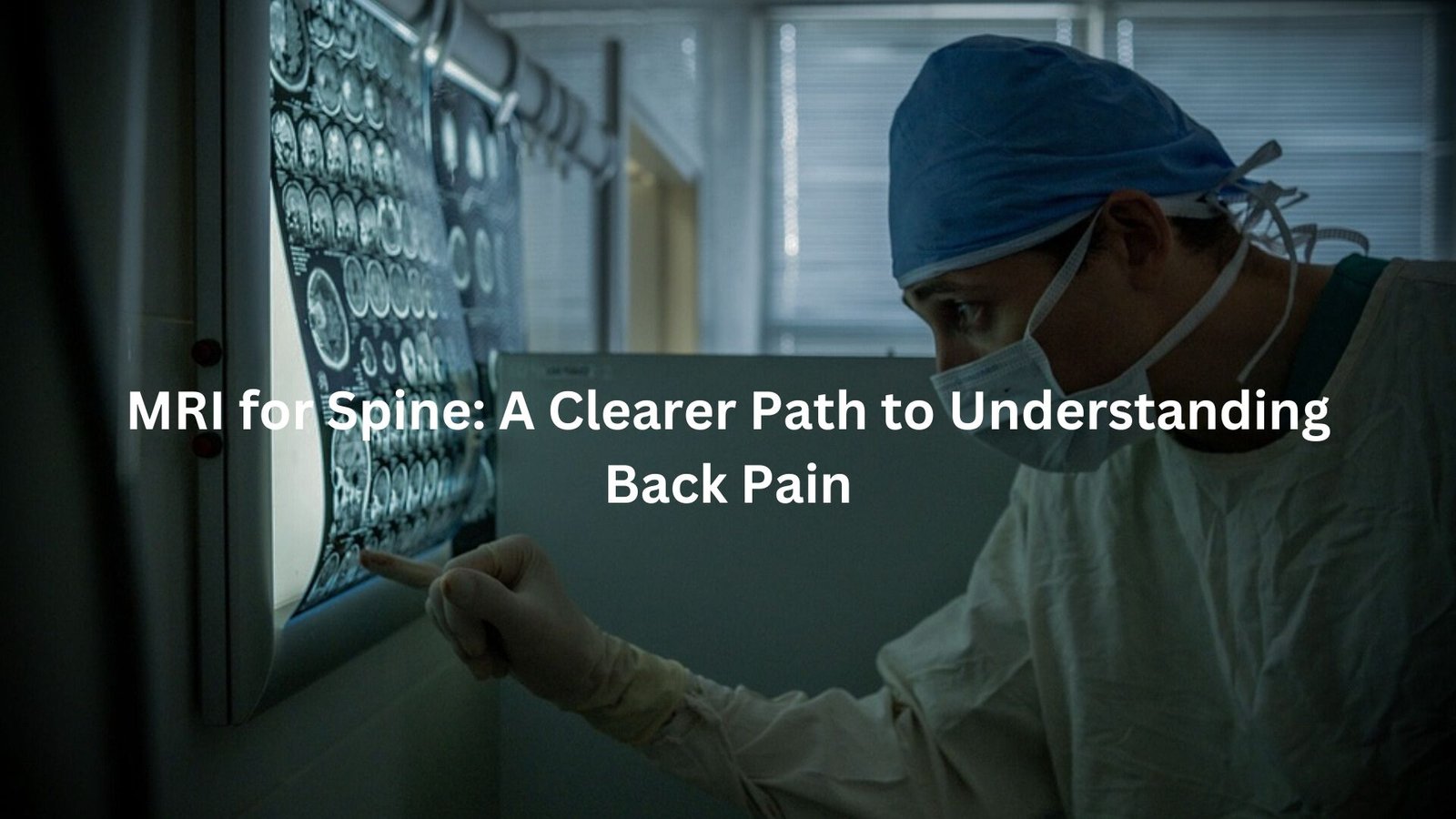
Learn how MRI provides detailed spinal images to help diagnose and treat your back pain.
MRI for the spine is a non-invasive diagnostic tool that offers clear, detailed images of spinal structures like vertebrae, discs, and surrounding tissues. Whether you’re dealing with chronic pain, recovering from an injury, or planning for surgery, MRI scans are essential for accurate diagnosis and effective treatment.
Key Takeaway
- MRI offers non-invasive, high-quality images to diagnose spinal conditions like disc herniation and nerve compression.
- Vital for identifying injuries, tumours, infections, and degenerative diseases affecting the spine.
- MRI scans are safe and reliable, with guidelines in place to protect patients, particularly those with metal implants or medical conditions.
When is MRI for the Spine Recommended?
You’ve probably heard people talk about MRIs before, but you might be wondering when exactly they’re recommended, especially for your spine. Well, it’s not just for those moments when you’ve hurt your back lifting a heavy box or you’re dealing with an aching neck that just won’t go away. Here’s the rundown:
- Chronic Pain with No Clear Cause: If you’ve been battling back or neck pain for ages and the reason isn’t obvious, an MRI might be the next step. It helps your doctor figure out what’s going on inside, even when everything seems fine on the surface.
- Injuries from Trauma: Whether it’s from a car accident, a fall, or some other impact, MRIs can detect damage to the bones, discs, or even the spinal cord itself. This helps to assess how serious the injury is and the best way to treat it.
- For Tumours, Infections, or Degeneration: MRIs are also fantastic for identifying things like tumours or infections that might be causing trouble. Plus, they can spot degenerative conditions, such as arthritis or damage caused by ageing, that might be affecting the spine.
- Pre-Surgical Planning: If surgery is in the cards, especially for something like spinal fusion, doctors use MRIs to get a clear picture of the area that’s about to be worked on. This ensures they know exactly what they’re dealing with before making any decisions. (1)
How MRI Works for Spinal Imaging
When you step into the MRI room, it might feel a bit strange. The machine looks like a big, noisy tunnel, and it’s all about getting you in the right position for clear images of your spine.
- Preparation: Before you get started, you’ll need to take off any metal items, like jewellery or even some body piercings. There’s also a safety questionnaire that’s part of the process – they just want to make sure you’re good to go and safe.
- Positioning: Depending on what’s being scanned, you’ll either go in head-first or feet-first. It’s a bit like lying down for a nap, except you need to stay completely still for the scan. The table you’re on slides into the big MRI machine, and your comfort is key here.
- Scanning Process: The actual scan takes about 20 to 25 minutes. It might sound like a long time, but you’ll be asked to stay as still as possible. Any movement can affect the quality of the images, and that’s what they’re counting on for a clear picture of your spine.
- Contrast Use: Sometimes, a contrast dye is injected before or during the scan to help highlight certain tissues, like blood vessels or soft tissue around your spine. It makes everything stand out better, allowing the radiologist to see any issues more clearly. (2)
MRI Safety Guidelines
Credits: Back In Shape Program
Now, let’s talk about the safety side of things. MRIs are generally very safe, but there are some guidelines that need to be followed to keep things running smoothly.
- Strong Magnetic Field: The MRI machine uses a powerful magnetic field, and because of that, strict access control is necessary. The room where the machine is located (called Zone IV) is restricted, and only trained personnel should be allowed to enter.
- Precautions for Metal: If you’ve got metal implants, body piercings, or certain medical conditions (like pacemakers), you’ll need to have a chat with your doctor beforehand. The magnetic field can affect metal objects in the body, so it’s crucial to disclose any relevant information.
- Pregnancy and MRI: If you’re pregnant or suspect you might be, it’s always a good idea to tell your doctor before having an MRI. While MRIs don’t use radiation like X-rays, they do have powerful magnets that might raise some safety concerns for pregnant women, particularly in the first trimester.
What Conditions Can MRI Diagnose?
MRI scans are incredibly useful for diagnosing a wide range of conditions affecting the spine. Here are just a few of the things an MRI can help spot:
- Disc Herniation: This is when one of the discs in your spine gets damaged, potentially causing pain, numbness, or weakness in the legs.
- Nerve Root Compression: If a nerve is pinched in the spine, it can lead to pain that radiates down the back or limbs. An MRI can pinpoint the exact location of the compression.
- Spinal Stenosis: A narrowing of the spaces within the spine, which can put pressure on the spinal cord and nerves.
- Degenerative Conditions: These include things like osteoarthritis, spinal degeneration, and other wear-and-tear issues. MRIs can identify these problems before they become serious enough to cause lasting damage.
- Injuries: Whether it’s a broken vertebra or damage to the spinal cord, MRIs help doctors get a good look at how bad the injury is.
- Tumours and Infections: MRIs can help detect growths or infections within the spine that might otherwise go unnoticed.
Preparing for Your MRI Scan
Getting ready for an MRI is a pretty simple process, but there are a few things you’ll need to do:
- Remove Metal Objects: Anything metal, like jewellery, keys, and even some body piercings, will need to be taken off. The strong magnetic field in the MRI machine can interfere with metals, so you’ll need to make sure there’s nothing on you that could be affected.
- Dress Comfortably: You’ll be lying down for the scan, so wear something comfortable. You may be asked to change into a hospital gown, but most of the time, comfy clothes will work.
- Talk About Your Medical History: If you’ve had surgery or have metal implants, it’s important to let the radiology staff know. They need to be aware of anything that might affect the scan.
Understanding MRI Reports and Results
Once your scan is over, you might be wondering what happens next. Here’s a bit of insight into how things usually go:
- MRI Report: Your doctor will receive a detailed MRI report that interprets the images. This helps them determine the cause of your pain or injury and decide on the best course of treatment. If you want to check out your report, many clinics offer online access so you can get the details yourself.
- How Doctors Read the Results: Doctors use the MRI images to assess the condition of your spine. They’ll look for things like swelling, damage, and abnormalities. Once they have the full picture, they’ll explain your options for treatment and next steps.
Common Concerns and Misconceptions about MRI
It’s normal to have some concerns about an MRI, but most of them are just misunderstandings. Here’s the truth:
- Metal Implants: You might have heard that metal implants or prosthetic joints can interfere with an MRI. While it’s true that certain metal objects might need to be checked carefully, most modern implants are MRI-safe. Be sure to tell your doctor if you have any implants.
- Contrast Dye Allergies: Some people worry about having an allergic reaction to the contrast dye. While it’s rare, it’s always a good idea to talk to your doctor if you’ve had any reactions to contrast agents in the past.
- MRI Safety: Many people are concerned about MRIs because they don’t understand how they work. The good news is that MRIs are completely non-invasive, and there’s no radiation involved. They’re safe, quick, and reliable.
Advancements in MRI Technology
MRI technology has come a long way in recent years, and things are only getting better.
- Better Image Quality: Modern MRIs provide much higher resolution images than older machines. This means doctors can see more detail and make more accurate diagnoses.
- Faster Scans: With advancements in technology, many MRIs can now be completed more quickly, sometimes in under 20 minutes. That’s great news if you’re not a fan of being inside that noisy tube.
- Future of MRI: Looking ahead, we can expect even faster scans, more detailed images, and better tools for diagnosing spinal conditions. As technology continues to improve, MRI scans will become even more effective and efficient.]
Conclusion
In conclusion, MRI scans are an invaluable tool for diagnosing spine-related issues, from injuries to chronic conditions. By providing detailed, non-invasive images, they help doctors make accurate diagnoses and guide treatment. If you’re dealing with persistent back or neck pain, an MRI could be the key to finding relief.
FAQ
What is Magnetic Resonance Imaging (MRI) used for in diagnosing back pain?
Magnetic Resonance Imaging (MRI) is a non-invasive procedure that creates detailed images of the spine and surrounding soft tissues. MRI uses a strong magnetic field, pulses of radio wave energy, and radio waves to produce high-quality images, helping doctors assess issues like disc herniation, nerve root compression, or degenerative diseases.
Can MRI scans detect spinal injuries?
Yes, MRI scans can detect various types of spinal injuries, including those to the cervical spine, lumbar spine, or spinal column. It can identify conditions like spinal cord injuries, compression fractures, and damage to spinal discs. This diagnostic tool provides a clearer image of any issues affecting the spinal cord or spinal canal.
Are there any safety concerns with MRI scans?
MRI scans are generally considered safe for most people. However, patients with certain medical conditions or metal implants, such as prosthetic joints, surgical clips, or aneurysm clips, should inform their doctor beforehand. The strong magnetic field in the scanner may interact with these objects. Pregnant women should also consult their doctor before undergoing MRI, as imaging during pregnancy may require special precautions.
How does MRI show soft tissues and blood vessels?
MRI uses radio wave energy and strong magnetic fields to create detailed images of soft tissues and blood vessels. It provides a clearer image of these areas compared to other imaging tests. The images can show important details, like nerve root compression, spinal discs, intervertebral discs, and the spinal canal.
Can MRI be used to diagnose degenerative diseases?
Yes, MRI is an effective tool for diagnosing degenerative diseases in the spine, such as spinal stenosis or degeneration of the spinal discs. MRI scans provide detailed images that help doctors identify and evaluate conditions affecting the soft tissues, cerebrospinal fluid, and spinal cord.
References
- https://www.racgp.org.au/afp/2012/november/making-sense-of-mri-of-the-lumbar-spine
- https://sportandspinalphysio.com.au/understanding-your-mri-results-for-lower-back-pain/


